A faster vessel for charting the brain (Nature Communications)

Mouse neuron
Mouse neuron expressing GCaMP3. (Image source: Nature.)

By Morgan Kelly, Office of Communications

Princeton University researchers have created “souped up” versions of the calcium-sensitive proteins that for the past decade or so have given scientists an unparalleled view and understanding of brain-cell communication.

Reported July 18 in the journal Nature Communications, the enhanced proteins developed at Princeton respond more quickly to changes in neuron activity, and can be customized to react to different, faster rates of neuron activity. Together, these characteristics would give scientists a more precise and comprehensive view of neuron activity.

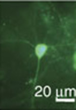
The researchers sought to improve the function of proteins known as green fluorescent protein/calmodulin protein (GCaMP) sensors, an amalgam of various natural proteins that are a popular form of sensor proteins known as genetically encoded calcium indicators, or GECIs. Once introduced into the brain via the bloodstream, GCaMPs react to the various calcium ions involved in cell activity by glowing fluorescent green. Scientists use this fluorescence to trace the path of neural signals throughout the brain as they happen.

GCaMPs and other GECIs have been invaluable to neuroscience, said corresponding author Samuel Wang, a Princeton associate professor of molecular biology and the Princeton Neuroscience Institute. Scientists have used the sensors to observe brain signals in real time, and to delve into previously obscure neural networks such as those in the cerebellum. GECIs are necessary for the BRAIN Initiative President Barack Obama announced in April, Wang said. The estimated $3 billion project to map the activity of every neuron in the human brain cannot be done with traditional methods, such as probes that attach to the surface of the brain. “There is no possible way to complete that project with electrodes, so you have to do it with other tools — GECIs are those tools,” he said.

Despite their value, however, the proteins are still limited when it comes to keeping up with the fast-paced, high-voltage ways of brain cells, and various research groups have attempted to address these limitations over the years, Wang said.

“GCaMPs have made significant contributions to neuroscience so far, but there have been some limits and researchers are running up against those limits,” Wang said.

One shortcoming is that GCaMPs are about one-tenth of a second slower than neurons, which can fire hundreds of times per second, Wang said. The proteins activate after neural signals begin, and mark the end of a signal when brain cells have (by neuronal terms) long since moved on to something else, Wang said. A second current limitation is that GCaMPs can only bind to four calcium ions at a time. Higher rates of cell activity cannot be fully explored because GCaMPs fill up quickly on the accompanying rush of calcium.

The Princeton GCaMPs respond more quickly to changes in calcium so that changes in neural activity are seen more immediately, Wang said. By making the sensors a bit more sensitive and fragile — the proteins bond more quickly with calcium and come apart more readily to stop glowing when calcium is removed — the researchers whittled down the roughly 20 millisecond response time of existing GCaMPs to about 10 milliseconds, Wang said.

The researchers also tweaked certain GCaMPs to be sensitive to different types of calcium ion concentrations, meaning that high rates of neural activity can be better explored. “Each probe is sensitive to one range or another, but when we put them together they make a nice choir,” Wang said.

The researchers’ work also revealed the location of a “bottleneck” in GCaMPs that occurs when calcium concentration is high, which poses a third limitation of the existing sensors, Wang said. “Now that we know where that bottle neck is, we think we can design the next generation of proteins to get around it,” Wang said. “We think if we open up that bottleneck, we can get a probe that responds to neuronal signals in one millisecond.”

The faster protein that the Princeton researchers developed could pair with work in other laboratories to improve other areas of GCaMP function, Wang said. For instance, a research group out of the Howard Hughes Medical Institute reported in Nature July 17 that it developed a GCaMP with a brighter fluorescence. Such improvements on existing sensors gradually open up more of the brain to exploration and understanding, said Wang, adding that the Princeton researchers will soon introduce their sensor into fly and mammalian brains.

“At some level, what we’ve done is like taking apart an engine, lubing up the parts and putting it back together. We took what was the best version of the protein at the time and made changes to the letter code of the protein,” Wang said. “We want to watch the whole symphony of thousands of neurons do their thing, and we think this variant of GCaMPs will help us do that better than anyone else has.”

Read the abstract.

Sun, Xiaonan R., Aleksandra Badura, Diego A. Pacheco, Laura A. Lynch, Eve R. Schneider, Matthew P. Taylor, Ian B. Hogue, Lynn W. Enquist, Mala Murthy and Samuel S.-H. Wang. 2013. Fast GCaMPs for improved tracking of neuronal activity. Nature Communications. Article first published online: July 18, 2013. DOI: 10.1038/ncomms3170

This work was supported by NIH R01 NS045193, (S.S.-H.W.) RC1 NS068414 (L.W.E./S.S.-H.W.), and P40 RR18604 and NS060699 (L.W.E.), a McKnight Technological Innovations Award (S.S.-H.W.), a W.M. Keck Foundation Distinguished Young Investigator award (S.S.-H.W.), an Alfred P. Sloan Research Fellowship, Klingenstein, McKnight, and NSF CAREER Young Investigator awards (M.M.), and an American Cancer Society Postdoctoral Research Fellowship (M.P.T./I.B.H.).

Pupil study reveals learning styles, brain activity (Nature Neuroscience)

Test of learning styles experiment
To test people’s learning styles, participants were presented with a choice between two images (objects or words) and rewarded according to their choices. In this exercise, to maximize reward, participants had to learn by trial and error that office-related images provide a higher reward than food-related images (semantic, or related to meaning), and that grayscale images provide a higher reward than color images (visual features). Image credit: Nature Neuroscience.

By Catherine Zandonella, Office of the Dean for Research

People are often said to have “learning styles” – for example, some people pay attention to visual details while others grab onto abstract concepts and meanings. A new study from Princeton University researchers found that changes in pupil size can reveal whether people are learning using their dominant learning style, or whether they are learning in modes outside of that style.

The researchers found that pupil dilation was smaller when people learned using their usual style and larger when people diverged from their normal style. The study was published in the journal Nature Neuroscience.

The study compared brain activity in individuals with two different learning styles – those who learn best by absorbing concrete visual details and those who are better at learning abstract concepts or meanings.

“We showed that changes in pupil dilation are associated with the degree to which learners use the style for which they have a predisposition,” said Eldar Eran, a graduate student in the Princeton Neuroscience Institute, who led the study.

The researchers used changes in pupil size as an indicator of variations in “neural gain,” which can be thought of as an amplifier of neural communication: when gain is increased, excited neurons become even more active and inhibited neurons become even less active. Smaller pupil dilation indicates more neural gain and larger pupil dilation indicates less neural gain.

The team showed that neural gain was correlated with different modes of communication between parts of the brain. In studies of human volunteers undergoing brain scans, when neural gain was high, communication tended to be tightly concentrated in certain regions of the brain that govern specific tasks, whereas low neural gain is associated with communication across wider regions of the brain.

“We showed that the brain has different modes of communication,” said Yael Niv, assistant professor of psychology and the Princeton Neuroscience Institute, “one mode where everything talks to everything else, and another mode where communication is more segregated into areas that don’t talk to each other.” The study also involved Jonathan Cohen, Princeton’s Robert Bendheim and Lynn Bendheim Thoman Professor in Neuroscience.

These modes are linked to the level of neural gain and to learning style, Niv said. Neural gain can be thought of as a contrast amplifier that increases intensity of both activation and inhibition of communication among brain areas. “If one area is trying to activate another, or trying to inhibit another – both effects are stronger, everything is more potent,” she said. “This is correlated with communication being segregated into clusters of activation in the brain, so each network is talking to itself loudly, but connections across networks are inhibited. In situations of lower gain, however, the areas can talk to each other across networks, so information flows more globally.”

“These two modes [of communication in the brain] seem to be associated with different constraints on learning,” she said. “According to our study, in the mode where everything talks to everything, learning is very flexible. In contrast, in the mode where communication is stronger and more focused, but also more segregated between brain areas, subjects were more true to their personal learning style. Neither of these modes are better than the other – in both cases participants were equally successful in the task, but in different ways.”

“We interpreted these results to mean that although we tend to have a dominant learning style, we are not a slave to that style, and when operating in the proper mode, we can overcome dominant styles to learn in other ways,” she said.

This research was funded by NIH grants R03 DA029073 and R01 MH098861, a Howard Hughes Medical Institute International Student Research fellowship, and a Sloan Research Fellowship. The authors also wish to thank the generous support of the Regina and John Scully Center for the Neuroscience of Mind and Behavior within the Princeton Neuroscience Institute.

Read the article

Eldar, Eran, Jonathan D Cohen & Yael Niv. 2013. The effects of neural gain on attention and learning.  Nature Neuroscience. Published online June 16, 2013 doi:10.1038/nn.3428

New imaging technique provides improved insight into controlling the plasma in fusion experiments (Plasma Physics and Controlled Fusion)

Graphic of fluctuating electron temperatures
Graphic representation of 2D images of fluctuating electron temperatures in a cross-section of a confined fusion plasma. (Image source: Plasma Physics and Controlled Fusion)

By John Greenwald, Office of Communications, Princeton Plasma Physics Laboratory

A key issue for the development of fusion energy to generate electricity is the ability to confine the superhot, charged plasma gas that fuels fusion reactions in magnetic devices called tokamaks. This gas is subject to instabilities that cause it to leak from the magnetic fields and halt fusion reactions.

Now a recently developed imaging technique can help researchers improve their control of instabilities. The new technique, developed by physicists at the U.S. Department of Energy’s Princeton Plasma Physics Laboratory (PPPL), the University of California-Davis and General Atomics in San Diego, provides new insight into how the instabilities respond to externally applied magnetic fields.

This technique, called Electron Cyclotron Emission Imaging (ECEI) and successfully tested on the DIII-D tokamak at General Atomics, uses an array of detectors to produce a 2D profile of fluctuating electron temperatures within the plasma. Standard methods for diagnosing plasma temperature have long relied on a single line of sight, providing only a 1D profile. Results of the ECEI technique, recently reported in the journal Plasma Physics and Controlled Fusion, could enable researchers to better model the response of confined plasma to external magnetic perturbations that are applied to improve plasma stability and fusion performance.

PPPL is managed by Princeton University.

Read the abstract.

B.J. Tobias; L.Yu; C.W. Domier; N.C. Luhmann, Jr; M.E. Austin; C. Paz-Soldan; A.D. Turnbull; I.G.J. Classen; and the DIII-D Team. 2013. Boundary perturbations coupled to core 3/2 tearing modes on the DIII-D tokamak. Plasma Physics and Controlled Fusion. Article first published online: July 5, 2013. DOI:10.1088/0741-3335/55/9/095006

This work was supported in part by the US Department of Energy under DE-AC02- 09CH11466, DE-FG02-99ER54531, DE-FG03-97ER54415, DE-AC05-00OR23100, DE- FC02-04ER54698, and DE-FG02-95ER54309.