“Kurly” protein keeps cilia moving, oriented in the right direction (Cell Reports)

Cilia with a mutant form of the Kurly protein are wild and disorganized.

By Catherine Zandonella, Office of the Dean for Research

A new study of a protein found in cilia – the hair-like projections on the cell surface – may help explain how genetic defects in cilia play a role in developmental abnormalities, kidney disease and a number of other disorders.

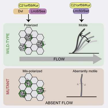
The researchers at Princeton University and Northwestern University found that the protein, which goes by the name C21orf59 or “Kurly,” is needed for cilia to undulate to keep fluid moving over the surface of cells. They also found that the protein is needed during development to properly orient the cilia so that they are facing the right direction to move the fluid.

“It’s extremely exciting that we’ve found a single protein that is responsible for these two functions – orientation and motility – in cilia,” said Rebecca Burdine, an associate professor of molecular biology at Princeton University. “Despite their importance in human disease, very little is known about how cilia motility and orientation are coordinated, so this protein will provide an important gateway into looking at this process.” The finding is published online and in the March 1 issue of the journal Cell Reports.

Kurly panels
Caption: Staining of cilia (hair-like projections in green and nuclei in blue) in zebrafish kidney tubules show cilia are disorganized and oriented incorrectly in fish with mutated Kurly protein (bottom panel) versus normal Kurly (top panel). Image courtesy of the Burdine lab.

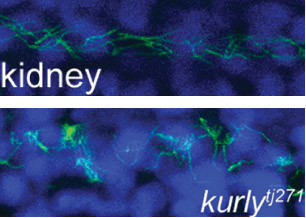
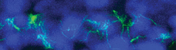
The studies were conducted in zebrafish at Princeton and in African clawed frogs (Xenopus laevis) at Northwestern. In the zebrafish kidney, the researchers found that the Kurly protein enabled cilia to orient themselves in a uniform direction, and most importantly, in the proper direction to facilitate the flow of fluid along the narrow channels in the kidney. In frogs, the cilia on skin cells help move fluid along the surface of the animal during its larval stage. In both cases, knocking out the gene for Kurly caused the cilia to orient incorrectly thereby losing their ability to move in the waving fashion that helps push fluid along.

The discovery of Kurly’s role in cilia movement and orientation stemmed from work in the Burdine lab on fetal organ development, specifically an investigation of mutations that alter the left-right asymmetric orientation of the heart. Such mutations can result in an organ that is working properly but is an exact mirror image of a normal heart. During a search for genes involved in this left-right patterning, the Burdine team discovered that mutations in a gene they called kur, which codes for the Kurly protein, were linked to errors in left-right orientation in zebrafish heart.

When the kurly protein is mutated, the cilia cannot orient and move properly.
Image credit: Burdine lab

As the team investigated kur, they noted that the mutation also affected the function of cilia. It has been known for some time that cilia are important for a number of jobs, from sensing the environment to facilitating fluid flow, to ensuring that the lungs excrete inhaled contaminants. Cilia genetic defects are linked to a number of human diseases, including polycystic kidney disease, respiratory distress, hearing loss, infertility, and left-right patterning disorders such as the one Burdine studies.

Researchers in Burdine’s laboratory found that Kurly’s role in cilia movement stems from its ability to ensure proteins called dynein arms are correctly located in the cilia. When the researchers knocked out the kur gene, the dynein proteins failed to form in the proper location.

The finding that a single protein is involved in both movement and orientation is surprising, said co-first author Daniel Grimes, a postdoctoral research associate in the Burdine lab. “These are two aspects that are both required to generate fluid flow, and we’d like to know how they are linked molecularly. This work adds a new gene that aids this discovery.”

The gene for Kurly has also been detected in relation to human cilia disorders, so the work may have an impact on understanding the mechanisms of human disease, Grimes added. The researchers also found that the mutation they discovered rendered the Kurly protein sensitive to temperature, and used this trait to find that the Kurly protein may be involved in initiating movement rather than keeping the cilia moving once they’ve started.

The team also explored proteins that interact with Kurly. The Northwestern team showed that when the kur gene was inactivated using a gene-editing technique called CRISPR-Cas9, the lack of a functioning Kurly protein led to the mis-positioning of a second protein on the cell surface called Prickle2, which helps cells know which direction they face. Without proper Prickle2 positioning, the cilia pushed fluid in the wrong direction.

The study of the Kurly protein involved Grimes as well as two additional co-authors, Kimberly Jaffe and Jodi Schottenfeld-Roames, a former postdoctoral researcher and graduate student respectively, in the Burdine lab. The initial studies on the Kurly protein were conducted as part of an undergraduate research project by Tse-shuen (Jade) Ku, Class of 2007. Additional work was contributed by Nicholas Morante and José Pelliccia, graduate students in the Burdine lab.

The work at Northwestern University was performed in the laboratory of Brian Mitchell with the assistance of Michael Werner and Sun Kim.

The research was supported by a National Institutes of Health (NIH) Ruth L. Kirschstein Institutional National Research Service Award grant to K. Jaffe (#1F32HD060396-01A1), an NIH National Institute of General Medical Sciences grant to B. Mitchell (#2R01GM089970), and an NIH Eunice Kennedy Shriver National Institute of Child Health and Human Development grant to R. Burdine (#2R01HD048584).

Read the article.

Kimberly M. Jaffe, Daniel T. Grimes, Jodi Schottenfeld-Roames, Michael E. Werner, Tse-Shuen J. Ku, Sun K. Kim, Jose L. Pelliccia, Nicholas F.C. Morante, Brian J. Mitchell, Rebecca D. Burdine.c21orf59/kurly controls both cilia motility and polarization. Cell Reports (2016), http://dx.doi.org/10.1016/j.celrep.2016.01.069. In Press Corrected Proof.


Study reveals mechanism behind enzyme that tags unneeded DNA (Nature Chem. Bio.)

Designer chromatin experiments
Graphical representation of designer chromatin experiments. Image courtesy of the Muir lab.

By Tien Nguyen, Department of Chemistry

Researchers have discovered the two-step process that activates an essential human enzyme, called Suv39h1, which is responsible for organizing large portions of the DNA found in every living cell.

For any particular cell, such as a skin or brain cell, much of this genetic information is extraneous and must be packed away to allow sufficient space and resources for more important genes. Failure to properly pack DNA jeopardizes the stability of chromosomes and can result in severe diseases. Suv39h1 is one of the main enzymes that chemically mark the irrelevant regions of DNA to be compacted by cellular machinery, but little is known about how it installs its tag.

Now, scientists at Princeton have used ‘designer chromatin’ templates – highly customized replicas of cellular DNA and histone proteins, the scaffolding proteins around which DNA is wrapped – to reveal new details about Suv39h1’s mechanism. The researchers investigated how Suv39h1 employs a positive feedback loop to chemically tag thousands of adjacent histones, thus signaling the cell to stow away these underlying, unnecessary DNA sequences. The work was published in in the journal Nature Chemical Biology.

“One of the things that has always fascinated me about feedback loops is that they’re super dangerous. If you make a mistake once, you end up getting reinforcement through the feedback loop,” said Manuel Müller, a postdoctoral researcher in the Muir lab and lead author on the study. “So how does Suv39h1 keep itself in check?”

Suv39h1 had been known to possess two distinct parts, but the new research revealed how they work together in order to ‘switch on’ the enzyme. One part of the enzyme, known as the chromodomain, is constantly exposed and seeks out specific chemical tags, known as a methyl groups, located at predetermined sites on histones. When the chromodomain finds these groups in the genome, it locks onto the spot and allows the other part, the enzymatic core, to install more methyl tags at adjacent histones.

“The second, anchoring step wasn’t really known before. It provides an extra level of control and allows the process to be extremely fine-tuned,” Müller said. A similar mechanism may be employed by many other enzymes operating on chromatin, given that they contain similar components of a feedback loop.

To understand how the enzyme carries out this process, the researchers synthesized complex chromatin templates that were three times larger than previously reported models. They divided the template into three blocks that could each be manipulated in various ways. For example, a block could be prepared with the chemical tag present, absent or mutated such that tagging can’t occur. “The different blocks should signal to the enzyme either start here or feel free to spread here or absolutely stop here,” said Glen Liszczak, a co-author and postdoctoral researcher in the Muir lab.

By rearranging the various domains, the research team observed where the enzyme spread its mark across the genome. They found that Suv39h1 preferred to spread across small distances, but that it could reach sequences further along if chromatin folding decreased the physical distance in space.

“We’ve learned something new about this enzyme, something that we couldn’t have without the pinpoint precision that the designer chromatin offers,” Liszczak said. “There are a lot of questions that our lab has been interested in that we can now start to answer.”

The research was funded by the Swiss National Science Foundation (postdoctoral fellowships) and the US National Institutes of Health (R01-GM107047).

Read the abstract or full article.

Müller, M. M.; Fierz, B.; Bittova, L.; Liszczak, G.; Muir, T. W. “A two-state activation mechanism controls the histone methyltransferase Suv39h1.” Nature Chem. Bio. Available online January 25, 2016.


Antibiotic’s killer strategy revealed (PNAS)

Marine algae
Satellite image of a E. huxleyi marine algae bloom. (Image: NASA)

By Tien Nguyen, Department of Chemistry

Using a special profiling technique, scientists at Princeton have determined the mechanism of action of a potent antibiotic, known as tropodithietic acid (TDA), leading them to uncover its hidden ability as a potential anticancer agent.

TDA is produced by marine bacteria belonging to the roseobacter family, which exist in a unique symbiosis with microscopic algae. The algae provide food for the bacteria, and the bacteria provide protection from the many pathogens of the open ocean.

“This molecule keeps everything out,” said Mohammad Seyedsayamdost, an assistant professor of chemistry at Princeton and corresponding author on the study published in the Proceedings of the National Academy of Science. “How could something so small be so broad spectrum? That’s what got us interested,” he said.

In collaboration with researchers in the laboratory of Zemer Gitai, an associate professor of molecular biology at Princeton, the team used a laboratory technique referred to as bacterial cytological profiling to investigate the mode of action of TDA. This method involves destroying bacterial cells with the antibiotic in the presence of a set of dyes, and then visually assessing the aftermath. “The key assumption is that dead cells that look the same probably died by the same mechanism,” he said.

marine algae
Scanning electron microscope image of E. huxleyi (Image credit. M. Seyedsayamdost)

The team used three dyes to evaluate 13 different features of the deceased cells, such as cell membrane thickness and nucleoid area, comprising TDA’s cytological profile. By comparing to profiles of known drugs, the researchers found a match with a class of compounds called polyethers, which possess anticancer activity.

Given their similar profiles, Seyedsayamdost and coworkers hypothesized that TDA might exhibit anticancer properties as well, and indeed observed its strong anticancer activity in a screen against 60 different cancer cell lines. “The strength of this profiling technique is that it tells you how to repurpose molecules,” Seyedsayamdost said.

The researchers were surprised by the compounds’ shared mode of action because unlike the small sized TDA, polyether compounds are quite large. But through different chemical reactions, they are both able to cause chemical disruptions in the cell membrane that render the bacterium unable to produce the energy needed to perform critical tasks, such as cell division and making proteins.

In addition to TDA’s killing mechanism, the researchers were interested in understanding the mechanism by which a bacterial strain could become resistant to the antibiotic. Particularly, they wondered how the marine roseobacter kept itself safe from the deadly antibiotic weapon that it produced.

The research team approached the task by probing the genes in roseobacter that synthesize TDA as well as the surrounding genes. They identified three nearby genes responsible for transport in and out of the cell, and upon transferring these specific genes to E. coli, were able to produce an elusive TDA resistant bacterial strain.

“We often look at natural products as black boxes,” said Seyedsayamdost, “but these molecules have evolved for millennia to fulfill a certain function. By linking the unusual structural features of TDA to its mode of action, we have begun to explain why TDA looks the way it does.”

Read the abstract:

Wilson, M. Z.; Wang, R.; Gitai, Z.; Seyedsayamdost, M. R. “Tropodithietic Acid: Mode of Action and Mechanism of Resistance.” Proc. Natl. Acad. Sci. 2016, Published online on January 22, 2016.

This work was supported by grants from the National Institutes of Health (GM 098299 and 1DPOD004389).

Fruit flies adjust their courtship song based on distance (Neuron)

A fly runs on an air-supported ball. The audio traces of the fly’s courtship song are shown.

Article courtesy of Joseph Caputo, Cell Press

Outside of humans, the ability to adjust the intensity of acoustic signals with distance has only been identified in songbirds. Research published February 3 in Neuron now demonstrates that the male fruit fly also displays this complex behavior during courtship, adjusting the amplitude of his song depending on how far away he is from a female. Studying this process in the fruit fly can help shed light on the building blocks for social interactions across the animal kingdom.

Mala Murthy, of Princeton University, and her colleagues have revealed an unanticipated level of control in insect acoustic communication by analyzing each stage of the neuronal pathway underlying male fruit flies’ ability to adjust their courtship song—from the visual cues that help estimate distance to the signals that pass through nervous system and cause changes in muscle activity that drive softer or louder song. The complexity is remarkable considering that the fruit fly has only 100,000 neurons, one-millionth that of a human brain.

During courtship, males chase females, extending and vibrating one wing at a time to produce a courtship song. Songs, which consist primarily of two modes: sine and pulse, are extremely quiet and must be recorded on sensitive microphones, then amplified more than 1 million times in order to be heard by humans. When amplified, the sine song sounds like the whine of an approaching mosquito, while the pulse song is more akin to a cat’s purr.

“Females listen to many minutes of male song before deciding whether to accept him,” says Murthy, of the Princeton Neuroscience Institute and Department of Molecular Biology. “There is thus enormous evolutionary pressure for males to optimize their song to match the female’s preference while simultaneously minimizing the energetic cost of singing for long periods of time.” Adjusting the amplitude of song to compensate for female distance allows males to conserve energy and thereby court for longer periods of time and better compete with other males.

“While the precise neural mechanisms underlying the generation and patterning of fly song may be distinct from humans or even songbirds, the fundamental problem is the same: how can a neural network produce such a complex and dynamic signal?” Murthy says. “For this reason, we anticipate that similar neural mechanisms will be employed in all systems, and the genetic model system of the fruit fly is an ideal starting point from which to dissect them.”

The researchers were funded by the Howard Hughes Medical Institute, the DAAD (German Academic Exchange Foundation), the Alfred P. Sloan Foundation, the Human Frontiers Science Program, a National Science Foundation CAREER award, a NIH New Innovator Award, the NSF BRAIN Initiative, an EAGER award, the McKnight Foundation, and the Klingenstein-Simons Foundation.

Read the abstract

Philip Coen, Marjorie Xie, Jan Clemens and Mala Murthy. Sensorimotor Transformations Underlying Variability in Song Intensity during Drosophila Courtship. Neuron. Vol. 89, Issue 3, p629–644, 3 February 2016.