Birds and beans: Which type of coffee is best for bird diversity?

green coffee beans

Article courtesy of Stephen Sautner, Wildlife Conservation Society

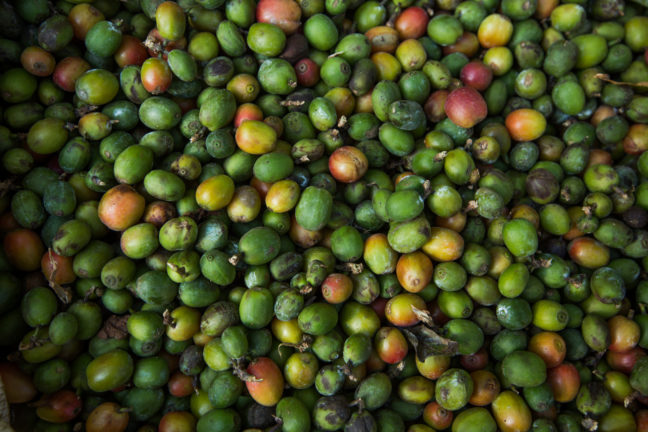
It’s an age-old debate for coffee lovers.  Which is better: Arabica beans with their sweeter, softer taste, or the bold, deep flavor of Robusta beans? A new study by the Wildlife Conservation Society (WCS), Princeton University, and the University of Wisconsin-Madison appearing in the journal Scientific Reports has taken the question to unlikely coffee aficionados: birds.

Green and red parakeet
Alexandrine parakeet (Psittacula eupatria). Credit Manish Kumar

The researchers, led by WCS Associate Conservation Scientist Krithi Karanth, surveyed bird diversity in coffee agroforests in India’s Western Ghats region. Previous research has demonstrated that shade-grown coffee (typically Arabica) can harbor substantial levels of biodiversity. But coffee production is globally shifting toward Robusta, which uses a more intensive full-sun agricultural systems, which may pose deleterious impacts for forest wildlife.

What the researchers found was surprising: although Arabica avian assemblages were more species rich, Robusta nevertheless offered substantial biodiversity benefits, and supported higher densities of several sensitive avian populations such as frugivores, birds that primarily eat fruit. In addition, farmers use less pesticides in the more disease-resistant Robusta farmlands.

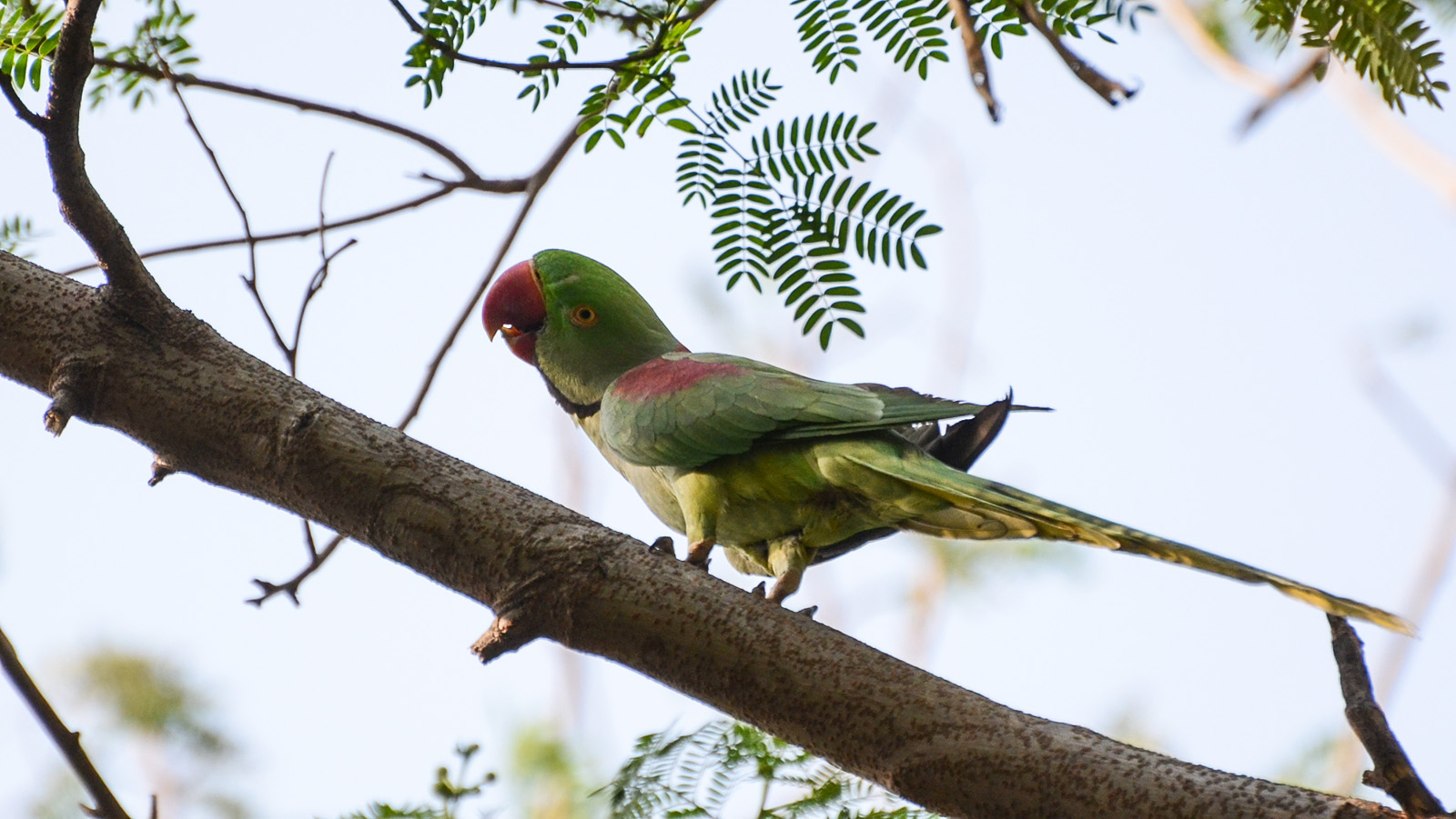
The authors found a total of 79 forest-dependent species living in the coffee plantations they surveyed, including three species listed by the International Union for Conservation of Nature as threatened: Alexandrine parakeet (Psittacula eupatria), grey-headed bulbul (Pycnonotus priocephalus) and the Nilgiri wood pigeon (Columba elphinstonii). Plantations can harbor a diversity of mammals, amphibians and tree species, too.

The study has important implications as coffee production is an increasingly important driver of landscape transformation, and shifts between different coffee bean species are a major dimension of agroforestry trends. The authors say that coffee certification efforts should prioritize maintaining native canopy shade trees to ensure that coffee landscapes can continue providing biodiversity benefits.

Said Dr. Karanth, “Coffee farms already play a complementary role to protected areas in a country like India where less than four percent of land is formally protected. Therefore, building partnerships with largely private individual and corporate land holders will provide much needed safe-passage and additional habitats for birds and other species.”

Indian robusta has relatively high “cup scores” (flavor ratings) by coffee experts, is disease-resistant, and commands a price premium.

Said lead author Charlotte Chang, who analyzed the data while a graduate student in the Department of Ecology and Evolutionary Biology at Princeton University and is now a postdoctoral researcher at the National Institute for Mathematical and Biological Synthesis at the University of Tennessee, Knoxville: “An encouraging result of the study is that coffee production in the Western Ghats, a global biodiversity hotspot, can be a win-win for birds and farmers.”

Chang’s advisers at Princeton were Simon Levin,the James S. McDonnell Distinguished University Professor in Ecology and Evolutionary Biology, and David Wilcove, professor of ecology and evolutionary biology and public affairs and the Princeton Environmental Institute. The senior author on the paper is Paul Robbins, director of the Nelson Institute for Environmental Studies at the University of Wisconsin-Madison.

Funding for this project was provided by the National Science Foundation (grant number 1265223 and a Graduate Research Fellowship), Oracle, and Graduate Research Opportunities Worldwide with USAID.

The study, “Birds and beans: Comparing avian richness and endemism in arabica and robusta agroforests in India’s Western Ghats,” by Charlotte H. Chang, Krithi K. Karanth and Paul Robbins, was published in the journal Scientific Reports online on Feb. 16, 2018. DOI 10.1038/s41598-018-21401-1.

Genetic instructions from mom set the pattern for embryonic development

Micrograph of a zebrafish organ called the Kupffer's vesicle

By the Department of Molecular Biology

A new study indicates an essential role for a maternally inherited gene in embryonic development. The study found that zebrafish that failed to inherit specific genetic instructions from mom developed fatal defects earlier in development, even if the fish could make their own version of the gene. The study by researchers at Princeton University was published Nov. 15 in the journal eLife.

When female animals form egg cells inside their ovaries, they deposit messenger RNAs (mRNAs) – a sort of genetic instruction set – in the egg cell cytoplasm. After fertilization, these maternally supplied mRNAs can be translated into proteins required for the early stages of embryonic development, before the embryo is able to produce mRNAs and proteins of its own.

More than thirty years ago, researchers discovered that mRNAs encoding a protein called Vg1 are deposited in the cytoplasm of frog eggs. “vg1 is famous for being one of the first recognized maternal mRNAs,” said Rebecca Burdine, associate professor of molecular biology at Princeton. “Many papers have been written on how this RNA is localized and regulated, but it was never clear what the Vg1 protein actually does in the developing embryo.”

Two zebrafish embryos
Compared to a normal zebrafish embryo (right), an embryo lacking gdf3 (left) inherited from mom shows major defects resulting from its inability to form mesoderm and endoderm cells early in development. Credit: Pelliccia et al., 2017.

In the study, Burdine and two graduate students Jose Pelliccia and Granton Jindal used CRISPR/Cas9 gene editing to remove Vg1, known as Gdf3 in zebrafish. Embryos that couldn’t produce any Gdf3 of their own–but received a healthy portion of the gdf3 mRNA from their mothers–developed perfectly normally. But embryos that didn’t receive maternal gdf3 mRNA showed major defects early on in their development, dying just three days after fertilization.

“If gdf3 is not supplied to the egg by the mother, the fertilized egg cannot produce two of the three major types of cells required for development,” Burdine said. “The embryos lack all [cell types known as] mesoderm and endoderm and are left with skin and some neural tissue, [which derive from the third major cell type, the ectoderm].”

Vg1/Gdf3 is a member of the TGF-beta family of cell-signaling molecules. Two other members of this family, Ndr1 and Ndr2, are required to form the mesoderm and endoderm early in zebrafish development. Embryos lacking maternally supplied gdf3 look very similar to embryos lacking both of these proteins, which are analogous to the Nodal 1 and 2 proteins in mammals.

The researchers found that maternal gdf3 is required for Ndr1 and Ndr2 to signal at the levels necessary to properly induce the formation of mesoderm and endoderm cells in early zebrafish embryos. In the absence of gdf3, Ndr1 and Ndr2 signaling is dramatically reduced and embryonic development goes awry.

Nodal signaling is also required later in zebrafish development when it helps to establish differences between the left and right sides of the developing embryo. It does this, in part, by directing the formation of an organ known as Kupffer’s vesicle, whose asymmetric shape helps determine the embryo’s left and right sides. Subsequently, Nodal signaling induces the expression of a third Nodal protein, called southpaw, in a group of mesoderm cells on the left-hand side of the embryo.

To investigate whether maternally supplied gdf3 mRNA also plays a role in left-right patterning, the researchers used a series of experimental tricks to supply embryos with enough Gdf3 protein to form the mesoderm and endoderm and survive until the later stages of embryonic development.

As predicted, these embryos showed defects in left-right patterning. Their Kupffer’s vesicles were abnormally symmetric in shape, and southpaw expression was greatly reduced, suggesting that gdf3 is also required for optimal Nodal signaling during later stages of embryonic development. At this stage, however, embryonic gdf3 seems to be capable of doing the job if maternally supplied gdf3 is absent.

Nodal and Vg1 proteins are known to bind to each other in other species. “Thus, we hypothesize that Gdf3 combines with Ndr1 and Ndr2 to facilitate Nodal signaling during zebrafish development, acting as an essential factor in embryonic patterning,” said Pelliccia, a graduate student in molecular biology. Co-author Jindal earned his Ph.D. in chemical and biological engineering in 2017.

At the same time as Burdine and colleagues, two other research groups, led by Joe Yost at the University of Utah and Alex Schier at Harvard University, made similar findings on the role of gdf3 during zebrafish development. “All three groups worked together to co-submit and co-publish in eLife, allowing the students involved to all get credit for their hard work,” Burdine said. “It’s a great example of how science should be done.”

###

The research was funded by the Eunice Kennedy Shriver National Institute of Child Health and Human Development (grant R01HD048584) and the National Science Foundation (graduate research fellowship DGE 1148900).

Citation: Pelliccia, J.L., G.A. Jindal, and R.D. Burdine. Gdf3 is required for robust Nodal signaling during germ layer formation and left-right patterning. eLife. 6: e28635 (2017). DOI: 10.7554/eLife.28635

Spotting the spin of the Majorana fermion under the microscope

Majorana detection
The figure shows a schematic of the experiment. A magnetized scanning tunneling microscope tip was used to probe the spin property of the quantum wave function of the Majorana fermion at the end of a chain of iron atoms on the surface of a superconductor made of lead. Image courtesy of Yazdani Lab, Princeton University.

By Catherine Zandonella, Office of the Dean for Research

Researchers at Princeton University have detected a unique quantum property of an elusive particle notable for behaving simultaneously like matter and antimatter. The particle, known as the Majorana fermion, is prized by researchers for its potential to open the doors to new quantum computing possibilities.

In the study published this week in the journal Science, the research team described how they enhanced an existing imaging technique, called scanning tunneling microscopy, to capture signals from the Majorana particle at both ends of an atomically thin iron wire stretched on the surface of a crystal of lead. Their method involved detecting a distinctive quantum property known as spin, which has been proposed for transmitting quantum information in circuits that contain the Majorana particle.

“The spin property of Majoranas distinguishes them from other types of quasi-particles that emerge in materials,” said Ali Yazdani, Princeton’s Class of 1909 Professor of Physics.  “The experimental detection of this property provides a unique signature of this exotic particle.”

The finding builds on the team’s 2014 discovery, also published in Science, of the Majorana fermion in a single atom-wide chain of iron atoms atop a lead substrate. In that study, the scanning tunneling microscope was used to visualize Majoranas for the first time, but provided no other measurements of their properties.

“Our aim has been to probe some of the specific quantum properties of Majoranas. Such experiments provide not only further confirmation of their existence in our chains, but open up possible ways of using them.” Yazdani said.

First theorized in the late 1930s by the Italian physicist Ettore Majorana, the particle is fascinating because it acts as its own antiparticle. In the last few years, scientists have realized that they can engineer one-dimensional wires, such as the chains of atoms on the superconducting surface in the current study, to make Majorana fermions emerge in solids.  In these wires, Majoranas occur as pairs at either end of the chains, provided the chains are long enough for the Majoranas to stay far enough apart that they do not annihilate each other. In a quantum computing system, information could be simultaneously stored at both ends of the wire, providing a robustness against outside disruptions to the inherently fragile quantum states.

Previous experimental efforts to detect Majoranas have used the fact that it is both a particle and an antiparticle. The telltale signature is called a zero-bias peak in a quantum tunneling measurement. But studies have shown that such signals could also occur due to a pair of ordinary quasiparticles that can emerge in superconductors. Professor of Physics Andrei Bernevig and his team, who with Yazdani’s group proposed the atomic chain platform, developed the theory that showed that spin-polarized measurements made using a scanning tunneling microscope can distinguish between the presence of a pair of ordinary quasi-particles and a Majorana.

Typically, scanning tunneling microscopy (STM) involves dragging a fine-tipped electrode over a structure, in this case the chain of iron atoms, and detecting its electronic properties, from which an image can be constructed.  To perform spin-sensitive measurements, the researchers create electrodes that are magnetized in different orientations. These “spin-polarized” STM measurements revealed signatures that agree with the theoretical calculations by Bernevig and his team.

“It turns out that, unlike in the case of a conventional quasi-particle, the spin of the Majorana cannot be screened out by the background. In this sense it is a litmus test for the presence of the Majorana state,” Bernevig said.

The quantum spin property of Majorana may also make them more useful for applications in quantum information. For example, wires with Majoranas at either end can be used to transfer information between far away quantum bits that rely on the spin of electrons. Entanglement of the spins of electrons and Majoranas may be the next step in harnessing their properties for quantum information transfer.

The STM studies were conducted by three co-first authors in the Yazdani group: scientist Sangjun Jeon, graduate student Yonglong Xie, and former postdoctoral research associate Jian Li (now a professor at Westlake University in Hangzhou, China).  The research also included contributions from postdoctoral research associate Zhijun Wang in Bernevig’s group.

This work has been supported by the Gordon and Betty Moore Foundation as part of the EPiQS initiative (grant GBMF4530), U.S. Office of Naval Research (grants ONR-N00014-14-1-0330, ONR-N00014-11-1-0635, and ONR- N00014-13-1-0661) , the National Science Foundation through the NSF-MRSEC program (grants DMR-142054 and DMR-1608848) and an EAGER Award (grant NOA -AWD1004957), the U.S. Army Research Office MURI program (grant W911NF-12-1-046), the U.S. Department of Energy Office of Basic Energy Sciences, the Simons Foundation, the David and Lucile Packard Foundation, and the Eric and Wendy Schmidt Transformative Technology Fund at Princeton.

The study, “Distinguishing a Majorana zero mode using spin resolved measurements,” was published in the journal Science on Thursday, October 12, 2017.

Researchers find an alternative mode of bacterial quorum sensing

By the Department of Molecular Biology

Whether growing in a puddle of dirty water or inside the human body, large groups of bacteria must coordinate their behavior to perform essential tasks that they would not be able to carry out individually. Bacteria achieve this coordination through a process called quorum sensing in which the microorganisms produce and secrete small molecules called autoinducers that can be detected by neighboring bacterial cells. Only when a large number of bacteria are present can the levels of secreted autoinducer build up to the point where the community can detect them and respond as a coordinated group.

In a paper published last month in PLoS Pathogens, a team of researchers led by postdoctoral research associate Sampriti Mukherjee and professor Bonnie Bassler of the Department of Molecular Biology at Princeton University revealed the existence of a new quorum-sensing molecule that increases the virulence of the pathogenic bacterium Pseudomonas aeruginosa. The finding could help researchers develop new antimicrobial drugs to treat the serious infections caused by this bacterium.

P. aeruginosa is an incredibly adaptable organism that can grow in environments ranging from soil and freshwater to the tissues of plants and animals. It thrives on the surfaces of medical equipment and is therefore a major cause of hospital-acquired infections, causing life-threatening conditions such as pneumonia and sepsis in vulnerable patients. The bacterium has become resistant to commonly used antibiotics, making the development of new antimicrobials a priority for both the Centers for Disease Control and Prevention and the World Health Organization.

Quorum sensing is crucial for P. aeruginosa‘s adaptability. The process regulates the development of biofilms, the three-dimensional structures formed by large bacterial communities that promote their ability to establish and maintain infections. “P. aeruginosa strains harboring mutations in the quorum-sensing machinery are attenuated for virulence, and thus, interfering with quorum sensing holds promise for the development of novel anti-microbial therapies,” said Bassler, the Squibb Professor in Molecular Biology at Princeton University and a Howard Hughes Medical Institute Investigator.

P. aeruginosa possesses similar quorum-sensing machinery to other species of bacteria. For example, it produces an enzyme called RhlI that synthesizes an autoinducer molecule known as N-butanoyl-L-homoserine lactone, or C4-HSL. This molecule can then bind and activate a protein called RhlR that regulates the expression of multiple genes that P. aeruginosa needs to form a biofilm and/or infect a host.

Biofilms
Caption: Compared to a typical, or wild-type (WT) colony (left), P. aeruginosa cells lacking RhlR form a much more wrinkled biofilm (middle), while cells lacking RhlI form a biofilm that is abnormally smooth (right). Credit: Mukherjee et al., 2017.

In theory, removing RhlI or RhlR should have the same effect on P. aeruginosa cells, since the latter protein shouldn’t be able to work without the autoinducer produced by the former. But the researchers, led by postdoctoral fellow Sampriti Mukherjee, noticed that bacterial colonies lacking RhlI formed unusually smooth biofilms, whereas strains lacking RhlR formed biofilms that were much more wrinkled than normal.

The researchers went on to show that in biofilms, many genes only depended on RhlR, not on RhlI. “That suggested that RhlR can be activated by an alternative molecule, in addition to C4-HSL,” Bassler said.

The researchers found that bacteria lacking RhlI, which are therefore unable to synthesize the C4-HSL autoinducer, still secrete a molecule capable of activating RhlR. Bassler and colleagues don’t yet know what this molecule is, but it seems to be quite different from C4-HSL. “We are currently working to purify and identify this molecule,” Bassler said.

Researchers at Princeton discovered that a molecule called RhlR, which is important for the ability of P. aeruginosa to infect animals, can be activated by an alternative molecule, in addition to C4-HSL. Image credit: Mukherjee et al., 2017.

Crucially, the activation of RhlR by this unknown molecule may be important for P. aeruginosa‘s ability to infect animals. Mukherjee and the team found that bacteria lacking RhlI were just as effective as wild-type bacteria in infecting both roundworms and mice. But bacteria lacking RhlR were much less virulent and far less able to grow inside these animals. “Targeting RhlR with small-molecule inhibitors could provide an exciting path forward for the development of novel antimicrobial drugs,” Bassler said.

The ability of RhlR to be activated by distinct molecules might also help explain P. aeruginosa‘s adaptability. Bassler and colleagues speculate that different environments could stimulate discrete levels of production of the different autoinducer molecules, each of which could activate RhlR, or a related protein called LasR, to induce expression of the specific genes the bacteria need to thrive in that particular location.

The work was supported by the Howard Hughes Medical Institute, the National Institutes of Health (grant 2R37GM065859), the National Science Foundation (grant MCB-0948112), and a Life Science Research Foundation Postdoctoral Fellowship through the Gordon and Betty Moore Foundation (grant GBMF2550.06).

In addition to Mukherjee and Bassler, the team consisted of postdoctoral fellow Dina Moustafa and professor Joanna Goldberg in the Department of Pediatrics at Emory University School of Medicine, and Chari Smith, a research consultant at Princeton University.

The study, “The RhlR quorum-sensing receptor controls Pseudomonas aeruginosa pathogenesis and biofilm development independently of its canonical homoserine lactone autoinducer,” by Sampriti Mukherjee, Dina Moustafa, Chari D. Smith, Joanna B. Goldberg, and Bonnie L. Bassler, was published in the journal PLoS Pathogens on July 17, 2017. DOI:10.1371/journal.ppat.1006504.

An immune signaling pathway for control of Yellow Fever Virus infection

By the Department of Molecular Biology

Princeton University researchers have uncovered a critical role for a new immune signaling pathway in controlling infection by the flavivirus Yellow Fever Virus (YFV).  The paper describing this discovery was published today in the journal mBio.

Infection with YFV causes a devastating illness with a mortality rate of up to 50%.  Like other members of its viral family—which includes West Nile Virus, Dengue Virus and Zika Virus—YFV is transmitted to humans by mosquitos that are expanding into new areas across the globe, exposing more people to these dangerous viruses. Fortunately, there is an effective vaccine for YFV: a live-attenuated strain of the virus, called YFV-17D, which differs by only a few amino acids from the virulent viral strain YFV-Asibi, but nonetheless provokes a potent and durable protective immune response in humans.

“An improved understanding of the complex mechanisms regulating YFV-17D attenuation will provide insights into key viral-host interactions that regulate host immune responses and infection outcomes, [and] open novel avenues for the development of innovative vaccine strategies,” said Alexander Ploss, assistant professor at Princeton’s Department of Molecular Biology, who led the study with first author Florian Douam, a postdoctoral research associate. However, research efforts have been hampered due to the fact that mice, which are used in the study of viral infections, are resistant to YFV infection. Nonetheless, recent mouse experiments have pointed to an important role for cytokines called interferons (IFN) in controlling the virus.

Mice, like humans, possess three types of interferons, molecules produced by the immune system during infection: type I interferons, which signal through the widely distributed IFN-α/β receptor; type II interferons that act on IFN-γ receptors present in most tissues; and type III interferons, which activate signaling by IFN-λ receptors found on epithelial cells. Mice lacking type I receptors die after infection by YFV-Asibi, but survive YFV-17D infection despite extensive viral replication at early stage of infection.Type II IFN signaling has also been shown to be important for clearing up late stage YFV-Asibi and YFV-17D infection when type I IFN signaling is defective. By contrast, the contribution of type III IFN signaling to control of YFV infection was unknown.

To address this question, Douam and colleagues studied YFV-17D infection in mice lacking the type III receptor. Initial experiments showed that these mice were able to control viral replication and rapidly cleared YFV-17D, indicating that type III signaling alone wasn’t necessary for resistance to YFV-17D. However, mice lacking both type I and type III receptors succumbed after YFV-17D infection, suggesting type III signaling does contribute to the antiviral immune response.

To find out more, the authors examined YFV-17D levels in various tissues. Early in the infection, the virus was present in every tissue of each mouse model examined. However, although viral loads were low in wild-type mice and type III receptor-deficient mice, they were much higher in type I and type I/III receptor-deficient mice. Surprisingly, the viral loads in brains of type I/III receptor-deficient mice increased over time in comparison to type I receptor-deficient mice, showing that loss of type III IFN signaling enhances the susceptibility of type I-receptor deficient animals to brain infection. This is significant because the presence of viruses in the brain can cause brain damage such as spongiosis or encephalitis. The low level of YFV-17FD brain invasion in wild-type mice caused mild spongiosis, whereas type I/III receptor-deficient mice had severe spongiosis—potentially explaining YFV-17D lethality in those animals.  However, this raised the question of why YFV-17 was present at such high levels in the animals’ brains.

Brain tissues
Left panel: Loss of Type I IFN signaling leads to active replication of the attenuated YFV strain (YFV-17D), which is accompanied by viral invasion of the brain and damage to brain tissues (spongiosis). Right panel: The additional loss of Type III IFN signaling in Type I IFN-deficient mice impairs the integrity of the blood-brain-barrier and alters immune cell function, which aggravates spongiosis and is ultimately lethal. Image Credit: Florian Douam and Alexander Ploss

Another study recently showed that type III IFN signaling affects the epithelial cells that make up the blood brain barrier (BBB), and modulates BBB integrity during infection by another flavivirus, West Nile Virus. Consistent with this, Ploss’s group observed that the BBB of type I/III receptor-deficient mice was especially leaky to a blue dye. But this wasn’t the only way that loss of type III IFN signaling impaired the body’s response to YFV; the researchers also found evidence that type III receptor deficiency provokes strong imbalances in several different kinds of immune cells during YFV-17D infection. In particular, type I/III receptor-deficient mice were defective in the activation of T cells, critical immune cells that control YFV-17D infection.

“We uncovered a critical role of type III IFN-mediated signaling in preserving the integrity of the blood brain barrier and preventing viral brain invasion,” Ploss said. More work is needed to explore how type III IFN signaling affects YFV infection in primates, but this study already provides important new insights about a poorly understood immune signaling pathway.

The study was supported by grants from National Institutes of Health (R01 AI107301, R21AI117213 to Alexander Ploss and R01 AI104669 to Sergei Kotenko). Additional funding included a Research Scholar Award from the American Cancer Society (RSG-15-048-01-MPC), the Princeton Environmental Institute‘s Grand Health Challenge program from Princeton University, and an Investigator in Pathogenesis Award by the Burroughs Wellcome Fund (all to Alexander Ploss).

The study, “Type III Interferon-mediated signaling is critical for controlling live attenuated Yellow Fever Virus infection in vivo.,” by Florian Douam, Yentil E. Soto-Albrecht, Gabriela Hrebikova, Evita Sadimin, Christian Davidson, Sergei V. Kotenko and Alexander Ploss was published in the journal mBio.  (2017). doi:10.1128/mBio.00819-17

‘Acidic patch’ regulates access to genetic information

Histone image

By Pooja Makhijani for the Department of Chemistry

Chromatin remodelers — protein machines that pack and unpack chromatin, the tightly wound DNA-protein complex in cell nuclei — are essential and powerful regulators for critical cellular processes, such as replication, recombination and gene transcription and repression. In a new study published Aug. 2 in the journal Nature, a team led by researchers from Princeton University unravels more details on how a class of ATP-dependent chromatin remodelers, called ISWI, regulate access to genetic information.

The researchers reported that ISWI remodelers use a structural feature of the nucleosome, known as the “acidic patch,” to remodel chromatin. The nucleosome is the fundamental structural subunit of chromatin, and is often compared to thread wrapped around a spool.

Geoffrey Dann
Geoffrey Dann. Photo by C. Todd Reichart

“The acidic patch is a negatively charged surface, presented on each face of the nucleosome disc, that is formed by amino acids contributed by two different histone proteins, H2A and H2B,” said Geoffrey Dann, a graduate student in the Department of Molecular Biology at Princeton and the study’s lead author. “Histone proteins are overall very positively charged, which makes the negatively charged acidic patch region of the nucleosome very unique. Recognition of the acidic patch has never before been implicated in chromatin remodeling.”

The research was conducted in the laboratory of Tom Muir, the Van Zandt Williams Jr. Class of 1965 Professor of Chemistry and chair of the Department of Chemistry. Research in the Muir group centers on elucidating the physiochemical basis of protein functions in biomedically relevant systems.

Because ISWI remodelers are known to interact extensively with nucleosomes, the researchers hypothesized that signals, in the form of chemical modifications on histone proteins embedded within nucleosomes, communicate to the remodelers on which nucleosome to act. Using high throughput screening technology, an assay process often used in drug discovery, allowed the researchers to quickly conduct tens of thousands of biochemical measurements to test their assumptions. “The number of chromatin modifications known to exist in vivo is astronomical,” Dann said.

Not only did the experiments reveal that ISWI remodelers use the “acidic patch” to remodel chromatin, but also determined that remodeling enzymes outside the family of ISWI remodelers also use this structural feature, “suggesting that this feature may be a general requirement for chromatin remodeling to occur,” Dann said.

Certain chemical modifications that act on histone proteins that are adjacent to the acidic patch also have the ability to enhance or inhibit ISWI remodeling activity, he explained. “A handful of other proteins are known to engage the acidic patch in their interaction with chromatin as well, and we also found that the biochemistry of several of these proteins was affected by such modifications. Interestingly, each protein tested had its own signature response to this collection of modifications.”

The high throughput screening technology method also generated a vast library of data to drive the design of future studies geared toward further understanding ISWI regulation. “This study generated an immense amount of data pointing to many other novel regulatory inputs, in the form of chromatin modifications, into ISWI remodeling activity,” Dann said. “A long-term goal in our lab is to use this data resource as a launch pad for additional studies investigating how chromatin modifications affect ISWI remodeling, and how this plays into the various roles ISWI remodelers assume in the cell.”

histone diagram
Diagram depicting all histone modifications, mutants, and variants present in the 115-member nucleosome library used in this study. Residues modified or mutated were mapped on to the nucleosome in black. H2A (light yellow), H2B (light red), H3 (light blue), and H4 (light green) modification and mutation locations are indicated by boxes and lines. For clarity, connections are only shown to a single copy of each histone protein.

Their findings may also identify a new instrument in cells’ molecular repertoire of chromatin-remodeling tools and spur investigations into potential cancer therapeutic targets. “Mutations in the acidic patch are known to occur in certain types of human cancers, which underscores the emerging importance of the acidic patch in chromatin biology,” Dann said.

The study, “ISWI chromatin remodellers sense nucleosome modifications to determine substrate preference,” was published Aug. 2 by Nature. doi:10.1038/nature23671

The authors at Princeton University were Geoffrey P. Dann, Glen Liszczak, John D. Bagert, Manuel M. Müller, Uyen T. T. Nguyen, Felix Wojcik, Zachary Z. Brown, Jeffrey Bos, Rasmus Pihl, Samuel B. Pollock, Katharine L. Diehl and Tom W. Muir. Also contributing to the study were Tatyana Panchenko & C. David Allis at The Rockefeller University.

The research was funded in part by the German Research Foundation and the National Institutes of Health (GM112365, R01 GM107047).

Read the full article here: https://www.nature.com/nature/journal/vaap/ncurrent/full/nature23671.html


Princeton researchers report new system to study chronic hepatitis B

A co-culture of human hepatocytes
A co-culture of human hepatocytes and non-parenchymal stromal cells self-assembles into liver-like structures that can be infected for extended periods with the hepatitis B virus. Image courtesy of Benjamin Winer and Alexander Ploss, Princeton University Department of Molecular Biology.

By the Department of Molecular Biology

Scientists from Princeton University‘s Department of Molecular Biology have successfully tested a cell-culture system that will allow researchers to perform laboratory-based studies of long-term hepatitis B virus (HBV) infections. The technique, which is described in a paper published July 25 in the journal Nature Communications, will aid the study of viral persistence and accelerate the development of antiviral drugs to cure chronic hepatitis B, a condition that affects over 250 million people worldwide and can cause severe liver disease, including liver cancer.

HBV specifically infects the liver by binding to a protein called sodium-taurocholate co-transporting polypeptide (NTCP) that is only present on the surface of liver cells. Once inside the cell, HBV hijacks its host’s cellular machinery to convert the virus’s DNA into a stable “mini-chromosome.” This allows the virus to establish persistent, long-term infections that can ultimately cause liver fibrosis, cirrhosis and hepatocellular carcinoma. The World Health Organization estimates that 600,000 people die every year as a result of HBV infection.

Researchers have so far failed to develop drugs that can cure chronic HBV infections, partly because they have not been able to study the long-term infection of liver cells grown in the laboratory. Liver cells—also known as hepatocytes—lose their function within days of being isolated from donor livers, preventing researchers from studying anything other than the acute stage of HBV infection. Hepatocytes can be maintained for longer when they are co-cultured with other, supportive cells.

“In previous studies using hepatocytes and cells known as fibroblasts grown on micro-patterned surfaces, HBV infections worked with only a few donors, and infection lasted for no longer than 14-19 days and required the suppression of antiviral cell signaling pathways, which poses problems for studying host-cell responses to HBV and for antiviral drug testing,” said Alexander Ploss, an assistant professor of molecular biology at Princeton University.

Dr. Ploss and colleagues at Princeton and the Hurel Corporation, led by graduate student Benjamin Winer, tested a different system, in which primary human hepatocytes are co-cultured with non-parenchymal stromal cells, which are cells that support the function of the parenchymal hepatocytes in the liver. When plated in collagen-coated labware, the co-cultures self-assemble into liver-like structures. These self-assembling liver-like cultures could be persistently infected with HBV for over 30 days, without the aid of antiviral signaling inhibitors. Moreover, the system worked with hepatocytes grown from a variety of donors and with viruses isolated from chronically-infected patients, which are harder to work with than lab-grown strains of HBV.

“The establishment of a co-culturing system of human primary hepatocytes and non-parenchymal stromal cells for extended HBV infection is a valuable addition to the armamentarium of cell culture model systems for the study of HBV biology and therapeutic development, which has been hampered by a relative lack of efficient infectious cell culture systems,” said T. Jake Liang, a senior investigator at the National Institute of Diabetes and Digestive and Kidney Diseases, who was not involved in the research.

Ploss and colleagues were able to scale down their co-culture infections to volumes as small as a few hundred microliters. This will be important for future high-throughput screens for anti-HBV drug candidates. As a proof-of-principle for these screens, the researchers found that they could block HBV infections in their co-culture system using drugs that either prevent the virus’ entry into hepatocytes or inhibit a viral enzyme that is essential for the virus’ replication. “The platform presented here may aid the identification and testing of novel therapeutic regimens,” Ploss said.

This study is supported in part by grants from the National Institutes of Health (R21AI117213 to Alexander Ploss and R37GM086868 to Tom W. Muir), a Burroughs Wellcome Fund Award for Investigators in Pathogenesis (to Alexander Ploss) and funds from Princeton University (to Alexander Ploss). Benjamin Y. Winer is a recipient of F31 NIH/NRSA Ruth L. Kirschstein Predoctoral awarded from the National Institute of Allergy and Infectious Diseases. Felix Wojcik is supported by a German Research Foundation (DFG) postdoctoral fellowship.

The study, “Long-term hepatitis B infection in a scalable hepatic co-culture system,” by Benjamin Y. Winer, Tiffany S. Huang, Eitan Pludwinski, Brigitte Heller, Felix Wojcik, Gabriel E. Lipkowitz, Amit Parekh, Cheul Cho, Anil Shrirao, Tom W. Muir, Eric Novik, Alexander Ploss, was published in Nature Communications on July 25, 2017. DOI: 10.1038/s41467-017-00200-8.

Read more in this commentary in Nature Microbiology.

Study reveals ways in which cells feel their surroundings

Model of fibrin network
Researchers used computer modeling to show how cells can feel their way through their surroundings, important when, for example, a tumor cell invades a new tissue or organ. This computer simulation depicts collagen fibers that make up the extracellular matrix in which cells live. Local arrangements of these fibers are extremely variable in their flexibility, with some fibers (blue) responding strongly to the cell and others (red) responding hardly at all. The surprising amount of variability in a local area makes it difficult for cells (represented by green arrows) to determine the overall amount of stiffness in a local area, and suggests that cells need to move or change shape to sample more of the surrounding area.

By Catherine Zandonella, Office of the Dean for Research

Cells push out tiny feelers to probe their physical surroundings, but how much can these tiny sensors really discover? A new study led by Princeton University researchers and colleagues finds that the typical cell’s environment is highly varied in the stiffness or flexibility of the surrounding tissue, and that to gain a meaningful amount of information about its surroundings, the cell must move around and change shape. The finding aids the understanding of how cells respond to mechanical cues and may help explain what happens when migrating tumor cells colonize a new organ or when immune cells participate in wound healing.

“Our study looks at how cells literally feel their way through an environment, such as muscle or bone,” said Ned Wingreen, Princeton’s Howard A. Prior Professor in the Life Sciences and professor of molecular biology and the Lewis-Sigler Institute for Integrative Genomics. “These tissues are highly disordered on the cellular scale, and the cell can only make measurements in the immediate area around it,” he said. “We wanted to model this process.” The study was published online on July 18 in the journal Nature Communications.

The organs and tissues of the body are enmeshed in a fiber-rich structure known as the extracellular matrix, which provides a scaffold for the cells to live, move and differentiate to carry out specific functions. Cells interact with this matrix by extending sticky proteins out from the cell surface to pull on nearby fibers. Previous work, mostly employing artificial flat surfaces, has shown that cells can use this tactile feedback to determine the elasticity or stiffness in a process called mechanosensing. But because the fibers of the natural matrix are all interconnected in a jumbled, three-dimensional network, it was not clear how much useful information the cell could glean from feeling its immediate surroundings.

To find out, the researchers built a computer simulation that mimicked a typical cell in a matrix made of collagen protein, which is found in skin, bones, muscles and connective tissue. The team also modeled a cell in a network of fibrin, the strong, stringy protein that makes up blood clots. To accurately capture the composition of these networks, the researchers worked with Chase Broedersz, a former Princeton Lewis-Sigler Fellow who is now professor of physics at Ludwig-Maximilians-University of Munich, and his colleagues Louise Jawerth and Stefan Münster to first create physical models of the matrices, using approaches originally developed in the group of collaborator David Weitz, a systems biologist at Harvard University. Princeton graduate student Farzan Beroz then used those models to recreate virtual versions of the collagen and fibrin networks in computer models.

With these virtual networks, Beroz, Broedersz and Wingreen could then ask the question: can cells glean useful information about the elasticity or stiffness of their environment by feeling their surroundings? If the answer is yes, then the finding would shed light on how cells can change in response to those surroundings. For example, the work might help explain how cancer cells are able to detect that they’ve arrived at an organ that has the right type of scaffold to support tumor growth, or how cells that arrive at a wound know to start secreting proteins to promote healing.

Using mathematics, the researchers calculated how the networks would deform when nearby fibers are pulled on by cells. They found that both the collagen and fibrin networks contained configurations of fibers with remarkably broad ranges of collective stiffness, from rather bendable to very rigid, and that these regions could be immediately next to each other. As a result, the cell could have two nearby probes whereby one detects hardness and the other detects softness, making it difficult for a cell to learn by mechanosensing what type of tissue it inhabits. “We were surprised to find that the cell’s environment can vary quite a lot even across a small distance,” Wingreen said.

The researchers concluded that to obtain an accurate assessment of its environment, a cell must move around and also change shape, for example elongating to cover a different area of the matrix. “What we found in our simulation conforms to what experimentalists have found,” Wingreen said, “and reveals new, ‘intelligent’ strategies that cells could employ to feel their way through tissue environments.”

The study was supported in part by the National Science Foundation (grants DMR-1310266, DMR-1420570, PHY-1305525 and PHY-1066293) the German Excellence Initiative, and the Deutsche Forschungsgemeinschaft.

The study, “Physical limits to biomechanical sensing in disordered fiber networks,” by Farzan Beroz, Louise Jawerth, Stefan Münster, David Weitz, Chase Broedersz, and Ned Wingreen, was published in the journal Nature Communications on July 18, 2017. DOI 10.1038/NCOMMS16096.

New model projects an increase in dust storms in the US

Drifting dust burying farm abandoned farm equipment.
Drifting dust burying farm abandoned farm equipment in 1935. Image courtesy of NOAA.

By Pooja Makhijani for the Office of Communications

Could the storms that once engulfed the Great Plains in clouds of black dust in the 1930’s once again wreak havoc in the U.S.? A new statistical model developed by researchers at Princeton University and the National Oceanic and Atmospheric Administration (NOAA)’s Geophysical Fluid Dynamics Laboratory (GFDL) predicts that climate change will amplify dust activity in parts of the U.S. in the latter half of the 21st century, which may lead to the increased frequency of spectacular dust storms that have far-reaching impacts on public health and infrastructure.

The model, detailed in a study published July 17 in the journal Scientific Reports, eliminates some of the uncertainty found in previous dust activity models by using present-day satellite data such as dust optical depth, which measures to what extent dust particles block sunlight, as well as leafy green coverage over land and other factors.

“Few existing climate models have captured the magnitude and variability of dust across North America,” said Bing Pu, the study’s lead author and an associate research scholar in the Program in Atmospheric and Oceanic Sciences (AOS), a collaboration between Princeton and GFDL.

Dust storms happen when wind blows soil particles into the atmosphere. Dust storms are most frequent and destructive in arid climates with loose soil — especially on lands affected by drought and deforestation. Certain regions of the U.S., such as the southwestern deserts and the central plains, are dust-prone. Most importantly, existing climate models predict “unprecedented” dry conditions in the late-21st century due to an increase in greenhouse gases in these very areas.

It is this “perfect storm” of geography and predicted drought and drought-like conditions that led Pu and her colleague Paul Ginoux, a physical scientist at GFDL, to examine the influence of climate change on dust. They analyzed satellite data about the frequency of dust events and the land’s leafy green coverage over the contiguous U.S., as well as precipitation and surface wind speed, and reported that climate change will increase dust activity in the southern Great Plains from spring to fall in the late half of the 21st century due to reduced rainfall, increased land surface bareness and increased surface wind speed. Conversely, they predicted reduced dust activity in the northern Great Plains in spring during the same time period due to increased precipitation and increased surface vegetation.

Although it is still unclear if rising temperatures themselves trigger the release of yet more dust into the atmosphere, this research offers a glimpse of what the future might hold. “This is an early attempt to project future changes in dust activity in parts of the United States caused by increasing greenhouse gases,” Pu said. Nonetheless, these findings are important given the huge economic and health consequences of severe dust storms, as they can disrupt public transportation systems and trigger respiratory disease epidemics. “Our specific projections may provide an early warning on erosion control, and help improve risk management and resource planning,” she said.

The paper, “Projection of American dustiness in the late 21st century due to climate change,” was published July 17, 2017 in the journal Scientific Reports (doi 10.1038/s41598-017-05431-9 ) and is available online.

This research was supported by NOAA, Princeton University’s Cooperative Institute for Climate Science, and NASA grantNNH14ZDA001N-ACMAP.

Read more about the research in this GFDL Research Highlight.

How TPX2 helps microtubules branch out

By Staff, Department of Molecular Biology

Branching microtubules, which are structures involved in cell division, form in response to a protein known as TPX2, according to a study conducted at Princeton University in the laboratory of Sabine Petry, assistant professor of molecular biology. The image was featured on the cover of the Journal of Cell Biology. Image credit: Alfaro-Aco et al.

A new study has revealed insights into how new microtubules branch from the sides of existing ones. Researchers at Princeton University investigated proteins that control the formation of the thin, hollow tubes, which play an essential role in cellular structure and cell division. In a study published in the Journal of Cell Biology in March, the team found that one of these microtubule regulators—a protein called TPX2—controls the formation of new microtubule branches.

“TPX2 is often overexpressed in various cancers, and, in many cases, serves as a prognostic indicator,” said Raymundo Alfaro-Aco, a graduate student in the Department of Molecular Biology. Aco conducted the study with graduate student Akanksha Thawani in the Department of Chemical and Biological Engineering in the laboratory of Sabine Petry, assistant professor of molecular biology. “Therefore, elucidating the role of TPX2 in cell division in general can have important implications in our understanding of human diseases,” Alfaro-Aco said.

Microtubules are formed by the polymerization of two proteins, α- and ß-tubulin, but a third form of tubulin—γ-tubulin—helps to initiate (or “nucleate”) microtubule polymerization inside cells. γ-Tubulin combines with several other proteins to form γ-tubulin ring complexes (γ-TuRCs) that localize, for example, to the cell’s centrosomes, which nucleate and organize most of the microtubules that assemble into the mitotic spindle, the cellular structure that segregates chromosomes into newly forming daughter cells during cell division.

While a postdoc at the University of California-San Francisco, Petry demonstrated that spindles also contain microtubules that are nucleated from the sides of other microtubules (Petry et al., Cell. 152: 768-777, 2013). This “branching nucleation” process depends, in part, on a microtubule-binding protein called TPX2. Petry and Alfaro-Aco decided to investigate exactly how this protein stimulates branching microtubule nucleation.

To explore this question, the researchers used cell-free extracts prepared from frog eggs, which are capable of forming functional spindles in vitro, Alfaro-Aco said. “This powerful system allows us to easily add or remove factors, such as proteins or small molecules, to probe different aspects of spindle assembly,” he said. “Combining this extract system with a powerful imaging method — known as total internal reflection fluorescence microscopy — allows us to observe and measure microtubule events, such as nucleation, at the level of single microtubules.”

By adding different fragments of TPX2 to egg extracts and observing their effects on microtubules, Alfaro-Aco found that a fragment containing three of the protein’s seven alpha-helical domains was the smallest piece capable of stimulating branching microtubule nucleation.

This minimal fragment contained three short stretches of amino acids that are similar to sequences found in proteins that bind and activate γ-TuRC. The researchers found that deleting or mutating these sequences eliminated the TPX2 fragment’s capacity to stimulate microtubule branching, without affecting the protein’s ability to bind to microtubules.

The team also found that this region of TPX2 binds to γ-TuRC. Mutating the three sequences found in other γ-TuRC-binding proteins didn’t inhibit this interaction but, because these mutants no longer stimulate branching microtubule nucleation, Alfaro-Aco and colleagues think that the sequences are required to activate γ-TuRC. TPX2 may therefore bind to existing spindle microtubules and then bind and activate γ-TuRC to initiate the formation of a new microtubule branch. This process is crucial for spindle assembly and the accurate segregation of chromosomes.

This work was supported by the National Institutes of Health/National Institute of General Medical Sciences (grant # 4R00GM100013), the Pew Scholars Program in the Biomedical Sciences, the Sidney Kimmel Foundation, and the David and Lucile Packard Foundation. In addition, Alfaro-Aco received support from the Howard Hughes Medical Institute and the National Science Foundation.

Alfaro-Aco, R., A. Thawani, and S. Petry. Structural analysis of the role of TPX2 in branching microtubule nucleation. Journal of Cell Biology, 216: 983-997, 2017. DOI: 10.1083/jcb.201607060 | Published March 6, 2017.