Vaccine signatures in humanized mice point to better understanding of infectious diseases

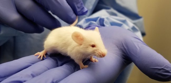
white laboratory mouse

Infectious diseases kill millions of people each year, but the search for treatments is hampered by the fact that laboratory mice are not susceptible to some human viruses, including killers like human immunodeficiency virus (HIV). For decades, researchers have turned to mice whose immune systems have been “humanized” to respond in a manner similar to humans.

Now a team at Princeton University has developed a comprehensive way to evaluate how immune responses of humanized mice measure up to actual humans. The research team looked at the mouse and human immune responses to one of the strongest vaccines known, a yellow fever vaccine called YFV-17D. The comparison of these “vaccine signatures” showed that a newly developed humanized mouse developed at Princeton shares significant immune-system responses with humans. The study was published in the journal Nature Communications.

“Understanding immune responses to human pathogens and potential vaccines remains challenging due to differences in the way our human immune system responds to stimuli, as compared to for example that of conventional mice, rats or other animals,” said Alexander Ploss, associate professor of molecular biology at Princeton. “Until now a rigorous method for testing the functionality of the human immune system in such a model has been missing. Our study highlights an experimental paradigm to address this gap.”

Humanized mice have been used in infectious disease research since the late 1980s. Yet without rigorous comparisons, researchers know little about how well the mice predict human responses such as the production of infection-fighting cells and antibodies.

To address this issue, researchers exposed the mice to the YFV-17D vaccine, which is made from a weakened, or attenuated, living yellow fever virus. Vaccines protect against future infection by provoking the production of antibodies and immune-system cells.

In previous work, the researchers explored the effect of YFV-17D on conventional humanized mice. But the researchers found that the mice responded only weakly. This led them to develop a mouse with responses that are more similar to those of humans.

To do so, the researchers introduced additional human genes for immune system components — such as molecules that detect foreign invaders and chemical messengers called cytokines — so that the complexity of the engrafted human immune system reflected that of humans. They found that the new mice have responses to YFV-17D that are very similar to the responses seen in humans. For example, the pattern of gene expression that occurs in response to YFV-17D in the mice shared significant similarities to that of humans. This signature gene expression pattern, reflected in the “transcriptome,” or total readout of all of the genes of the organism, translated into better control of the yellow fever virus infection and to immune responses that were more specific to yellow fever.

The researchers also looked at two other types of immune responses: the cellular responses, involving production of cytotoxic T cells and natural killer cells that attack and kill infected cells, and the production of antibodies specific to the virus. By evaluating these three types of responses – transcriptomic, cellular, and antibody – in both mice and humans, the researchers produced a reliable platform for evaluating how well the mice can serve as proxies for humans.

Florian Douam, a postdoctoral research associate and the first author on the study, hopes that the new testing platform will help researchers explore exactly how vaccines induce immunity against pathogens, which in many cases is not well understood.

“Many vaccines have been generated empirically without profound knowledge of how they induce immunity,” Douam said. “The next generation of mouse models, such as the one we introduced in our study, offer unprecedented opportunities for investigating the fundamental mechanisms that define the protective immunity induced by live-attenuated vaccines.”

Mice bearing human cells or human tissues have the potential to aid research on treatments for many diseases that infect humans but not other animals, such as – in addition to HIV – Epstein Barr Virus, human T-cell leukemia virus, and Karposi sarcoma-associated herpes virus.

“Our study highlights the importance of human biological signatures for guiding the development of mouse models of disease,” said Ploss. “It also highlights a path toward developing better models for human immune responses.”

The study involved contributions from Florian Douam, Gabriela Hrebikova, Jenna Gaska, Benjamin Winer and Brigitte Heller in Princeton University’s Department of Molecular Biology; Robert Leach, Lance Parsons and Wei Wang in Princeton University’s Lewis Sigler Institute for Integrative Genomics; Bruno Fant at the University of Pennsylvania; Carly G. K. Ziegler and Alex K. Shalek of Massachusetts Institute of Technology and Harvard Medical School; and Alexander Ploss in Princeton University’s Department of Molecular Biology.

The research was supported the National Institutes of Health (NIH, R01AI079031 and R01AI107301, to A.P) and an Investigator in Pathogenesis Award by the Burroughs Wellcome Fund (to A.P.). Additionally, A.K.S. was supported by the Searle Scholars Program, the Beckman Young Investigator Program, the NIH (1DP2OD020839, 5U24AI118672, 1U54CA217377, 1R33CA202820, 2U19AI089992, 1R01HL134539, 2RM1HG006193, 2R01HL095791, P01AI039671), and the Bill & Melinda Gates Foundation (OPP1139972). C.G.K.Z. was supported by a grant from the National Institute of General Medical Sciences (NIGMS, T32GM007753). J.M.G. and B.Y.W. were supported by a pre-doctoral training grant from the NIGMS (T32GM007388). B.Y.W. was also a recipient of a pre-doctoral fellowship from the New Jersey Commission on Cancer Research.

By Catherine Zandonella, Office of the Dean for Research

‘Radiolabeling’ lets scientists track the breakdown of drugs (Nature)

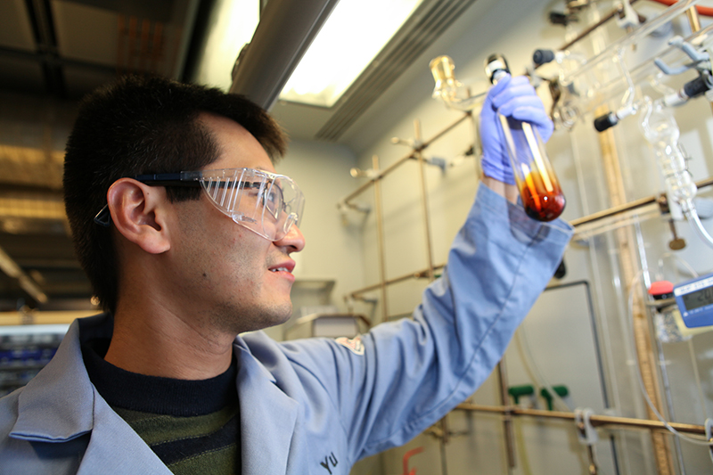
Graduate student Renyuan Pony Yu
Renyuan Pony Yu, a graduate student working with Princeton Professor Paul Chirik, has discovered a new way to radiolabel compounds for use in drug development.

By Tien Nguyen, Department of Chemistry

A new method for labeling molecules with radioactive elements could let chemists more easily track how drugs under development are metabolized in the body.

Chemists consider thousands of compounds in the search for a new drug, and a candidate’s metabolism is a key factor that must be evaluated carefully and quickly. Researchers at Princeton University and pharmaceutical company Merck & Co., Inc. report in the journal Nature that scientists can selectively replace hydrogen atoms in molecules with tritium atoms — a radioactive form of hydrogen that possesses two extra neutrons — to “radiolabel” compounds. This technique can be done in a single step while preserving the biological properties of the parent compound.

While current state-of-the-art techniques are quite reliable, they only work when dissolved in specific solvents, ones that aren’t always capable of dissolving the drug compound of interest. The researchers’ method, however, used an iron-based catalyst that is tolerant to a wider variety of solvents, and it labels the molecules at the opposite positions as compared to existing methods.

“The fact that you can access other positions is what makes this reaction really special,” said corresponding author Paul Chirik, the Edwards S. Sanford Professor of Chemistry at Princeton. Previous methods only incorporate radioactive tritium atoms into the molecule directly next to an atom or a group of atoms called a directing group. The new iron-catalyzed method does not require a directing group, and instead places tritium at whatever positions in the molecules are the least crowded.

“Radiolabeled compounds help medicinal chemists get a better picture of what actually happens to the drug by showing how the drug is metabolized and cleared,” said David Hesk, a collaborator at Merck and co-author on the work. By rapidly assessing the compounds’ metabolism early on, scientists can shorten the time it takes to develop and bring a drug to market. “Having another labeling reaction is very powerful because it gives radiochemists another tool in the toolbox,” he said.

This unique reactivity was actually discovered unexpectedly. Renyuan Pony Yu, a graduate student in the Chirik lab, had originally set out to use their iron catalyst for a different reaction that they were collaborating on with Merck. To study the iron catalyst’s capabilities, Yu subjected it to a technique called proton nuclear magnetic resonance spectroscopy (NMR), which allows chemists to deduce the positions of hydrogen atoms in molecules.

“We started seeing this beautiful, very systematic pattern of signals in the NMR, but we didn’t really know what they were,” said Yu, who is first author on the new study. Particularly puzzling was the fact that the pattern of signals would disappear over time.

The researchers turned to Istvan Pelczer, Director of the NMR Facility at Princeton chemistry and co-author on the work, who developed a special technique that helped them analyze the signals with much greater confidence. Using this method, they realized that the iron catalyst was reacting with the liquid solvent used to dissolve the NMR sample. The solvent’s deuterium atoms, another form of hydrogen that has one extra neutron and is not radioactive, were replacing the hydrogen atoms.

It wasn’t until Yu presented his findings to Matt Tudge, the Princeton authors’ collaborator at Merck, that the catalyst’s potential to introduce tritium atoms into radiolabeled molecules was recognized. “This is a classic example where you really need both partners,” Chirik said. “We were the catalyst experts, but they were the applications experts.”

Though tritium-labeled compounds are used mostly in metabolism studies, they can also be helpful at the very outset of a drug-discovery project to identify a biological target that the potential drugs can be tested against. The biological target could be an enzyme or protein associated with a certain disease. For example, statins are a well-known class of cholesterol-lowering drugs that target a specific enzyme in the body called HMG-CoA reductase.

To explore the scope of the reaction, Yu first optimized the reaction to incorporate deuterium atoms, which is commonly accepted as a model system for tritium. He found that the iron catalyst was surprisingly robust and successfully labeled many different types of compounds, including some from Merck’s library of past drug candidates.

“It was a very exciting project for me because I got to work with real drugs that are fully functionalized and useful,” Yu said. One of their test substrates was Claritin, which Yu bought from a local store; he extracted its active ingredient back in the lab.

Finally, Yu traveled to Merck’s campus in Rahway, where he received radioactivity training — Chirik’s laboratory isn’t equipped to handle radioactivity — and performed the reactions using tritium gas. The reactions were run in a special apparatus that looks like a steel-lined box and releases radioactive tritium gas. The apparatus can capture any unspent gas to limit the amount of radioactive waste produced.

Chemists take care to handle radioactive compounds and waste very carefully, but tritium’s radioactivity is so weak that the particles it emits cannot penetrate simple glassware. For this reason, tritium-labeled compounds can’t be used in any human imaging studies such as PET scans, which require radiolabeled compounds that emit high-energy particles.

This past summer, Yu presented the preliminary results of the iron-catalyzed reaction at the 2015 International Isotope Society Symposia to researchers in the radiolabeling and pharmaceutical community. They were very excited about the research and eager to use the catalyst in their own studies, Yu said.

But the major challenge for the researchers is that the iron catalyst is extremely air and moisture sensitive, and it can only be handled inside a glovebox, a special chamber in which oxygen and water vapor have been excluded. The Chirik group is working to develop a more stable catalyst that can be made commercially available, and have recently entered into a partnership with Green Center Canada, a company that helps bring academic research to market.

In the meantime, the Chirik group has found that the iron catalyst can replace hydrogen atoms with other groups besides deuterium and tritium atoms and is extending this chemistry into many other projects in the lab.

“This project is always going to be a special one for me because it’s kind of a pivot point for the type of chemistry that our group can do,” Chirik said, “and there’s this really cool application.”

Read the abstract.

Yu, R. P.; Hesk, D.; Rivera, N.; Pelczer, I.; Chirik, P. J. “Iron-Catalyzed Tritiation of Pharmaceuticals.” Nature, 2016, DOI: 10.1038/nature16464.

This work was supported by Merck & Co. and Princeton University’s Intellectual Property Accelerator Fund.