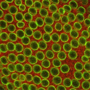
Movie caption: Researchers at Princeton studied the temperature dependence of the formation of the nucleolus, a cellular organelle. The movie shows the nuclei of intact fly cells as they are subjected to temperature changes in the surrounding fluid. As the temperature is shifted from low to high, the spontaneously assembled proteins dissolve, as can be seen in the disappearance of the bright spots.
By Catherine Zandonella, Office of the Dean for Research
Researchers at Princeton found that the nucleolus, a cellular organelle involved in RNA synthesis, assembles in part through the passive process of phase separation – the same type of process that causes oil to separate from water. The study, published in the journal Proceedings of the National Academy of Sciences, is the first to show that this happens in living, intact cells.
Understanding how cellular structures form could help explain how organelles change in response to diseases. For example, a hallmark of cancer cells is the swelling of the nucleolus.
To explore the role of passive processes – as opposed to active processes that involve energy consumption – in nucleolus formation, Hanieh Falahati, a graduate student in Princeton’s Lewis-Sigler Institute for Integrative Genomics, looked at the behavior of six nucleolus proteins under different temperature conditions. Phase separation is enhanced at lower temperatures, which is why salad dressing containing oil and vinegar separates when stored in the refrigerator. If phase separation were driving the assembly of proteins, the researchers should see the effect at low temperatures.
Falahati showed that four of the six proteins condensed and assembled into the nucleolus at low temperatures and reverted when the temperature rose, indicating that the passive process of phase separation was at work. However, the assembly of the other two proteins was irreversible, indicating that active processes were in play.
“It was kind of a surprising result, and it shows that cells can take advantage of spontaneous processes for some functions, but for other things, active processes may give the cell more control,” said Falahati, whose adviser is Eric Wieschaus, Princeton’s Squibb Professor in Molecular Biology and a professor of molecular biology and the Lewis-Sigler Institute for Integrative Genomics, and a Howard Hughes Medical Institute researcher.
The research was funded in part by grant 5R37HD15587 from the National Institute of Child Health and Human Development (NICHD), and by the Howard Hughes Medical Institute.
By Marisa Sanders for the Office of the Dean for Research
A new study by researchers at Princeton University suggests that sporadic bursts of gene activity may be important features of genetic regulation rather than just occasional mishaps. The researchers found that snippets of DNA called enhancers can boost the frequency of bursts, suggesting that these bursts play a role in gene control.
The researchers analyzed videos of Drosophila fly embryos undergoing DNA transcription, the first step in the activation of genes to make proteins. In a study published on July 14 in the journal Cell, the researchers found that placing enhancers in different positions relative to their target genes resulted in dramatic changes in the frequency of the bursts.
“The importance of transcriptional bursts is controversial,” said Michael Levine, Princeton’s Anthony B. Evnin ’62 Professor in Genomics and director of the Lewis-Sigler Institute for Integrative Genomics. “While our study doesn’t prove that all genes undergo transcriptional bursting, we did find that every gene we looked at showed bursting, and these are the critical genes that define what the embryo is going to become. If we see bursting here, the odds are we are going to see it elsewhere.”
The transcription of DNA occurs when an enzyme known as RNA polymerase converts the DNA code into a corresponding RNA code, which is later translated into a protein. Researchers were puzzled to find about ten years ago that transcription can be sporadic and variable rather than smooth and continuous.
In the current study, Takashi Fukaya, a postdoctoral research fellow, and Bomyi Lim, a postdoctoral research associate, both working with Levine, explored the role of enhancers on transcriptional bursting. Enhancers are recognized by DNA-binding proteins to augment or diminish transcription rates, but the exact mechanisms are poorly understood.
Until recently, visualizing transcription in living embryos was impossible due to limits in the sensitivity and resolution of light microscopes. A new method developed three years ago has now made that possible. The technique, developed by two separate research groups, one at Princeton led by Thomas Gregor, associate professor of physics and the Lewis-Sigler Institute for Integrative Genomics, and the other led by Nathalie Dostatni at the Curie Institute in Paris, involves placing fluorescent tags on RNA molecules to make them visible under the microscope.
The researchers used this live-imaging technique to study fly embryos at a key stage in their development, approximately two hours after the onset of embryonic life where the genes undergo fast and furious transcription for about one hour. During this period, the researchers observed a significant ramping up of bursting, in which the RNA polymerase enzymes cranked out a newly transcribed segment of RNA every 10 or 15 seconds over a period of perhaps 4 or 5 minutes per burst. The genes then relaxed for a few minutes, followed by another episode of bursting.
The team then looked at whether the location of the enhancer – either upstream from the gene or downstream – influenced the amount of bursting. In two different experiments, Fukaya placed the enhancer either upstream of the gene’s promoter, or downstream of the gene and saw that the different enhancer positions resulted in distinct responses. When the researchers positioned the enhancer downstream of the gene, they observed periodic bursts of transcription. However when they positioned the enhancer upstream of the gene, the researchers saw some fluctuations but no discrete bursts. They found that the closer the enhancer is to the promoter, the more frequent the bursting.
To confirm their observations, Lim applied further data analysis methods to tally the amount of bursting that they saw in the videos. The team found that the frequency of the bursts was related to the strength of the enhancer in upregulating gene expression. Strong enhancers produced more bursts than weak enhancers. The team also showed that inserting a segment of DNA called an insulator reduced the number of bursts and dampened gene expression.
In a second series of experiments, Fukaya showed that a single enhancer can activate simultaneously two genes that are located some distance apart on the genome and have separate promoters. It was originally thought that such an enhancer would facilitate bursting at one promoter at a time—that is, it would arrive at a promoter, linger, produce a burst, and come off. Then, it would randomly select one of the two genes for another round of bursting. However, what was instead observed was bursting occurring simultaneously at both genes.
“We were surprised by this result,” Levine said. “Back to the drawing board! This means that traditional models for enhancer-promoter looping interactions are just not quite correct,” Levine said. “It may be that the promoters can move to the enhancer due to the formation of chromosomal loops. That is the next area to explore in the future.”
The study was funded by grants from the National Institutes of Health (U01EB021239 and GM46638).
Access the paper here:
Takashi Fukaya, Bomyi Lim & Michael Levine. Enhancer Control of Transcriptional Bursting, Cell (2016), Published July 14. EPub ahead of print June 9. http://dx.doi.org/10.1016/j.cell.2016.05.025
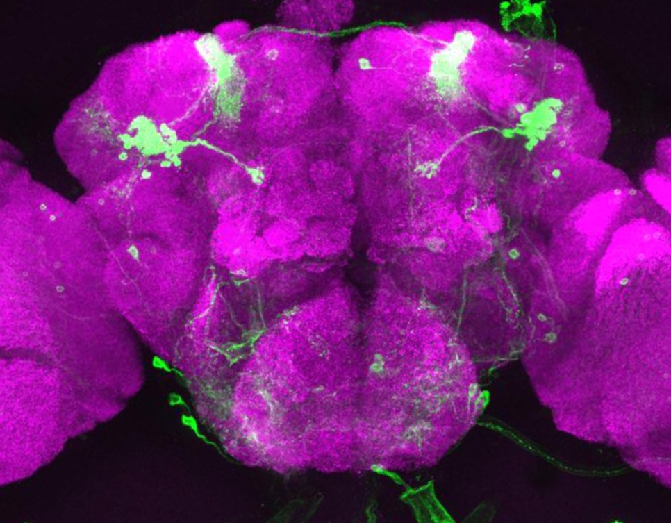
Image of the Drosophila brain (magenta) with a subset of mushroom body neurons expressing green fluorescent protein (GFP) via a genetic marker. (Credit: Janelia Farm/HHMI – FlyLight)
By Kristin Qian for the Office of the Dean for Research
Researchers at Princeton University have developed a highly sensitive and precise method to explore genes important for memory formation within single neurons of the Drosophila fly brain. With this method, the researchers found an unexpected result: certain genes involved in creating long-term memories in the brain are the same ones that the eye uses for sensing light.
The study, published in the May 17 issue of the journal Cell Reports, demonstrated the utility of the new method and also identified new patterns of gene expression that drive long-term memory formation.
“Ultimately, to understand the brain, we want to know what individual neurons are doing,” said Mala Murthy, assistant professor in the Princeton Neuroscience Institute and the Department of Molecular Biology. “We found that single neurons can be defined by their pattern of their gene expression, even if they’re all in the same brain network.”
To their surprise, the researchers found that many of the active genes in these neurons produce proteins that are best known for their roles in detecting light in the fly’s eye or sensing odor in the fly’s nose. “It is possible that these sensory proteins have been repurposed by the brain for a different function,” Murthy said.
“Even though the paper is focused on the methodology, which I think will be impactful for the field, there is this new science here—a whole new class of molecules we found that is in the central brain and seems to be involved in memory formation,” Murthy said.
Researchers have known that genes “turn on,” or start making proteins, during the formation of long-term memories in Drosophila, a widely used organism in studies of neurobiology, but they didn’t know exactly which genes in which neurons were involved.
To investigate this question, the researchers first trained flies to form long-term memories. Then they extracted single neurons from the fly brains and evaluated all of the gene readouts, or transcripts, which encode proteins. By comparing the transcripts of the memory-trained flies to those of non-trained flies, researchers were able to identify genes involved in long-term memory formation.
The task was complicated by the tiny size of the fly’s head, which is just one millimeter across, and contains fewer than 100,000 neurons. Murthy’s team focused on neuron types in one part of the brain, the mushroom body, named for its distinctive shape.
First author Amanda Crocker, a former postdoctoral fellow in Murthy’s lab and now an assistant professor of neuroscience at Middlebury College, conducted the experiments in collaboration with co-authors Xiao-Juan Guan, a senior research specialist in the Princeton Neuroscience Institute; Coleen Murphy, professor of molecular biology and the Lewis-Sigler Institute for Integrative Genomics; and Murthy.
“Our work opens up the ability to use Drosophila as a way to study how gene expression in single neurons relates to brain function,” Crocker said. “This has been a challenge because the fly brain is very small and contains fewer neurons than other organisms that neuroscientists study. The advantage of using flies is that they have significantly less redundancy in the neurons that they do have. We can look at specific neurons and gene expression, and ask what the genes are doing in that cell to cause the behavior.”
The researchers trained the flies to form long-term memories by exposing them to an odor – either an earthy, mushroom-like smell (3-octanol) or a menthol-like smell (4-methylcyclohexanol) – while simultaneously delivering a negative stimulus in the form of an electric shock.
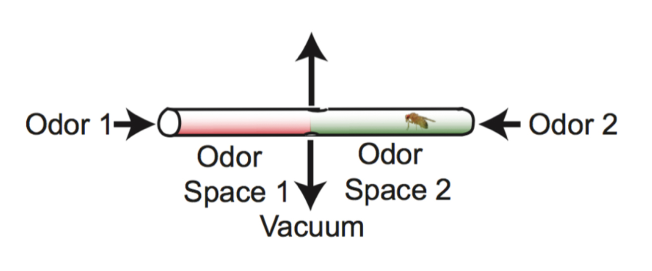
Flies experience two odor spaces in each tube. If neither odor has been paired with electric shock, flies spend an equal amount of time on both sides of the tube (control). If one of the odors is paired with electric shock, flies avoid that side of the tube. For example, flies trained to associate the odor 3-OCT with electric shock avoided the red side (containing 3-OCT) of the tube. (Credit: Murthy lab, Princeton University)
The training took place in a tube containing the two odors, one at each end of the tube. Researchers paired one of the odors with the electric shock, and as a result the fly avoided that end of the tube. The assay was conducted in the dark, so that the flies could use only their sense of smell, not their vision, to navigate the tube.
A second group of flies received the electric shock and the odor, but not at the same time, so they did not form the memory that linked odor to shock.
The researchers then isolated single neurons from the fly brains using tiny glass tubes to suction out the cells. Harvesting neurons using this technique is not common, Murthy said, and it had not been combined with a complete analysis of gene activity in fly neurons before. With this novel method, they were able to use only 10 to 90 femtograms – a quintillionth of a kilogram – of genetic material.
They evaluated gene activity by looking at the production of messenger ribonucleic acid (mRNA), an intermediary between DNA and proteins. The result is a “transcriptome,” or readout of all of the genetic messages that the cell uses to produce proteins. The researchers then read the transcriptome to see which genes produced proteins in the memory-trained flies versus the non-trained flies, and found that some of the active genes in memory-trained flies were the same as ones used in the sensory organs to detect light, odors and taste.
To follow-up, the researchers bred mutant flies that lacked genes for some of the light-sensing proteins and thus could not see. The same memory experiments as before were carried out, and the researchers confirmed that the flies lacking light-sensing proteins were both unable to see and unable to form long-term memories.
The discovery of the expression of genes for classical ‘light-sensing’ proteins, such as rhodopsin, as well as other sensory-related proteins for odor and taste detection, was unexpected because these proteins were not known to be utilized in mushroom bodies, Murthy said. Although studies in other organisms, including humans, have detected sensory genes in areas of the brain unrelated to the sensory organ itself, this may be the first study to link these genes to memory formation.
The study was funded by a National Institutes of Health Ruth L. Kirschstein Institutional National Research Service Award, the Alfred P. Sloan Foundation, the Human Frontier Science Program, a National Science Foundation (NSF) CAREER award, the McKnight Endowment Fund for Neuroscience, the Klingenstein Foundation, a National Institutes of Health New Innovator award, and an NSF BRAIN Initiative EAGER award. The study was also funded in part through Princeton’s Glenn Center for Quantitative Aging Research, directed by Coleen Murphy.
The paper, “Cell-Type-Specific Transcriptome Analysis in the Drosophila Mushroom Body Reveals Memory-Related Changes in Gene Expression,” was published in the May 17 issue of Cell Reports.
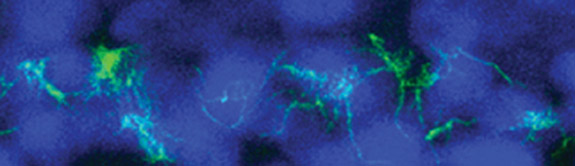
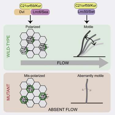
Cilia with a mutant form of the Kurly protein are wild and disorganized.
By Catherine Zandonella, Office of the Dean for Research
A new study of a protein found in cilia – the hair-like projections on the cell surface – may help explain how genetic defects in cilia play a role in developmental abnormalities, kidney disease and a number of other disorders.
The researchers at Princeton University and Northwestern University found that the protein, which goes by the name C21orf59 or “Kurly,” is needed for cilia to undulate to keep fluid moving over the surface of cells. They also found that the protein is needed during development to properly orient the cilia so that they are facing the right direction to move the fluid.
“It’s extremely exciting that we’ve found a single protein that is responsible for these two functions – orientation and motility – in cilia,” said Rebecca Burdine, an associate professor of molecular biology at Princeton University. “Despite their importance in human disease, very little is known about how cilia motility and orientation are coordinated, so this protein will provide an important gateway into looking at this process.” The finding is published online and in the March 1 issue of the journal Cell Reports.
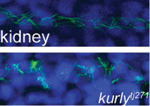
Caption: Staining of cilia (hair-like projections in green and nuclei in blue) in zebrafish kidney tubules show cilia are disorganized and oriented incorrectly in fish with mutated Kurly protein (bottom panel) versus normal Kurly (top panel). Image courtesy of the Burdine lab.
The studies were conducted in zebrafish at Princeton and in African clawed frogs (Xenopus laevis) at Northwestern. In the zebrafish kidney, the researchers found that the Kurly protein enabled cilia to orient themselves in a uniform direction, and most importantly, in the proper direction to facilitate the flow of fluid along the narrow channels in the kidney. In frogs, the cilia on skin cells help move fluid along the surface of the animal during its larval stage. In both cases, knocking out the gene for Kurly caused the cilia to orient incorrectly thereby losing their ability to move in the waving fashion that helps push fluid along.
The discovery of Kurly’s role in cilia movement and orientation stemmed from work in the Burdine lab on fetal organ development, specifically an investigation of mutations that alter the left-right asymmetric orientation of the heart. Such mutations can result in an organ that is working properly but is an exact mirror image of a normal heart. During a search for genes involved in this left-right patterning, the Burdine team discovered that mutations in a gene they called kur, which codes for the Kurly protein, were linked to errors in left-right orientation in zebrafish heart.
Image credit: Burdine lab
As the team investigated kur, they noted that the mutation also affected the function of cilia. It has been known for some time that cilia are important for a number of jobs, from sensing the environment to facilitating fluid flow, to ensuring that the lungs excrete inhaled contaminants. Cilia genetic defects are linked to a number of human diseases, including polycystic kidney disease, respiratory distress, hearing loss, infertility, and left-right patterning disorders such as the one Burdine studies.
Researchers in Burdine’s laboratory found that Kurly’s role in cilia movement stems from its ability to ensure proteins called dynein arms are correctly located in the cilia. When the researchers knocked out the kur gene, the dynein proteins failed to form in the proper location.
The finding that a single protein is involved in both movement and orientation is surprising, said co-first author Daniel Grimes, a postdoctoral research associate in the Burdine lab. “These are two aspects that are both required to generate fluid flow, and we’d like to know how they are linked molecularly. This work adds a new gene that aids this discovery.”
The gene for Kurly has also been detected in relation to human cilia disorders, so the work may have an impact on understanding the mechanisms of human disease, Grimes added. The researchers also found that the mutation they discovered rendered the Kurly protein sensitive to temperature, and used this trait to find that the Kurly protein may be involved in initiating movement rather than keeping the cilia moving once they’ve started.
The team also explored proteins that interact with Kurly. The Northwestern team showed that when the kur gene was inactivated using a gene-editing technique called CRISPR-Cas9, the lack of a functioning Kurly protein led to the mis-positioning of a second protein on the cell surface called Prickle2, which helps cells know which direction they face. Without proper Prickle2 positioning, the cilia pushed fluid in the wrong direction.
The study of the Kurly protein involved Grimes as well as two additional co-authors, Kimberly Jaffe and Jodi Schottenfeld-Roames, a former postdoctoral researcher and graduate student respectively, in the Burdine lab. The initial studies on the Kurly protein were conducted as part of an undergraduate research project by Tse-shuen (Jade) Ku, Class of 2007. Additional work was contributed by Nicholas Morante and José Pelliccia, graduate students in the Burdine lab.
The work at Northwestern University was performed in the laboratory of Brian Mitchell with the assistance of Michael Werner and Sun Kim.
The research was supported by a National Institutes of Health (NIH) Ruth L. Kirschstein Institutional National Research Service Award grant to K. Jaffe (#1F32HD060396-01A1), an NIH National Institute of General Medical Sciences grant to B. Mitchell (#2R01GM089970), and an NIH Eunice Kennedy Shriver National Institute of Child Health and Human Development grant to R. Burdine (#2R01HD048584).
Kimberly M. Jaffe, Daniel T. Grimes, Jodi Schottenfeld-Roames, Michael E. Werner, Tse-Shuen J. Ku, Sun K. Kim, Jose L. Pelliccia, Nicholas F.C. Morante, Brian J. Mitchell, Rebecca D. Burdine.c21orf59/kurly controls both cilia motility and polarization. Cell Reports (2016), http://dx.doi.org/10.1016/j.celrep.2016.01.069. In Press Corrected Proof.
A fly runs on an air-supported ball. The audio traces of the fly’s courtship song are shown.
Article courtesy of Joseph Caputo, Cell Press
Outside of humans, the ability to adjust the intensity of acoustic signals with distance has only been identified in songbirds. Research published February 3 in Neuron now demonstrates that the male fruit fly also displays this complex behavior during courtship, adjusting the amplitude of his song depending on how far away he is from a female. Studying this process in the fruit fly can help shed light on the building blocks for social interactions across the animal kingdom.
Mala Murthy, of Princeton University, and her colleagues have revealed an unanticipated level of control in insect acoustic communication by analyzing each stage of the neuronal pathway underlying male fruit flies’ ability to adjust their courtship song—from the visual cues that help estimate distance to the signals that pass through nervous system and cause changes in muscle activity that drive softer or louder song. The complexity is remarkable considering that the fruit fly has only 100,000 neurons, one-millionth that of a human brain.
During courtship, males chase females, extending and vibrating one wing at a time to produce a courtship song. Songs, which consist primarily of two modes: sine and pulse, are extremely quiet and must be recorded on sensitive microphones, then amplified more than 1 million times in order to be heard by humans. When amplified, the sine song sounds like the whine of an approaching mosquito, while the pulse song is more akin to a cat’s purr.
“Females listen to many minutes of male song before deciding whether to accept him,” says Murthy, of the Princeton Neuroscience Institute and Department of Molecular Biology. “There is thus enormous evolutionary pressure for males to optimize their song to match the female’s preference while simultaneously minimizing the energetic cost of singing for long periods of time.” Adjusting the amplitude of song to compensate for female distance allows males to conserve energy and thereby court for longer periods of time and better compete with other males.
“While the precise neural mechanisms underlying the generation and patterning of fly song may be distinct from humans or even songbirds, the fundamental problem is the same: how can a neural network produce such a complex and dynamic signal?” Murthy says. “For this reason, we anticipate that similar neural mechanisms will be employed in all systems, and the genetic model system of the fruit fly is an ideal starting point from which to dissect them.”
The researchers were funded by the Howard Hughes Medical Institute, the DAAD (German Academic Exchange Foundation), the Alfred P. Sloan Foundation, the Human Frontiers Science Program, a National Science Foundation CAREER award, a NIH New Innovator Award, the NSF BRAIN Initiative, an EAGER award, the McKnight Foundation, and the Klingenstein-Simons Foundation.
Philip Coen, Marjorie Xie, Jan Clemens and Mala Murthy. Sensorimotor Transformations Underlying Variability in Song Intensity during Drosophila Courtship. Neuron. Vol. 89, Issue 3, p629–644, 3 February 2016.
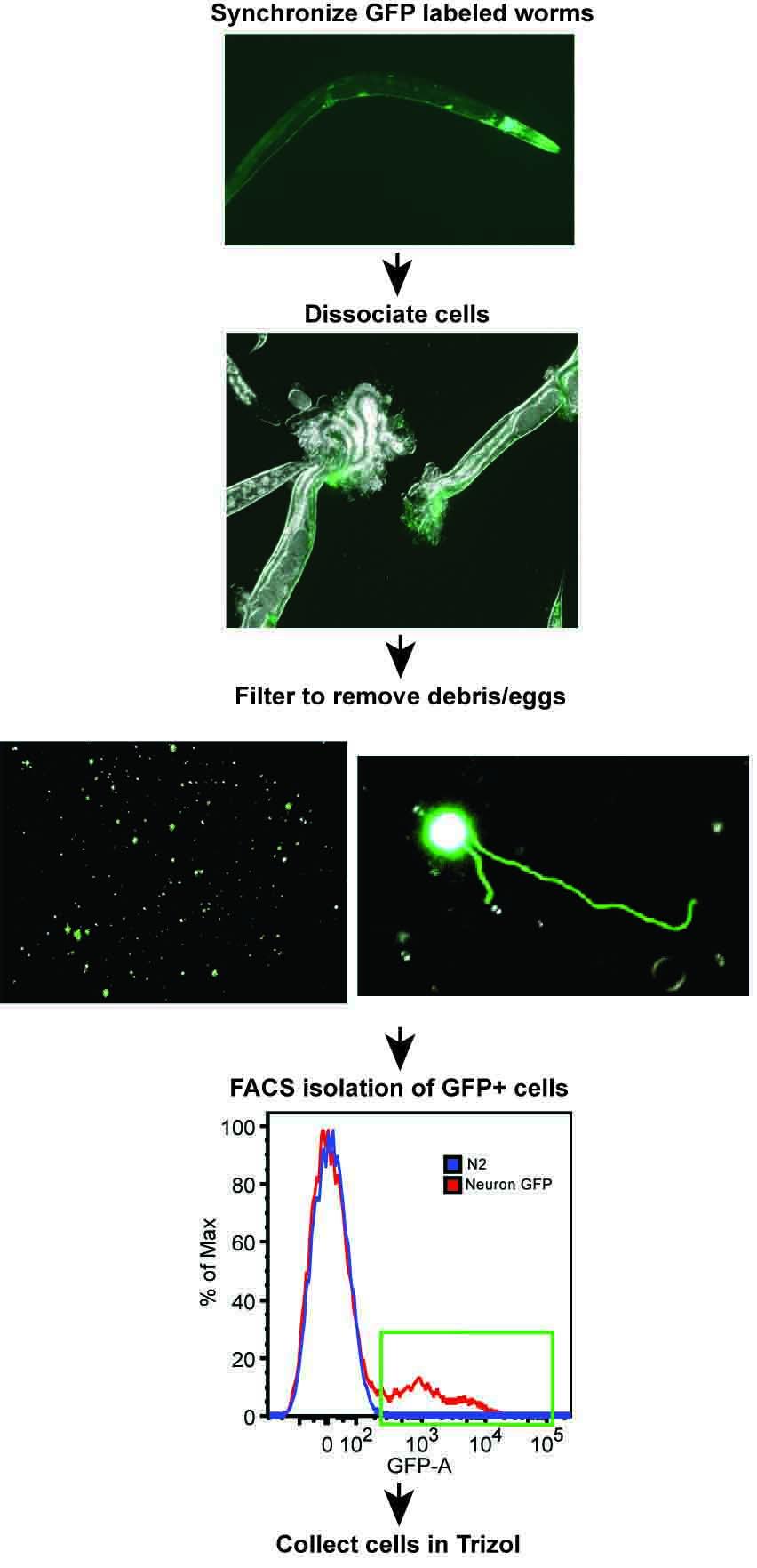
A research team from Princeton University led by Coleen Murphy, professor of Molecular Biology and the Lewis-Sigler Institute for Integrative Genomics, has developed a new method for isolating neurons from adult C. elegans worms. The first panel shows worms containing neurons labeled with green fluorescent protein (GFP). Using rapid, chilled chemomechanical disruption, the neuron cells were extracted and purified, then sorted using fluorescence-activated cell sorting (FACS).
Researchers from Princeton University have identified genes important for age-related cognitive declines in memory in adult worm neurons, which had not been studied previously. The research, published in the journal Nature, could eventually point the way toward therapies to extend life and enhance health in aging human populations.
“The newly discovered genes regulate enhanced short-term memory as well as the ability to repair damaged neurons, two factors that play an important role in healthy aging,” said Coleen Murphy, a professor of Molecular Biology and the Lewis-Sigler Institute for Integrative Genomics, director of the Glenn Center for Quantitative Aging Research at Princeton, and senior author on the study. “Identifying the individual factors involved in neuron health in the worm is the first step to understanding human neuronal decline with age.”
The small soil-dwelling roundworm, Caenorhabditis elegans, contains genes that determine the rate of aging and overall health during aging. Mutations in one of these genetic pathways, the insulin/IGF-1 signaling (IIS) pathway, can double worm lifespan. Similar mutations in humans have been found in long-lived humans.
But studying the IIS mutation in adult worm neurons was difficult because the adults have a thick, durable covering that protects the neurons.
Using a new technique they developed to break up the tough outer covering, researchers at Princeton succeeded in isolating adult neurons, which enabled the detection of the new set of genes regulated by the insulin/IGF-1 signaling pathway.
“Our technique enabled us to study gene expression in adult neurons, which are the cells that govern cognitive aspects such as memory and nerve-regeneration,” said Murphy, whose research on aging is funded in part by the National Institutes of Health. “Prior to this work, researchers were only able to examine gene regulation either using adult worms or individual tissues from young worms.”
The work allowed co-first authors Rachel Kaletsky and Vanisha Lakhina to explore why long-lived IIS mutants maintain memory and neuron-regeneration abilities with age. Until now, the known targets of the insulin longevity pathway were located mostly in the intestine and skin of the worm rather than the neurons. Kaletsky is a postdoctoral research fellow and Lakhina is a postdoctoral research associate in the Lewis-Sigler Institute.
Kaletsky worked out the new way to isolate neurons from adult worms, and with Lakhina, proceeded to profile the gene activity in adult C. elegans neurons for the first time. They discovered that the IIS mutant worms express genes that keep neurons working longer, and that these genes are completely different from the previously known longevity targets. They also discovered a new factor that is responsible for nerve cell (axon) regeneration in adult worms, which could have implications for human traumatic brain injury.
“Kaletsky and Lakhina developed a new technique that is going to be used by the entire worm community, so it really opens up new avenues of research even beyond the discoveries we describe in the paper,” Murphy said.
One of the newly identified genes, fkh-9, regulates both enhanced memory and neuronal regeneration in IIS mutants. Previous studies have detected only one other gene that regulates neuronal regeneration in the mutants, demonstrating the power of the technique to identify new gene regulators. The researchers also found that fkh-9 gene expression is required for long lifespan in many IIS mutants, but it did not play that role in neurons, suggesting the gene governs multiple outcomes in the worm.
Murphy’s lab is now working to understand how fkh-9 works to influence memory, axon regeneration, and lifespan. The gene codes for a protein, FKH-9, that acts as a transcription factor, meaning it controls the expression of other genes and is likely part of a larger regulatory network. FKH-9 also appears to regulate different processes in different tissues: It is required in neurons for memory and axon repair, but not for lifespan. Murphy’s group is working to figure out how FKH-9 acts in distinct tissues to regulate such different processes.
The study provides a more complete picture of how IIS mutants control gene expression in different tissues to promote healthy aging, Murphy said.
“fkh-9 is likely only one of the exciting genes that will emerge from using this technique,” Murphy said. “By identifying the suite of IIS-regulated neuronal genes, there are many candidates for follow-up, only a fraction of which have been characterized in any great detail,” she said.
Other contributors to the study included Rachel Arey, a postdoctoral research fellow; former graduate students April Williams and Jessica Landis; and Jasmine Ashraf, a research specialist in the Lewis-Sigler Institute.
Additional funding for the study was provided by the Keck Foundation, the Ruth L. Kirschstein National Research Service Awards, the National Science Foundation and the New Jersey Commission on Brain Injury Research.
The article, The C. elegans adult neuronal IIS/FOXO transcriptome reveals adult phenotype regulators, by Rachel Kaletsky, Vanisha Lakhina, Rachel Arey, April Williams, Jessica Landis, Jasmine Ashraf and Coleen T. Murphy, was published in the journal Nature online ahead of print on December 14, 2015. doi:10.1038/nature16483.
Researchers explore the shift from maternal genes to the embryo’s genes during development
By Catherine Zandonella, Office of the Dean for Research
Cells in an early-stage fruit fly embryo. (Image courtesy of NIGMS image gallery).
A new study from Princeton University researchers sheds light on the handing over of genetic control from mother to offspring early in development. Learning how organisms manage this transition could help researchers understand larger questions about how embryos regulate cell division and differentiation into new types of cells.
The study, published in the March 12 issue of the journal Cell, provides new insight into the mechanism for this genetic hand-off, which happens within hours of fertilization, when the newly fertilized egg is called a zygote.
“At the beginning, everything the embryo needs to survive is provided by mom, but eventually that stuff runs out, and the embryo needs to start making its own proteins and cellular machinery,” said Princeton postdoctoral researcher in the Department of Molecular Biology and first author Shelby Blythe. “We wanted to find out what controls that transition.”
Blythe conducted the study with senior author Eric Wieschaus, Princeton’s Squibb Professor in Molecular Biology, Professor of Molecular Biology and the Lewis-Sigler Institute for Integrative Genomics, a Howard Hughes Medical Institute investigator, and a Nobel laureate in physiology or medicine.
Researchers have known that in most animals, a newly fertilized egg cell divides rapidly, producing exact copies of itself using gene products supplied by the mother. After a short while, this rapid cell division pauses, and when it restarts, the embryonic DNA takes control and the cells divide much more slowly, differentiating into new cell types that are needed for the body’s organs and systems.
To find out what controls this maternal to zygotic transition, also called the midblastula transition (MBT), Blythe conducted experiments in the fruit fly Drosophila melanogaster, which has long served as a model for development in higher organisms including humans.
These experiments revealed that the slower cell division is a consequence of an upswing in DNA errors after the embryo’s genes take over. Cell division slows down because the cell’s DNA-copying machinery has to stop and wait until the damage is repaired.
Blythe found that it wasn’t the overall amount of embryonic DNA that caused this increase in errors. Instead, his experiments indicated that the high error rate was due to molecules that bind to DNA to activate the reading, or “transcription,” of the genes. These molecules stick to the DNA strands at thousands of sites and prevent the DNA copying machinery from working properly.
To discover this link between DNA errors and slower cell replication, Blythe used genetic techniques to create Drosophila embryos that were unable to repair DNA damage and typically died shortly after beginning to use their own genes. He then blocked the molecules that initiate the process of transcription of the zygotic genes, and found that the embryos survived, indicating that these molecules that bind to the DNA strands, called transcription factors, were triggering the DNA damage. He also discovered that a protein involved in responding to DNA damage, called Replication Protein A (RPA), appeared near the locations where DNA transcription was being initiated. “This provided evidence that the process of awakening the embryo’s genome is deleterious for DNA replication,” he said.
The study also demonstrates a mechanism by which the developing embryo ensures that cell division happens at a pace that is slow enough to allow the repair of damage to DNA during the switchover from maternal to zygotic gene expression. “For the first time we have a mechanistic foothold on how this process works,” Blythe said.
The work also enables researchers to explore larger questions of how embryos regulate DNA replication and transcription. “This study allows us to think about the idea that the ‘character’ of the DNA before and after the MBT has something to do with the DNA acquiring the architectural features of chromatin [the mix of DNA and proteins that make up chromosomes] that allow us to point to a spot and say ‘this is a gene’ and ‘this is not a gene’,” Blythe said. “Many of these features are indeed absent early in embryogenesis, and we suspect that the absence of these features is what allows the rapid copying of the DNA template early on. Part of what is so exciting about this is that early embryos may represent one of the only times when this chromatin architecture is missing or ‘blank’. Additionally, these early embryos allow us to study how the cell builds and installs these features that are so essential to the fundamental processes of cell biology.”
This work was supported in part by grant 5R37HD15587 from the Eunice Kennedy Shriver National Institute of Child Health and Human Development.
Blythe, Shelby A. & Eric R. Wieschaus. Zygotic Genome Activation Triggers the DNA Replication Checkpoint at the Midblastula Transition. Cell. Published online on March 5, 2015. doi:10.1016/j.cell.2015.01.050. http://www.sciencedirect.com/science/article/pii/S0092867415001282
Princeton University researchers found that the banana-like curve of the bacteria Caulobacter crescentus provides stability and helps them flourish as a group in the moving water they experience in nature. The findings suggest a new way of studying the evolution of bacteria that emphasizes using naturalistic settings. The illustration shows how C. crescentus divides asymmetrically into a “stalked” mother cell that anchors to a bacterium’s home surface, and an upper unattached portion that forms a new, juvenile cell known as a “swarmer.” Swarmer cells later morph into stalked cells and lay down roots nearby. They repeat the life cycle with their own swarmer cell and the bacterial colony grows. The Princeton researchers found that in moving water, curvature points the swarmer cell toward the surface to which it needs to attach. This ensures that the bacteria’s next generation does not stray too far from its progenitors. (Image by Laura Ancona)
By Morgan Kelly, Office of Communications
Drawing from his engineering background, Princeton University researcher Alexandre Persat had a notion as to why the bacteria Caulobacter crescentus are curved — a hunch that now could lead to a new way of studying the evolution of bacteria, according to research published in the journal Nature Communications.
Commonly used in labs to study cell division, C. crescentus naturally take on a banana-like curve, but they also can undergo a mutation in which they grow to be perfectly straight. The problem was that in a laboratory there was no apparent functional difference between the two shapes. So a question among biologists was, why would nature bother?
Then Persat, who is a postdoctoral researcher in the group of Associate Professor of Molecular BiologyZemer Gitai, considered that the bacteria dwell in large groups attached to surfaces in lakes, ponds and streams. That means that their curvature could be an adaptation that allows C. crescentus to better develop in the water currents the organisms experience in nature.
In the new paper, first author Persat, corresponding author Gitai and Howard Stone, Princeton’s Donald R. Dixon ’69 and Elizabeth W. Dixon Professor of Mechanical and Aerospace Engineering, report that curvature does more than just help C. crescentus hold their ground in moving fluid. The researchers monitored C. crescentus growth on surfaces in flow and found that the bacteria’s arched anatomy is crucial to flourishing as a group.
“It didn’t take a long time to figure out how flow brought out the advantages of curvature,” Persat said. “The obvious thing to me as someone with a fluid-dynamics background was that this shape had something to do with fluid flow.”
The findings emphasize the need to study bacteria in a naturalistic setting, said Gitai, whose group focuses on how bacterial shapes are genetically determined. While a petri dish generally suffices for this line of study, the functionality of bacterial genes and anatomy can be elusive in most lab settings, he said. For instance, he said, 80 percent of the genes in C. crescentus are seemingly disposable — but they might not be in nature.
“We now see there can be benefits to bacterial shapes that are only seen in a growth environment that is close to the bacteria’s natural environment,” Gitai said.
“For C. crescentus, the ecology was telling us there is an advantage to being curved, but nothing we previously did in the lab could detect what that was,” he said. “We need to not only think of the chemical environment of the bacteria — we also need to think of the physical environment. I think of this research as opening a whole new axis of studying bacteria.”
While most bacteria grow and divide as two identical “daughter” cells, C. crescentus divides asymmetrically. A “stalked” mother cell anchors to a bacterium’s home surface while the upper unattached portion forms a new, juvenile version of the stalked cell known as a “swarmer” cell. The swarmer cells later morph into stalked cells then eventually detach before laying down roots nearby. They repeat the life cycle with their own swarmer cell and the bacterial colony grows.
The Princeton researchers found that in moving water, curvature points the swarmer cell toward the surface to which it needs to attach. This ensures that the bacteria’s next generation does not stray too far from its progenitors, as well as from the nutrients that prompted cell division in the first place, Gitai said. On the other hand, the upper cells of straight bacteria — which are comparatively higher from the ground — are more likely to be carried far away as they are to stay near home.
But the advantage of curvature only goes so far. The researchers found that when the water current was too strong, both curved and straight bacteria were pressed flat against the surface, eliminating the curved cells’ colonization advantage.
These findings put some interesting boundaries on what is known about C. crescentus, starting with the upper limits of the current in which the organism can thrive, Gitai said. He and Persat also plan to pursue whether the bacteria are able to straighten out and cast offspring downstream when the home colony faces a decline in available nutrients.
At the same time, understanding why C. crescentus got its curve helps in figuring out the evolution of other bacteria, he said. Close relatives of the bacteria, for example, are not curved — could it have to do with the severity of their natural environment, such as the powerful turbulence of an ocean? Harmful bacteria such as Vibrio cholerae, strains of which cause cholera, are curved, though the reason is unclear. It’s possible this shape could be related to the organism’s environment in a way that might help treat those infected by it, Gitai said.
Whatever the reason for a specific bacteria’s shape, the Princeton research shows that exploring the influence of its natural habitat could be worthwhile, Gitai said.
“It was clear with C. crescentus that we needed to try something different,” Gitai said. “People didn’t really think of flow as a major driver of this bacteria’s evolution. That really is a new idea.”
Persat, Alexandre, Howard A. Stone, Zemer Gitai. 2014. The curved shape of Caulobacter crescentus enhances surface colonization in flow. Nature Communications. Article published online May 8, 2014. DOI: 10.1038/ncomms4824
The work was supported by the Gordon and Betty Moore Foundation (grant no. GBMF 2550.02), the National Science Foundation (grant no. CBET-1234500), and the National Institutes of Health Director’s New Investigator Innovator Award (grant no. 1DP2OD004389).
By Catherine Zandonella, Office of the Dean for Research
Researchers at Princeton have deciphered the 3D structure of RNase L, an enzyme that slices through RNA and is a first responder in the innate immune system. The structure contains two subunits, represented in red as two parts of a pair of scissors. Illustration by Sneha Rath, Inset courtesy of Science.
An essential weapon in the body’s fight against infection has come into sharper view. Researchers at Princeton University have discovered the 3D structure of an enzyme that cuts to ribbons the genetic material of viruses and helps defend against bacteria.
The discovery of the structure of this enzyme, a first-responder in the body’s “innate immune system,” could enable new strategies for fighting infectious agents and possibly prostate cancer and obesity. The work was published Feb. 27 in the journal Science.
Until now, the research community has lacked a structural model of the human form of this enzyme, known as RNase L, said Alexei Korennykh, an assistant professor of molecular biology and leader of the team that made the discovery.
“Now that we have the human RNase L structure, we can begin to understand the effects of carcinogenic mutations in the RNase L gene. For example, families with hereditary prostate cancers often carry genetic mutations in the region, or locus, encoding RNase L,” Korennykh said. The connection is so strong that the RNase L locus also goes by the name “hereditary prostate cancer 1.” The newly found structure reveals the positions of these mutations and explains why some of these mutations could be detrimental, perhaps leading to cancer, Korennykh said. RNase L is also essential for insulin function and has been implicated in obesity.
The Princeton team’s work has also led to new insights on the enzyme’s function.
The enzyme is an important player in the innate immune system, a rapid and broad response to invaders that includes the production of a molecule called interferon. Interferon relays distress signals from infected cells to neighboring healthy cells, thereby activating RNase L to turn on its ability to slice through RNA, a type of genetic material that is similar to DNA. The result is new cells armed for destruction of the foreign RNA.
The 3D structure uncovered by Korennykh and his team consists of two nearly identical subunits called protomers. The researchers found that one protomer finds and attaches to the RNA, while the other protomer snips it.
The initial protomer latches onto one of the four “letters” that make up the RNA code, in particular, the “U,” which stands for a component of RNA called uridine. The other protomer “counts” RNA letters starting from the U, skips exactly one letter, then cuts the RNA.
Although the enzyme can slice any RNA, even that of the body’s own cells, it only does so when activated by interferon.
“We were surprised to find that the two protomers were identical but have different roles, one binding and one slicing,” Korennykh said. “Enzymes usually have distinct sites that bind the substrate and catalyze reactions. In the case of RNase L, it appears that the same exact protein surface can do both binding and catalysis. One RNase L subunit randomly adopts a binding role, whereas the other identical subunit has no other choice but to do catalysis.”
To discover the enzyme’s structure, the researchers first created a crystal of the RNase L enzyme. The main challenge was finding the right combination of chemical treatments that would force the enzyme to crystallize without destroying it.
After much trial and error and with the help of an automated system, postdoctoral research associate Jesse Donovan and graduate student Yuchen Han succeeded in making the crystals.
Next, the crystals were bombarded with powerful X-rays, which diffract when they hit the atoms in the crystal and form patterns indicative of the crystal’s structure. The diffraction patterns revealed how the atoms of RNase L are arranged in 3D space.
At the same time Sneha Rath, a graduate student in Korennykh’s laboratory, worked on understanding the RNA cleavage mechanism of RNase L using synthetic RNA fragments. Rath’s results matched the structural findings of Han and Donovan, and the two pieces of data ultimately revealed how RNase L cleaves its RNA targets.
Han, Donovan and Rath contributed equally to the paper and are listed as co-first authors.
Finally, senior research specialist Gena Whitney and graduate student Alisha Chitrakar conducted additional studies of RNase L in human cells, confirming the 3D structure.
Now that the human structure has been solved, researchers can explore ways to either enhance or dampen RNase L activity for medical and therapeutic uses, Korennykh said.
“This work illustrates the wonderful usefulness of doing both crystallography and careful kinetic and enzymatic studies at the same time,” said Peter Walter, professor of biochemistry and biophysics at the University of California-San Francisco School of Medicine. “Crystallography gives a static picture which becomes vastly enhanced by studies of the kinetics.”
Han, Yuchen, Jesse Donovan, Sneha Rath, Gena Whitney, Alisha Chitrakar, and Alexei Korennykh. Structure of Human RNase L Reveals the Basis for Regulated RNA Decay in the IFN Response Science 1249845. Published online 27 February 2014 [DOI:10.1126/science.1249845]
By Catherine Zandonella, Office of the Dean for Research
Neurons firing in synchrony could be responsible for pain, itch in shingles and herpes infection. Click to view movie. (Source: PNAS)
The pain and itching associated with shingles and herpes may be due to the virus causing a “short circuit” in the nerve cells that reach the skin, Princeton researchers have found.
This short circuit appears to cause repetitive, synchronized firing of nerve cells, the researchers reported in the journal Proceedings of the National Academy of Sciences. This cyclical firing may be the cause of the persistent itching and pain that are symptoms of oral and genital herpes as well as shingles and chicken pox, according to the researchers.
These diseases are all caused by viruses of the herpes family. Understanding how these viruses cause discomfort could lead to better strategies for treating symptoms.
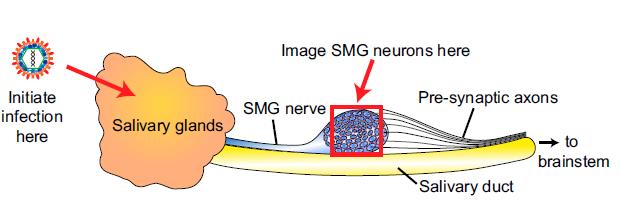
The team studied what happens when a herpes virus infects neurons. For research purposes the investigators used a member of the herpes family called pseudorabies virus. Previous research indicated that these viruses can drill tiny holes in neurons, which pass messages in the form of electrical signals along long conduits known as axons.
The researchers’ findings indicate that electrical current can leak through these holes, or fusion pores, and spread to nearby neurons that were similarly damaged, causing the neurons to fire all at once rather than as needed. The pores were likely created for the purpose of infecting new cells, the researchers said.
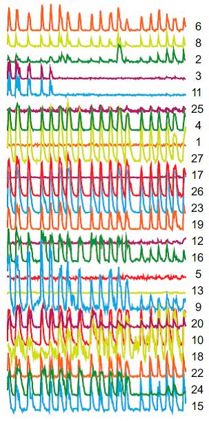
Researchers at Princeton University imaged the synchronized, repetitive firing of herpes-infected neurons in a region known as the submandibular ganglia (SMG) between the salivary glands and the brain in mice. (Source: PNAS)
The investigators observed the cyclical firing of neurons in a region called the submandibular ganglia between the salivary glands and the brain in mice using a technique called 2-photon microscopy and dyes that flash brightly when neurons fire. (Movie of synchronized firing of herpes-infected neurons.)
The team found that two viral proteins appear to work together to cause the simultaneous firing, according to Andréa Granstedt, who received her Ph.D. in molecular biology at Princeton in 2013 and is the first author on the article. The team was led by Lynn Enquist, Princeton’s Henry L. Hillman Professor in Molecular Biology and a member of the Princeton Neuroscience Institute.
Each colored line and number on the right represents an individual neuron. The overlapping peaks indicate synchronized firing of neurons, which occurs when electrical current is able to leak from one neuron to the next. (Source: PNAS)
The first of these two proteins is called glycoprotein B, a fusion protein that drills the holes in the axon wall. A second protein, called Us9, acts as a shuttle that sends glycoprotein B into axons, according to the researchers. “The localization of glycoprotein B is crucial,” Granstedt said. “If glycoprotein B is present but not in the axons, the synchronized flashing won’t happen.”
The researchers succeeded in stopping the short circuit from occurring in engineered viruses that lacked the gene for either glycoprotein B or Us9. Such genetically altered viruses are important as research tools, Enquist said.
Finding a way to block the activity of the proteins could be a useful strategy for treating the pain and itching associated with herpes viral diseases, Enquist said. “If you could block fusion pore formation, you could stop the generation of the signal that is causing pain and discomfort,” he said.
Granstedt conducted the experiments with Jens-Bernhard Bosse, a postdoctoral research associate in molecular biology. Assistance with 2-photon microscopy was provided by Stephan Thiberge, director of the Bezos Center for Neural Circuit Dynamics at the Princeton Neuroscience Institute.
The team previously observed the synchronized firing in laboratory-grown neurons (PLoS Pathogens, 2009), but the new study expands on the previous work by observing the process in live mice and including the contribution of Us9, Granstedt said.
Shingles, which is caused by the virus herpes zoster and results in a painful rash, will afflict almost one out of three people in the United States over their lifetime. Genital herpes, which is caused by herpes simplex virus-2, affects about one out of six people ages 14 to 49 years in the United States, according the Centers for Disease Control and Prevention.
This research was funded by National Institutes of Health (NIH) Grants NS033506 and NS060699. The Imaging Core Facility at the Lewis-Sigler Institute is funded by NIH National Institute of General Medical Sciences Center Grant PM50 GM071508.
Granstedt, Andréa E., Jens B. Bosse, Stephan Y. Thiberge, and Lynn W. Enquist. 2013. In vivo imaging of alphaherpesvirus infection reveals synchronized activity dependent on axonal sorting of viral proteins. PNAS 2013 ; published ahead of print August 26, 2013, doi:10.1073/pnas.1311062110


You must be logged in to post a comment.