Flexibility is key in mechanism of biological self-assembly

By Catherine Zandonella, Office of the Dean for Research

A new study has modeled a crucial first step in the self-assembly of cellular structures such as drug receptors and other protein complexes, and found that the flexibility of the structures has a dramatic impact on how fast they join together.

The study, published this week in the journal Proceedings of the National Academy of Sciences, explored what happens when two water-repelling surfaces connect to build more complex structures. Using molecular simulations, researchers at Princeton University illustrated the mechanism by which the process occurs and explored factors that favor self-assembly.

A surprise finding was the sensitivity with which the surfaces’ flexibility determined the rate at which the surfaces eventually came together, with more flexible surfaces favoring joining. “Flexibility is like a knob that nature can tune to control the self-assembly of molecules,” said Pablo Debenedetti, senior author on the study and Princeton’s Dean for Research. Debenedetti is the Class of 1950 Professor in Engineering and Applied Science and a professor of chemical and biological engineering.

Researchers have long been interested in how biological structures can self-assemble according to physical laws. Tapping the secrets of self-assembly could, for example, lead to new methods of building nanomaterials for future electronic devices. Self-assembled protein complexes are the basis not only of drug receptors but also many other cellular structures, including ion channels that facilitate the transmission of signals in the brain.

The study illustrated the process by which two water-repelling, or hydrophobic, structures come together. At the start of the simulation, the two surfaces were separated by a watery environment. Researchers knew from previous studies that these surfaces, due to their hydrophobic nature, will push water molecules away until only a very few water molecules remain in the gap. The evaporation of these last few molecules allows the two surfaces to snap together.

The new molecular simulation conducted at Princeton yielded a more detailed look at the mechanism behind this process. In the simulation, when the surfaces are sufficiently close to each other, their hydrophobic nature triggered fluctuations in the number of water molecules in the gap, causing the liquid water to evaporate and form bubbles on the surfaces. The bubbles grew as more water molecules evaporated. Eventually two bubbles on either surface connected to form a gap-spanning tube, which expanded and pushed away any remaining water until the two surfaces collided.

Biological surfaces, such as cellular membranes, are flexible, so the researchers explored how the surfaces’ flexibility affected the process. The researchers tuned the flexibility of the surfaces by varying the strength of the coupling between the surface atoms. The stronger the coupling, the less each atom can wiggle relative to its neighbors.

The researchers found that the speed at which the two surfaces snap together depended greatly on flexibility. Small changes in flexibility led to large changes in the rate at which the surfaces stuck together. For example, two very flexible surfaces adhered in just nanoseconds, whereas two inflexible surfaces fused incredibly slowly, on the order of seconds.

Another finding was that the last step in the process, where the vapor tube expands, was critical for ensuring that the surfaces came together. In simulations where the tube failed to expand, the surfaces never joined. Flexibility was key to ensuring that the tube expanded, the researchers found. Making the material more flexible lowered the barriers to evaporation and stabilized the vapor tube, increasing the chances that the tube would expand.

The molecular simulation provides a foundation for understanding how biological structures assemble and function, according to Elia Altabet, a graduate student in Debenedetti’s group, and first author on the study. “A deeper understanding of the formation and function of protein assemblies such as drug receptors and ion channels could inform the design of new drugs to treat diseases,” he said.

Funding for this study was provided by National Science Foundation grants CHE-1213343 and CBET-1263565. Computations were performed at the Terascale Infrastructure for Groundbreaking Research in Engineering and Science (TIGRESS) at Princeton University.

The study, “Effect of material flexibility on the thermodynamics and kinetics of hydrophobically induced evaporation of water,” by Y. Elia Altabet, Amir Haji-Akbari and Pablo Debenedetti, was published online in the journal Proceedings of the National Academy of Sciences the week of March 13, 2017. doi: 10.1073/pnas.1620335114

Scientists capture the elusive structure of essential digestive enzyme (JACS)

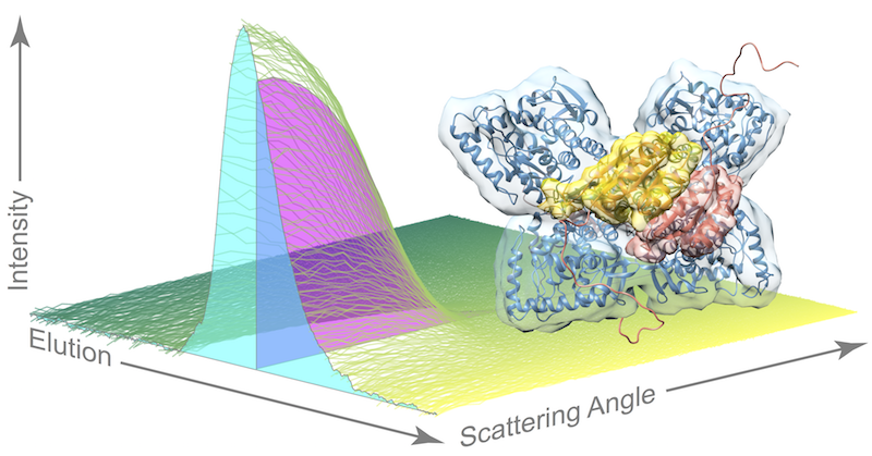
Stylized graphic of data on the structure of an active form of an important digestive enzyme, phenylalanine hydolase. The cyan cross-section shows the elution profile and magenta cross-section shows scattering profile. At right is the structure of the activated phenylalanine hydroxylase. Image source: Ando et al.
Stylized graphic of data on the structure of an important digestive enzyme, phenylalanine hydroxylase. At right is the structure of the activated enzyme. Image source: Ando et al.

By Tien Nguyen, Department of Chemistry

Using a powerful combination of techniques from biophysics to mathematics, researchers have revealed new insights into the mechanism of a liver enzyme that is critical for human health. The enzyme, phenylalanine hydroxylase, turns the essential amino acid phenylalanine – found in eggs, beef and many other foods and as an additive in diet soda —into tyrosine, a precursor for multiple important neurotransmitters.

“We need phenylalanine hydroxylase to control levels of phenylalanine in the blood because too much is toxic to the brain,” said Steve Meisburger, lead author on the study and a post-doctoral researcher in the Ando lab. Genetic mutations in phenylalanine hydroxylase can lead to disorders such as phenylketonuria, an inherited condition that can cause intellectual and behavioral disabilities unless detected at birth and managed through dietary restrictions.

Published earlier this month in the Journal of the American Chemical Society, the article presented detailed structural data on the enzyme’s active state – the shape it adopts when performing its chemical duties – that has eluded scientists for years.

“It’s a floppy enzyme which means it’s dynamic,” said Nozomi Ando, an assistant professor of chemistry at Princeton and corresponding author on the paper. “That also means it doesn’t like to crystallize,” she said. This is problematic for the classic method used to study enzymatic structure, known as x-ray crystallography, which requires solid crystal samples. Efforts to crystallize phenylalanine hydroxylase have just recently met success, but still only captured the enzyme in its inactive state.

The researchers in the Ando lab were able to bypass the tricky task of growing crystals of the active enzyme by using their expertise in a special technique akin to crystallography, called small angle x-ray scattering (SAXS), which allows scientists to study enzymes in a solution. And because the enzyme is susceptible to aggregation or clumping up in solution, the researchers coupled their scattering method with a purification technique called size exclusion chromatography (SEC), in which different species in a sample flow through a column at different speeds based on their size.

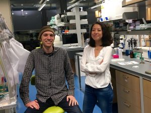
Steve Meisburger (left) and Nozomi Ando (right)

“Pairing SEC with SAXS is an emergent technique. Our contribution is that we saw a clever way to use it,” Ando said. The experiment is highly specialized and relies on powerful x-rays emitted by particles speeding around the circular track at a synchrotron facility. The research team traveled from Princeton to the Cornell High Energy Synchrotron Source in Ithaca, New York, for multiple intensive data-collection sessions. “Any time on the machine that is available, we use it. Not a single photon gets wasted,” Ando said.

As the enzyme solution passes through the purification technique, flowing across the path of the x-ray beam, researchers record snapshots of the x-ray scattering patterns. The resulting dataset is quite complex as the sample also contains phenylalanine, the compound that “turns on” phenylalanine hydroxylase so that researchers can catch the dynamic enzyme in action.

“Current approaches for analyzing this type of dataset are very crude,” Meisburger said. Essentially, these methods assume that each signal – known as an elution peak – represents a single species, when each peak is actually a mixture of species. In this work, the team used an advanced linear algebra method known as evolving factor analysis that allowed them to separate the scattering components. “We can use these linear algebra methods to ‘un-mix’ species that are overlapping,” Meisburger said, “That’s the piece that I think is really exciting.”

By applying their unique approach, the researchers were able to provide evidence for a model of the active structure of phenylalanine hydroxylase that builds upon recent work by their collaborators in Paul Fitzpatrick’s group at UT Health Science Center at San Antonio. In this model, two phenylalanine molecules dock to a pair of sites on the enzyme, bringing a pair of arms together and freeing up the active sites for doing chemistry once more phenylalanine molecules come along.

“I’m very proud that this is our first paper [published since Ando joined the faculty at Princeton]. We wanted it to be very quantitative and heavy on the biochemistry plus heavy on the physical chemistry. I’m really pleased with the way it turned out,” Ando said.

This work was supported by National Health Institutes grants GM100008 and GM098140 and Welch Foundation grant AQ-1245.

Access the paper here:

Meisburger, S. P.; Taylor, A. B.; Khan, C. A.; Zhang, S.; Fitzpatrick, P. F.; Ando, N. “Domain movements upon activation of phenylalanine hydroxylase characterized by crystallography and chromatography-coupled small-angle X-ray scattering.J. Am. Chem. Soc., 2016, 138 (20), pp 6506–6516.DOI: 10.1021/jacs.6b01563. Published online May 4, 2016.


Chemical tracers reveal oxygen-dependent switch in cellular pathway to fat (Nature Chemical Biology)

By Tien Nguyen, Department of Chemistry

Using tracer compounds, scientists have been able to track the cellular production of NADPH, a key coenzyme for making fat, through a pathway that has never been measured directly before.

By tracking this pathway, known as malic enzyme metabolism, which is one of a few recognized routes to make NADPH, researchers from Rabinowitz lab discovered a novel switch in the way fat cells make NADPH depending on the presence of oxygen. The findings were published in Nature Chemical Biology.

Ling Liu (left) and Joshua Rabinowitz (right)

“No one had ever shown an environmental dependent switch in any NADPH production pathway,” said Joshua Rabinowitz, Professor of Chemistry and the Lewis-Sigler Institute for Integrative Genomics at Princeton and principal investigator of the work. “No one had the tools to look,” he said.

NADPH is critical to not only fat synthesis, but also protein and DNA synthesis, and antioxidant defense, implicating it in many diseases such as cancer and diabetes. By understanding and monitoring the pathways through which NADPH is made, scientists can work towards influencing these processes using therapeutic compounds.

The Rabinowitz lab first applied their tracer method in 2014 to study the most well known NADPH production pathway, the oxidative pentose phosphate pathway (oxPPP). The method relied on compounds labeled with deuterium atoms, hydrogen’s heavier cousin, which can be deployed in the cell and measured by a technique called mass spectrometry.

In this work, the researchers extended their method to probe the lesser-known malic enzyme pathway by developing two new, orthogonal tracer compounds specific to this pathway. One tracer, a deuterated succinate compound, enters the cycle more directly but is somewhat challenging for the cell to uptake, while the other, a deuterated glucose molecule, is taken up by the cell readily but takes an extra step to enter the pathway.

The research team investigated the malic enzyme pathway under various concentrations of oxygen. Low oxygen environments, which are found in fat cells in obesity, are of particular clinical interest. They found that in a low oxygen environment, the oxidative pentose phosphate pathway produced more NADPH than the malic enzyme pathway, but in a higher oxygen environment, the pathway contributions completely flipped.

“It’s like the cells are quite clever. They choose the pathway depending on what they want to make, and what nutrients they can access,” said Ling Liu, a graduate student in the Rabinowitz lab and lead author on the work.

One advantage of this method is that it tracks NADPH made specifically in the cytosolic compartment of the cell, whereas the previous leading technique, which relied on tracer compounds with carbon-13 atoms, is unable to differentiate between malic enzyme activity in the cytosol and mitochondria.

NADPH involvement in essential cellular processes has a direct impact on diseases such as diabetes, obesity and cancer. “All of these central biomedical questions depend on an understanding of NADPH pathways, and if you can’t quantify how a metabolite is made and used, you can’t understand what’s going on,” Rabinowitz said. “Ultimately, we’re trying to understand the fundamental chemistry that’s leading to these important biological outcomes,” he said.

Read the full article or abstract:

Liu, L.; Shah, S.; Fan, J.; Park, J. O.; Wellen, K. E.; Rabinowitz, J. D. “Malic enzyme tracers reveal hypoxia-induced switch in adipocyte NADPH pathway usage.Nat. Chem. Bio. Published online March 21, 2016.

This work was supported by the US National Institutes of Health grants R01CA163591, R01AI097382 and P30DK019525 (to the University of Pennsylvania Diabetes Research Center).