‘Acidic patch’ regulates access to genetic information

Histone image

By Pooja Makhijani for the Department of Chemistry

Chromatin remodelers — protein machines that pack and unpack chromatin, the tightly wound DNA-protein complex in cell nuclei — are essential and powerful regulators for critical cellular processes, such as replication, recombination and gene transcription and repression. In a new study published Aug. 2 in the journal Nature, a team led by researchers from Princeton University unravels more details on how a class of ATP-dependent chromatin remodelers, called ISWI, regulate access to genetic information.

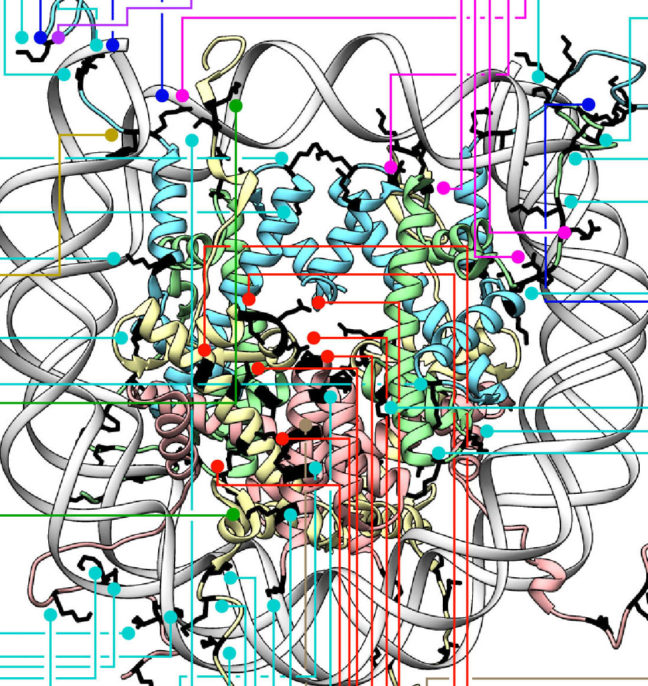
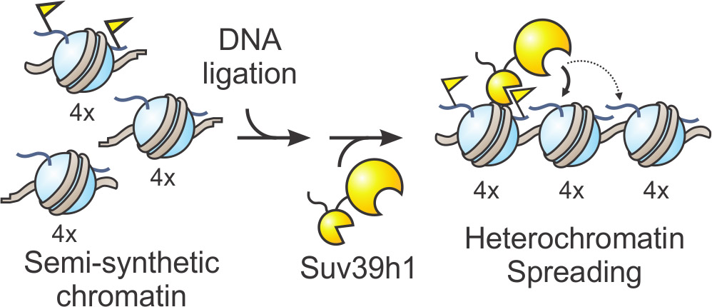
The researchers reported that ISWI remodelers use a structural feature of the nucleosome, known as the “acidic patch,” to remodel chromatin. The nucleosome is the fundamental structural subunit of chromatin, and is often compared to thread wrapped around a spool.

Geoffrey Dann
Geoffrey Dann. Photo by C. Todd Reichart

“The acidic patch is a negatively charged surface, presented on each face of the nucleosome disc, that is formed by amino acids contributed by two different histone proteins, H2A and H2B,” said Geoffrey Dann, a graduate student in the Department of Molecular Biology at Princeton and the study’s lead author. “Histone proteins are overall very positively charged, which makes the negatively charged acidic patch region of the nucleosome very unique. Recognition of the acidic patch has never before been implicated in chromatin remodeling.”

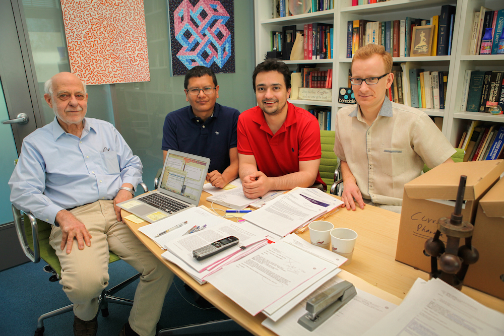
The research was conducted in the laboratory of Tom Muir, the Van Zandt Williams Jr. Class of 1965 Professor of Chemistry and chair of the Department of Chemistry. Research in the Muir group centers on elucidating the physiochemical basis of protein functions in biomedically relevant systems.

Because ISWI remodelers are known to interact extensively with nucleosomes, the researchers hypothesized that signals, in the form of chemical modifications on histone proteins embedded within nucleosomes, communicate to the remodelers on which nucleosome to act. Using high throughput screening technology, an assay process often used in drug discovery, allowed the researchers to quickly conduct tens of thousands of biochemical measurements to test their assumptions. “The number of chromatin modifications known to exist in vivo is astronomical,” Dann said.

Not only did the experiments reveal that ISWI remodelers use the “acidic patch” to remodel chromatin, but also determined that remodeling enzymes outside the family of ISWI remodelers also use this structural feature, “suggesting that this feature may be a general requirement for chromatin remodeling to occur,” Dann said.

Certain chemical modifications that act on histone proteins that are adjacent to the acidic patch also have the ability to enhance or inhibit ISWI remodeling activity, he explained. “A handful of other proteins are known to engage the acidic patch in their interaction with chromatin as well, and we also found that the biochemistry of several of these proteins was affected by such modifications. Interestingly, each protein tested had its own signature response to this collection of modifications.”

The high throughput screening technology method also generated a vast library of data to drive the design of future studies geared toward further understanding ISWI regulation. “This study generated an immense amount of data pointing to many other novel regulatory inputs, in the form of chromatin modifications, into ISWI remodeling activity,” Dann said. “A long-term goal in our lab is to use this data resource as a launch pad for additional studies investigating how chromatin modifications affect ISWI remodeling, and how this plays into the various roles ISWI remodelers assume in the cell.”

histone diagram
Diagram depicting all histone modifications, mutants, and variants present in the 115-member nucleosome library used in this study. Residues modified or mutated were mapped on to the nucleosome in black. H2A (light yellow), H2B (light red), H3 (light blue), and H4 (light green) modification and mutation locations are indicated by boxes and lines. For clarity, connections are only shown to a single copy of each histone protein.

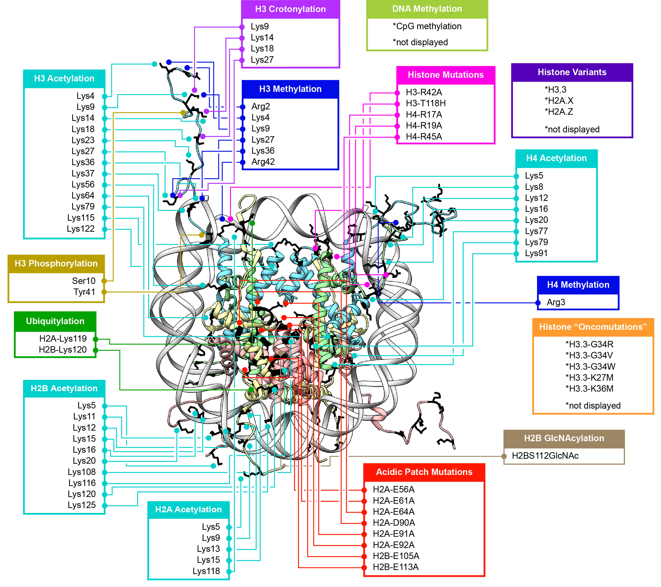
Their findings may also identify a new instrument in cells’ molecular repertoire of chromatin-remodeling tools and spur investigations into potential cancer therapeutic targets. “Mutations in the acidic patch are known to occur in certain types of human cancers, which underscores the emerging importance of the acidic patch in chromatin biology,” Dann said.

The study, “ISWI chromatin remodellers sense nucleosome modifications to determine substrate preference,” was published Aug. 2 by Nature. doi:10.1038/nature23671

The authors at Princeton University were Geoffrey P. Dann, Glen Liszczak, John D. Bagert, Manuel M. Müller, Uyen T. T. Nguyen, Felix Wojcik, Zachary Z. Brown, Jeffrey Bos, Rasmus Pihl, Samuel B. Pollock, Katharine L. Diehl and Tom W. Muir. Also contributing to the study were Tatyana Panchenko & C. David Allis at The Rockefeller University.

The research was funded in part by the German Research Foundation and the National Institutes of Health (GM112365, R01 GM107047).

Read the full article here: https://www.nature.com/nature/journal/vaap/ncurrent/full/nature23671.html


Flexibility is key in mechanism of biological self-assembly

By Catherine Zandonella, Office of the Dean for Research

A new study has modeled a crucial first step in the self-assembly of cellular structures such as drug receptors and other protein complexes, and found that the flexibility of the structures has a dramatic impact on how fast they join together.

The study, published this week in the journal Proceedings of the National Academy of Sciences, explored what happens when two water-repelling surfaces connect to build more complex structures. Using molecular simulations, researchers at Princeton University illustrated the mechanism by which the process occurs and explored factors that favor self-assembly.

A surprise finding was the sensitivity with which the surfaces’ flexibility determined the rate at which the surfaces eventually came together, with more flexible surfaces favoring joining. “Flexibility is like a knob that nature can tune to control the self-assembly of molecules,” said Pablo Debenedetti, senior author on the study and Princeton’s Dean for Research. Debenedetti is the Class of 1950 Professor in Engineering and Applied Science and a professor of chemical and biological engineering.

Researchers have long been interested in how biological structures can self-assemble according to physical laws. Tapping the secrets of self-assembly could, for example, lead to new methods of building nanomaterials for future electronic devices. Self-assembled protein complexes are the basis not only of drug receptors but also many other cellular structures, including ion channels that facilitate the transmission of signals in the brain.

The study illustrated the process by which two water-repelling, or hydrophobic, structures come together. At the start of the simulation, the two surfaces were separated by a watery environment. Researchers knew from previous studies that these surfaces, due to their hydrophobic nature, will push water molecules away until only a very few water molecules remain in the gap. The evaporation of these last few molecules allows the two surfaces to snap together.

The new molecular simulation conducted at Princeton yielded a more detailed look at the mechanism behind this process. In the simulation, when the surfaces are sufficiently close to each other, their hydrophobic nature triggered fluctuations in the number of water molecules in the gap, causing the liquid water to evaporate and form bubbles on the surfaces. The bubbles grew as more water molecules evaporated. Eventually two bubbles on either surface connected to form a gap-spanning tube, which expanded and pushed away any remaining water until the two surfaces collided.

Biological surfaces, such as cellular membranes, are flexible, so the researchers explored how the surfaces’ flexibility affected the process. The researchers tuned the flexibility of the surfaces by varying the strength of the coupling between the surface atoms. The stronger the coupling, the less each atom can wiggle relative to its neighbors.

The researchers found that the speed at which the two surfaces snap together depended greatly on flexibility. Small changes in flexibility led to large changes in the rate at which the surfaces stuck together. For example, two very flexible surfaces adhered in just nanoseconds, whereas two inflexible surfaces fused incredibly slowly, on the order of seconds.

Another finding was that the last step in the process, where the vapor tube expands, was critical for ensuring that the surfaces came together. In simulations where the tube failed to expand, the surfaces never joined. Flexibility was key to ensuring that the tube expanded, the researchers found. Making the material more flexible lowered the barriers to evaporation and stabilized the vapor tube, increasing the chances that the tube would expand.

The molecular simulation provides a foundation for understanding how biological structures assemble and function, according to Elia Altabet, a graduate student in Debenedetti’s group, and first author on the study. “A deeper understanding of the formation and function of protein assemblies such as drug receptors and ion channels could inform the design of new drugs to treat diseases,” he said.

Funding for this study was provided by National Science Foundation grants CHE-1213343 and CBET-1263565. Computations were performed at the Terascale Infrastructure for Groundbreaking Research in Engineering and Science (TIGRESS) at Princeton University.

The study, “Effect of material flexibility on the thermodynamics and kinetics of hydrophobically induced evaporation of water,” by Y. Elia Altabet, Amir Haji-Akbari and Pablo Debenedetti, was published online in the journal Proceedings of the National Academy of Sciences the week of March 13, 2017. doi: 10.1073/pnas.1620335114

Researchers’ Sudoku strategy democratizes powerful tool for genetics research (Nature Communications)

Princeton University researchers Buz Barstow (left), graduate student Kemi Adesina and undergraduate researcher Isao Anzai ’17,
Princeton University researchers Buz Barstow (left), graduate student Kemi Adesina and undergraduate researcher Isao Anzai, Class of 2017, with colleagues at Harvard Universiy, have developed a strategy called “Knockout Sodoku” for figuring out gene function.

By Tien Nguyen, Department of Chemistry

Researchers at Princeton and Harvard Universities have developed a way to produce the tools for figuring out gene function faster and cheaper than current methods, according to new research in the journal Nature Communications.

The function of sizable chunks of many organisms’ genomes is a mystery, and figuring out how to fill these information gaps is one of the central questions in genetics research, said study author Buz Barstow, a Burroughs-Wellcome Fund Research Fellow in Princeton’s Department of Chemistry. “We have no idea what a large fraction of genes do,” he said.

One of the best strategies that scientists have to determine what a particular gene does is to remove it from the genome, and then evaluate what the organism can no longer do. The end result, known as a whole-genome knockout collection, provides full sets of genomic copies, or mutants, in which single genes have been deleted or “knocked out.” Researchers then test the entire knockout collection against a specific chemical reaction. If a mutant organism fails to perform the reaction that means it must be missing the particular gene responsible for that task.

It can take several years and millions of dollars to build a whole-genome knockout collection through targeted gene deletion. Because it’s so costly, whole-genome knockout collections only exist for a handful of organisms such as yeast and the bacterium Escherichia coli. Yet, these collections have proven to be incredibly useful as thousands of studies have been conducted on the yeast gene-deletion collection since its release.

The Princeton and Harvard researchers are the first to create a collection quickly and affordably, doing so in less than a month for several thousand dollars. Their strategy, called “Knockout Sudoku,” relies on a combination of randomized gene deletion and a powerful reconstruction algorithm. Though other research groups have attempted this randomized approach, none have come close to matching the speed and cost of Knockout Sudoku.

“We sort of see it as democratizing these powerful tools of genetics,” said Michael Baym, a co-author on the work and a Harvard Medical School postdoctoral researcher. “Hopefully it will allow the exploration of genetics outside of model organisms,” he said.

Their approach began with steep pizza bills and a technique called transposon mutagenesis that ‘knocks out’ genes by randomly inserting a single disruptive DNA sequence into the genome. This technique is applied to large colonies of microbes to ensure the likelihood that every single gene is disrupted. For example, the team started with a colony of about 40,000 microbes for the bacterium Shewanella oneidensis, which has approximately 3,600 genes in its genome.

Barstow recruited undergraduates and graduate students to manually transfer 40,000 mutants out of laboratory Petri dishes into separate wells using toothpicks. He offered pizza as an incentive, but after a full day of labor, they only managed to move a couple thousand mutants. “I thought to myself, ‘Wait a second, this pizza is going to ruin me,’” Barstow said.

Instead, they decided to rent a colony-picking robot. In just two days, the robot was able to transfer each mutant microbe to individual homes in 96-well plates, 417 plates in total.

But the true challenge and opportunity for innovation was in identifying and cataloging the mutants that could comprise a whole-genome knockout collection in a fast and practical way.

DNA amplification and sequencing is a straightforward way to identify each mutant, but doing it individually quickly gets very expensive and time-consuming. So the researchers’ proposed a scheme in which mutants could be combined into groups that would only require 61 amplification reactions and a single sequencing run.

But still, after sequencing each of the pools, the researchers had an incredible amount of data. They knew the identities of all the mutants, but now they had to figure exactly where each mutant came from in the grid of plates. This is where the Sudoku aspect of the method came in. The researchers built an algorithm that could deduce the location of individual mutants through its repeated appearance in various row, column, plate-row and plate-column pools.

Knockout sodoku helps find genes' functions.

But there’s a problem. Because the initial gene-disruption process is random, it’s possible that the same mutant is formed more than once, which means that playing Sudoku wouldn’t be simple. To find a solution for this issue, Barstow recalled watching the movie, “The Imitation Game,” about Alan Turing’s work on the enigma code, for inspiration.

“I felt like the problem in some ways was very similar to code breaking,” he said. There are simple codes that substitute one letter for another that can be easily solved by looking at the frequency of the letter, Barstow said. “For instance, in English the letter A is used 8.2 percent of the time. So, if you find that the letter X appears in the message about 8.2 percent of the time, you can tell this is supposed to be decoded as an A. This is a very simple example of Bayesian inference.”

With that same logic, Barstow and colleagues developed a statistical picture of what a real location assignment should look like based on a mutant that only appeared once and used that to rate the likelihood of possible locations being real.

“One of the things I really like about this technique is that it’s a prime example of designing a technique with the mathematics in mind at the outset which lets you do much more powerful things than you could do otherwise,” Baym said. “Because it was designed with the mathematics built in, it allows us to get much, much more data out of much less experiments,” he said.

Using their expedient strategy, the researchers created a collection for microbe Shewanella oneidensis. These microbes are especially good at transferring electrons and understanding their powers could prove highly valuable for developing sustainable energy sources, such as artificial photosynthesis, and for environmental remediation in the neutralization of radioactive waste.

Using the resultant collection, the team was able to recapitulate 15 years of research, Barstow said, bolstering their confidence in their method. In an early validation test, they noticed a startlingly poor accuracy rate. After finding no fault with the math, they looked at the original plates to realize that one of the researchers had grabbed the wrong sample. “The least reliable part of this is the human,” Barstow said.

The work was supported by a Career Award at the Scientific Interface from the Burroughs Wellcome Fund and Princeton University startup funds and Fred Fox Class of 1939 funds.

Read the full article here:

Baym, M.; Shaker, L.; Anzai, I. A.; Adesina, O.; Barstow, B. “Rapid construction of a whole-genome transposon insertion collection for Shewanella oneidensis by Knockout Sudoku.” Nature Comm. Available online on Nov. 10, 2016.

Unconventional quasiparticles predicted in conventional crystals (Science)

Fermi arcs on the surface of uncoventional materials
Two electronic states known as Fermi arcs, localized on the surface of a material, stem out of the projection of a 3-fold degenerate bulk new fermion. This new fermion is a cousin of the Weyl fermion discovered last year in another class of topological semimetals. The new fermion has a spin-1, a reflection of the 3- fold degeneracy, unlike the spin-½ that the recently discovered Weyl fermions have.

By Staff

An international team of researchers has predicted the existence of several previously unknown types of quantum particles in materials. The particles — which belong to the class of particles known as fermions — can be distinguished by several intrinsic properties, such as their responses to applied magnetic and electric fields. In several cases, fermions in the interior of the material show their presence on the surface via the appearance of electron states called Fermi arcs, which link the different types of fermion states in the material’s bulk.

The research, published online this week in the journal Science, was conducted by a team at Princeton University in collaboration with researchers at the Donostia International Physics Center (DIPC) in Spain and the Max Planck Institute for Chemical Physics of Solids in Germany. The investigators propose that many of the materials hosting the new types of fermions are “protected metals,” which are metals that do not allow, in most circumstances, an insulating state to develop. This research represents the newest avenue in the physics of “topological materials,” an area of science that has already fundamentally changed the way researchers see and interpret states of matter.

The team at Princeton included Barry Bradlyn and Jennifer Cano, both associate research scholars at the Princeton Center for Theoretical Science; Zhijun Wang, a postdoctoral research associate in the Department of Physics, Robert Cava, the Russell Wellman Moore Professor of Chemistry; and B. Andrei Bernevig, associate professor of physics. The research team also included Maia Vergniory, a postdoctoral research fellow at DIPC, and Claudia Felser, a professor of physics and chemistry and director of the Max Planck Institute for Chemical Physics of Solids.

For the past century, gapless fermions, which are quantum particles with no energy gap between their highest filled and lowest unfilled states, were thought to come in three varieties: Dirac, Majorana and Weyl. Condensed matter physics, which pioneers the study of quantum phases of matter, has become fertile ground for the discovery of these fermions in different materials through experiments conducted in crystals. These experiments enable researchers to explore exotic particles using relatively inexpensive laboratory equipment rather than large particle accelerators.

In the past four years, all three varieties of gapless fermions have been theoretically predicted and experimentally observed in different types of crystalline materials grown in laboratories around the world. The Weyl fermion was thought to be last of the group of predicted quasiparticles in nature. Research published earlier this year in the journal Nature (Wang et al., doi:10.1038/nature17410) has shown, however, that this is not the case, with the discovery of a bulk insulator which hosts an exotic surface fermion.

In the current paper, the team predicted and classified the possible exotic fermions that can appear in the bulk of materials. The energy of these fermions can be characterized as a function of their momentum into so-called energy bands, or branches. Unlike the Weyl and Dirac fermions, which, roughly speaking, exhibit an energy spectrum with 2- and 4-fold branches of allowed energy states, the new fermions can exhibit 3-, 6- and 8-fold branches. The 3-, 6-, or 8-fold branches meet up at points – called degeneracy points – in the Brillouin zone, which is the parameter space where the fermion momentum takes its values.

“Symmetries are essential to keep the fermions well-defined, as well as to uncover their physical properties,” Bradlyn said. “Locally, by inspecting the physics close to the degeneracy points, one can think of them as new particles, but this is only part of the story,” he said.

Cano added, “The new fermions know about the global topology of the material. Crucially, they connect to other points in the Brillouin zone in nontrivial ways.”

During the search for materials exhibiting the new fermions, the team uncovered a fundamentally new and systematic way of finding metals in nature. Until now, searching for metals involved performing detailed calculations of the electronic states of matter.

“The presence of the new fermions allows for a much easier way to determine whether a given system is a protected metal or not, in some cases without the need to do a detailed calculation,” Wang said.

Verginory added, “One can just count the number of electrons of a crystal, and figure out, based on symmetry, if a new fermion exists within observable range.”

The researchers suggest that this is because the new fermions require multiple electronic states to meet in energy: The 8-branch fermion requires the presence of 8 electronic states. As such, a system with only 4 electrons can only occupy half of those states and cannot be insulating, thereby creating a protected metal.

“The interplay between symmetry, topology and material science hinted by the presence of the new fermions is likely to play a more fundamental role in our future understanding of topological materials – both semimetals and insulators,” Cava said.

Felser added, “We all envision a future for quantum physical chemistry where one can write down the formula of a material, look at both the symmetries of the crystal lattice and at the valence orbitals of each element, and, without a calculation, be able to tell whether the material is a topological insulator or a protected metal.”

Read the abstract.

Funding for this study was provided by the US Army Research Office Multidisciplinary University Research Initiative, the US Office of Naval Research, the National Science Foundation, the David and Lucile Packard Foundation, the W. M. Keck Foundation, and the Spanish Ministry of Economy and Competitiveness.

Scientists capture the elusive structure of essential digestive enzyme (JACS)

Stylized graphic of data on the structure of an active form of an important digestive enzyme, phenylalanine hydolase. The cyan cross-section shows the elution profile and magenta cross-section shows scattering profile. At right is the structure of the activated phenylalanine hydroxylase. Image source: Ando et al.
Stylized graphic of data on the structure of an important digestive enzyme, phenylalanine hydroxylase. At right is the structure of the activated enzyme. Image source: Ando et al.

By Tien Nguyen, Department of Chemistry

Using a powerful combination of techniques from biophysics to mathematics, researchers have revealed new insights into the mechanism of a liver enzyme that is critical for human health. The enzyme, phenylalanine hydroxylase, turns the essential amino acid phenylalanine – found in eggs, beef and many other foods and as an additive in diet soda —into tyrosine, a precursor for multiple important neurotransmitters.

“We need phenylalanine hydroxylase to control levels of phenylalanine in the blood because too much is toxic to the brain,” said Steve Meisburger, lead author on the study and a post-doctoral researcher in the Ando lab. Genetic mutations in phenylalanine hydroxylase can lead to disorders such as phenylketonuria, an inherited condition that can cause intellectual and behavioral disabilities unless detected at birth and managed through dietary restrictions.

Published earlier this month in the Journal of the American Chemical Society, the article presented detailed structural data on the enzyme’s active state – the shape it adopts when performing its chemical duties – that has eluded scientists for years.

“It’s a floppy enzyme which means it’s dynamic,” said Nozomi Ando, an assistant professor of chemistry at Princeton and corresponding author on the paper. “That also means it doesn’t like to crystallize,” she said. This is problematic for the classic method used to study enzymatic structure, known as x-ray crystallography, which requires solid crystal samples. Efforts to crystallize phenylalanine hydroxylase have just recently met success, but still only captured the enzyme in its inactive state.

The researchers in the Ando lab were able to bypass the tricky task of growing crystals of the active enzyme by using their expertise in a special technique akin to crystallography, called small angle x-ray scattering (SAXS), which allows scientists to study enzymes in a solution. And because the enzyme is susceptible to aggregation or clumping up in solution, the researchers coupled their scattering method with a purification technique called size exclusion chromatography (SEC), in which different species in a sample flow through a column at different speeds based on their size.

Steve Meisburger (left) and Nozomi Ando (right)

“Pairing SEC with SAXS is an emergent technique. Our contribution is that we saw a clever way to use it,” Ando said. The experiment is highly specialized and relies on powerful x-rays emitted by particles speeding around the circular track at a synchrotron facility. The research team traveled from Princeton to the Cornell High Energy Synchrotron Source in Ithaca, New York, for multiple intensive data-collection sessions. “Any time on the machine that is available, we use it. Not a single photon gets wasted,” Ando said.

As the enzyme solution passes through the purification technique, flowing across the path of the x-ray beam, researchers record snapshots of the x-ray scattering patterns. The resulting dataset is quite complex as the sample also contains phenylalanine, the compound that “turns on” phenylalanine hydroxylase so that researchers can catch the dynamic enzyme in action.

“Current approaches for analyzing this type of dataset are very crude,” Meisburger said. Essentially, these methods assume that each signal – known as an elution peak – represents a single species, when each peak is actually a mixture of species. In this work, the team used an advanced linear algebra method known as evolving factor analysis that allowed them to separate the scattering components. “We can use these linear algebra methods to ‘un-mix’ species that are overlapping,” Meisburger said, “That’s the piece that I think is really exciting.”

By applying their unique approach, the researchers were able to provide evidence for a model of the active structure of phenylalanine hydroxylase that builds upon recent work by their collaborators in Paul Fitzpatrick’s group at UT Health Science Center at San Antonio. In this model, two phenylalanine molecules dock to a pair of sites on the enzyme, bringing a pair of arms together and freeing up the active sites for doing chemistry once more phenylalanine molecules come along.

“I’m very proud that this is our first paper [published since Ando joined the faculty at Princeton]. We wanted it to be very quantitative and heavy on the biochemistry plus heavy on the physical chemistry. I’m really pleased with the way it turned out,” Ando said.

This work was supported by National Health Institutes grants GM100008 and GM098140 and Welch Foundation grant AQ-1245.

Access the paper here:

Meisburger, S. P.; Taylor, A. B.; Khan, C. A.; Zhang, S.; Fitzpatrick, P. F.; Ando, N. “Domain movements upon activation of phenylalanine hydroxylase characterized by crystallography and chromatography-coupled small-angle X-ray scattering.J. Am. Chem. Soc., 2016, 138 (20), pp 6506–6516.DOI: 10.1021/jacs.6b01563. Published online May 4, 2016.


Theorists smooth the way to solving one of quantum mechanics oldest problems: Modeling quantum friction (J. Phys. Chem. Letters)

Researchers at Princeton
From left to right: Herschel Rabitz, Renan Cabrera, Andre Campos and Denys Bondar. Photo credit: C. Todd Reichart

By: Tien Nguyen, Department of Chemistry

Theoretical chemists at Princeton University have pioneered a strategy for modeling quantum friction, or how a particle’s environment drags on it, a vexing problem in quantum mechanics since the birth of the field. The study was published in the Journal of Physical Chemistry Letters.

“It was truly a most challenging research project in terms of technical details and the need to draw upon new ideas,” said Denys Bondar, a research scholar in the Rabitz lab and corresponding author on the work.

Quantum friction may operate at the smallest scale, but its consequences can be observed in everyday life. For example, when fluorescent molecules are excited by light, it’s because of quantum friction that the atoms are returned to rest, releasing photons that we see as fluorescence. Realistically modeling this phenomenon has stumped scientists for almost a century and recently has gained even more attention due to its relevance to quantum computing.

“The reason why this problem couldn’t be solved is that everyone was looking at it through a certain lens,” Bondar said. Previous models attempted to describe quantum friction by considering the quantum system as interacting with a surrounding, larger system. This larger system presents an impossible amount of calculations, so in order to simplify the equations to the pertinent interactions, scientists introduced numerous approximations.

These approximations led to numerous different models that could each only satisfy one or the other of two critical requirements. In particular, they could either produce useful observations about the system, or they could obey the Heisenberg Uncertainty Principle, which states that there is a fundamental limit to the precision with which a particle’s position and momentum can be simultaneous measured. Even famed physicist Werner Heisenberg’s attempt to derive an equation for quantum friction was incompatible with his own uncertainty principle.

The researchers’ approach, called operational dynamic modeling (ODM) and introduced in 2012 by the Rabitz group, led to the first model for quantum friction to satisfy both demands. “To succeed with the problem, we had to literally rethink the physics involved, not merely mathematically but conceptually,” Bondar said.

Bondar and his colleagues focused on the two ultimate requirements for their model – that it should obey the Heisenberg principle and produce real observations – and worked backwards to create the proper model.

“Rather than starting with approximations, Denys and the team built in the proper physics in the beginning,” said Herschel Rabitz, the Charles Phelps Smyth ’16 *17 Professor of Chemistry and co-author on the paper. “The model is built on physical and mathematical truisms that must hold. This distinct approach creates a new rigorous and practical formulation for quantum friction,” he said.

The research team included research scholar Renan Cabrera and Ph.D. student Andre Campos as well as Shaul Mukamel, professor of chemistry at the University of California, Irvine.

Their model opens a way forward to understand not only quantum friction but other dissipative phenomena as well. The researchers are interested in exploring the means to manipulate these forces to their advantage. Other theorists are rapidly taking up the new paradigm of operational dynamic modeling, Rabitz said.

Reflecting on how they arrived at such a novel approach, Bondar recalled the unique circumstances under which he first started working on this problem. After he received the offer to work at Princeton, Bondar spent four months awaiting a US work visa (he is a citizen of the Ukraine) and pondering fundamental physics questions. It was during this time that he first thought of this strategy. “The idea was born out of bureaucracy, but it seems to be holding up,” Bondar said.

Read the full article here:

Bondar, D. I.; Cabrera, R.; Campos, A.; Mukamel, S.; Rabitz, H. A. “Wigner-Lindblad Equations for Quantum Friction.J. Phys. Chem. Lett. 2016, 7, 1632.

This work was supported by the US National Science Foundation CHE 1058644, the US Department of Energy DE-FG02-02ER-15344, and ARO-MURI W911NF-11-1-0268.

Chemical tracers reveal oxygen-dependent switch in cellular pathway to fat (Nature Chemical Biology)

By Tien Nguyen, Department of Chemistry

Using tracer compounds, scientists have been able to track the cellular production of NADPH, a key coenzyme for making fat, through a pathway that has never been measured directly before.

By tracking this pathway, known as malic enzyme metabolism, which is one of a few recognized routes to make NADPH, researchers from Rabinowitz lab discovered a novel switch in the way fat cells make NADPH depending on the presence of oxygen. The findings were published in Nature Chemical Biology.

Ling Liu (left) and Joshua Rabinowitz (right)

“No one had ever shown an environmental dependent switch in any NADPH production pathway,” said Joshua Rabinowitz, Professor of Chemistry and the Lewis-Sigler Institute for Integrative Genomics at Princeton and principal investigator of the work. “No one had the tools to look,” he said.

NADPH is critical to not only fat synthesis, but also protein and DNA synthesis, and antioxidant defense, implicating it in many diseases such as cancer and diabetes. By understanding and monitoring the pathways through which NADPH is made, scientists can work towards influencing these processes using therapeutic compounds.

The Rabinowitz lab first applied their tracer method in 2014 to study the most well known NADPH production pathway, the oxidative pentose phosphate pathway (oxPPP). The method relied on compounds labeled with deuterium atoms, hydrogen’s heavier cousin, which can be deployed in the cell and measured by a technique called mass spectrometry.

In this work, the researchers extended their method to probe the lesser-known malic enzyme pathway by developing two new, orthogonal tracer compounds specific to this pathway. One tracer, a deuterated succinate compound, enters the cycle more directly but is somewhat challenging for the cell to uptake, while the other, a deuterated glucose molecule, is taken up by the cell readily but takes an extra step to enter the pathway.

The research team investigated the malic enzyme pathway under various concentrations of oxygen. Low oxygen environments, which are found in fat cells in obesity, are of particular clinical interest. They found that in a low oxygen environment, the oxidative pentose phosphate pathway produced more NADPH than the malic enzyme pathway, but in a higher oxygen environment, the pathway contributions completely flipped.

“It’s like the cells are quite clever. They choose the pathway depending on what they want to make, and what nutrients they can access,” said Ling Liu, a graduate student in the Rabinowitz lab and lead author on the work.

One advantage of this method is that it tracks NADPH made specifically in the cytosolic compartment of the cell, whereas the previous leading technique, which relied on tracer compounds with carbon-13 atoms, is unable to differentiate between malic enzyme activity in the cytosol and mitochondria.

NADPH involvement in essential cellular processes has a direct impact on diseases such as diabetes, obesity and cancer. “All of these central biomedical questions depend on an understanding of NADPH pathways, and if you can’t quantify how a metabolite is made and used, you can’t understand what’s going on,” Rabinowitz said. “Ultimately, we’re trying to understand the fundamental chemistry that’s leading to these important biological outcomes,” he said.

Read the full article or abstract:

Liu, L.; Shah, S.; Fan, J.; Park, J. O.; Wellen, K. E.; Rabinowitz, J. D. “Malic enzyme tracers reveal hypoxia-induced switch in adipocyte NADPH pathway usage.Nat. Chem. Bio. Published online March 21, 2016.

This work was supported by the US National Institutes of Health grants R01CA163591, R01AI097382 and P30DK019525 (to the University of Pennsylvania Diabetes Research Center).

Electrons slide through the hourglass on surface of bizarre material (Nature)

An illustration of the hourglass fermion predicted to lie on the surface of crystals of potassium mercury antimony. (Bernevig et al., Princeton University)
An illustration of the hourglass fermion predicted to lie on the surface of crystals of potassium mercury antimony. (Image credit: Laura R. Park and Aris Alexandradinata)

By Staff

A team of researchers at Princeton University has predicted the existence of a new state of matter in which current flows only through a set of surface channels that resemble an hourglass. These channels are created through the action of a newly theorized particle, dubbed the “hourglass fermion,” which arises due to a special property of the material. The tuning of this property can sequentially create and destroy the hourglass fermions, suggesting a range of potential applications such as efficient transistor switching.

In an article published in the journal Nature this week, the researchers theorize the existence of these hourglass fermions in crystals made of potassium and mercury combined with either antimony, arsenic or bismuth. The crystals are insulators in their interiors and on their top and bottom surfaces, but perfect conductors on two of their sides where the fermions create hourglass-shaped channels that enable electrons to flow.

The research was performed by Princeton University postdoctoral researcher Zhi Jun Wang and former graduate student Aris Alexandradinata, now a postdoctoral researcher at Yale University, working with Robert Cava, Princeton’s Russell Wellman Moore Professor of Chemistry, and Associate Professor of Physics B. Andrei Bernevig.

The new hourglass fermion exists – theoretically for now, until detected experimentally – in a family of materials broadly called topological insulators, which were first observed experimentally in the mid-2000s and have since become one of the most active and interesting branches of quantum physics research. The bulk, or interior, acts as an insulator, which means it prohibits the travel of electrons, but the surface of the material is conducting, allowing electrons to travel through a set of channels created by particles known as Dirac fermions.

Fermions are a family of subatomic particles that include electrons, protons and neutrons, but they also appear in nature in many lesser known forms such as the massless Dirac, Majorana and Weyl fermions. After years of searching for these particles in high-energy accelerators and other large-scale experiments, researchers found that they can detect these elusive fermions in table-top laboratory experiments on crystals. Over the past few years, researchers have used these “condensed matter” systems to first predict and then confirm the existence of Majorana and Weyl fermions in a wide array of materials.

The next frontier in condensed matter physics is the discovery of particles that can exist in the so-called “material universe” inside crystals but not in the universe at large. Such particles come about due to the properties of the materials but cannot exist outside the crystal the way other subatomic particles do. Classifying and discovering all the possible particles that can exist in the material universe is just beginning. The work reported by the Princeton team lays the foundations of one of the most interesting of these systems, according to the researchers.

In the current study, the researchers theorize that the laws of physics prohibit current from flowing in the crystal’s bulk and top and bottom surfaces, but permit electron flow in completely different ways on the side surfaces through the hourglass-shaped channels. This type of channel, known more precisely as a dispersion, was completely unknown before.

The researchers then asked whether this dispersion is a generic feature found in certain materials or just a fluke arising from a specific crystal model.

It turned out to be no fluke.

A long-standing collaboration with Cava, a material science expert, enabled Bernevig, Wang, and Alexandradinata to uncover more materials exhibiting this remarkable behavior.

“Our hourglass fermion is curiously movable but unremovable,” said Bernevig. “It is impossible to remove the hourglass channel from the surface of the crystal.”

Bernevig explained that this robust property arises from the intertwining of spatial symmetries, which are characteristics of the crystal structure, with the modern band theory of crystals. Spatial symmetries in crystals are distinguished by whether a crystal can be rotated or otherwise moved without altering its basic character.

In a paper published in Physical Review X this week to coincide with the Nature paper, the team detailed the theory behind how the crystal structure leads to the existence of the hourglass fermion.

An illustration of the complicated dispersion of the surface fermion arising from a background of mercury and bismuth atoms (blue and red). (Image credit: Mingyee Tsang and Aris Alexandradinata)

“Our work demonstrates how this basic geometric property gives rise to a new topology in band insulators,” Alexandradinata said. The hourglass is a robust consequence of spatial symmetries that translate the origin by a fraction of the lattice period, he explained. “Surface bands connect one hourglass to the next in an unbreakable zigzag pattern,” he said.

The team found esoteric connections between their system and high-level mathematics. Origin-translating symmetries, also called non-symmorphic symmetries, are described by a field of mathematics called cohomology, which classifies all the possible crystal symmetries in nature. For example, cohomology gives the answer to how many crystal types exist in three spatial dimensions: 230.

In the cohomological perspective, there are 230 ways to combine origin-preserving symmetries with real-space translations, known as the “space groups.” The theoretical framework to understand the crystals in the current study requires a cohomological description with momentum-space translations.

“The hourglass theory is the first of its kind that describes time-reversal-symmetric crystals, and moreover, the crystals in our study are the first topological material class which relies on origin-translating symmetries,” added Wang.

Out of the 230 space groups in which materials can exist in nature, 157 are non-symmorphic, meaning they can potentially host interesting electronic behavior such as the hourglass fermion.

“The exploration of the behavior of these interesting fermions, their mathematical description, and the materials where they can be observed, is poised to create an onslaught of activity in quantum, solid state and material physics,” Cava said. “We are just at the beginning.”

The study was funded by the National Science Foundation, the Office of Naval Research, the David and Lucile Packard Foundation, the W. M. Keck Foundation, and the Eric and Wendy Schmidt Transformative Technology Fund at Princeton University.

The paper, “Hourglass fermions” by Zhijun Wang, A. Alexandradinata, R. J. Cava and B. Andrei Bernevig, was published in the April 14, 2016 issue of Nature, 532,189–194, doi:10.1038/nature17410. Read the preprint.

The paper, “Topological insulators from group cohomology” by A. Alexandradinata, Zhijun Wang, and B. Andrei Bernevig, was published in the April 15, 2016 issue of Phys. Rev. X 6, 021008.

How an artificial protein rescues dying cells (PNAS)

By Tien Nguyen, Department of Chemistry

A new study from Princeton has revealed how a synthetic protein revives E. coli cells that lack a life-sustaining gene, offering insight into how life can adapt to survive and potentially be reinvented.

Michael Hecht and Katie Digianantonio

Researchers in the Hecht lab discovered the unexpected way in which a synthetic protein called SynSerB promotes the growth of cells that lack the natural SerB gene, which encodes an enzyme responsible for the last step in the production of the essential amino acid serine. The findings were published in the Proceedings of the National Academy of Sciences.

The Hecht group first discovered SynSerB’s ability to rescue serine-depleted E. Coli cells in 2011. At that time, they also discovered several other de novo proteins capable of rescuing the deletions of three other essential proteins in E. coli. “These are novel proteins that have never existed on Earth, and aren’t related to anything on Earth yet they enable life to grow where it otherwise would not,” said Michael Hecht, professor of chemistry at Princeton and corresponding author on the article.

Natural proteins are complex molecular machines constructed from a pool of twenty different amino acids. Typically they range from several dozen to several hundred amino acids in length.  In principle, there are more possible protein sequences than atoms in the universe, but through evolution Nature has selected just a small fraction to carry out the cellular functions that make life possible.

“Those proteins must be really special,” Hecht said. “The driving question was, ‘Can we do that in the laboratory? Can we come up with non-natural sequences that are that special, from an enormous number of possibilities?”

To address this question, the Hecht lab developed a library of non-natural proteins guided by a concept called binary design. The idea was to narrow down the number of possible sequences by choosing from eleven select amino acids that were divided into two groups: polar and non-polar. By using only the polar or non-polar characteristics of those amino acids, the researchers could design a plethora of novel proteins to fold into a particular shape based on their affinity to and repulsion from water. Then, by allowing the specific positions to have different amino acids, the researchers were able to produce a diverse library of about one million proteins, each 102 amino acids long.

“We had to focus on certain subsets of proteins that we knew would fold and search there first for function,” said Katie Digianantonio, a graduate student in the Hecht lab and first author on the paper. “It’s like instead of searching the whole universe for life, we’re looking in specific solar systems.”

Having found several non-natural proteins that could rescue specific cell lines, this latest work details their investigation specifically into how SynSerB promotes cell growth. The most obvious explanation, that SynSerB simply catalyzed the same reaction performed by the deleted SerB gene, was discounted by an early experiment.

To discern SynSerB’s mechanism among the multitude of complex biochemical pathways in the cell, the researchers turned to a technique called RNA sequencing. This technique allowed them to take a detailed snapshot of the serine-depleted E. Coli cells with and without their synthetic protein and compare the differences.

“Instead of guessing and checking, we wanted to look at the overall environment to see what was happening,” Digianantonio said. The RNA sequencing experiment revealed that SynSerB induced overexpression of a protein called HisB, high levels of which have been shown to promote the key reaction normally performed by the missing gene. By enlisting the help of HisB, the non-natural protein was able to induce the production of serine, which ultimately allowed the cell to survive.

“Life is opportunistic. Some proteins are going to work by acting similarly to what they replaced and some will find another pathway,” Hecht said. “Either way it’s cool.”

Read the full articles here:

Digianantonio, K. M.; Hecht, M. H. “A protein constructed de novo enables cell growth by altering gene regulation.Proc. Natl. Acad. Sci. Published online before print on Feb. 16, 2016.

Fisher, M. A.; McKinley, K. L.; Bradley, L. H; Viola, S. R; Hecht, M. H. “De Novo designed proteins from a library of artificial sequences function in Escherichia Coli and enable cell growth.” PLoS One 2011, 6(1): e15364.

This research was funded by the US National Science Foundation (NSF) Grant MCB-1050510.

Study reveals mechanism behind enzyme that tags unneeded DNA (Nature Chem. Bio.)

Designer chromatin experiments
Graphical representation of designer chromatin experiments. Image courtesy of the Muir lab.

By Tien Nguyen, Department of Chemistry

Researchers have discovered the two-step process that activates an essential human enzyme, called Suv39h1, which is responsible for organizing large portions of the DNA found in every living cell.

For any particular cell, such as a skin or brain cell, much of this genetic information is extraneous and must be packed away to allow sufficient space and resources for more important genes. Failure to properly pack DNA jeopardizes the stability of chromosomes and can result in severe diseases. Suv39h1 is one of the main enzymes that chemically mark the irrelevant regions of DNA to be compacted by cellular machinery, but little is known about how it installs its tag.

Now, scientists at Princeton have used ‘designer chromatin’ templates – highly customized replicas of cellular DNA and histone proteins, the scaffolding proteins around which DNA is wrapped – to reveal new details about Suv39h1’s mechanism. The researchers investigated how Suv39h1 employs a positive feedback loop to chemically tag thousands of adjacent histones, thus signaling the cell to stow away these underlying, unnecessary DNA sequences. The work was published in in the journal Nature Chemical Biology.

“One of the things that has always fascinated me about feedback loops is that they’re super dangerous. If you make a mistake once, you end up getting reinforcement through the feedback loop,” said Manuel Müller, a postdoctoral researcher in the Muir lab and lead author on the study. “So how does Suv39h1 keep itself in check?”

Suv39h1 had been known to possess two distinct parts, but the new research revealed how they work together in order to ‘switch on’ the enzyme. One part of the enzyme, known as the chromodomain, is constantly exposed and seeks out specific chemical tags, known as a methyl groups, located at predetermined sites on histones. When the chromodomain finds these groups in the genome, it locks onto the spot and allows the other part, the enzymatic core, to install more methyl tags at adjacent histones.

“The second, anchoring step wasn’t really known before. It provides an extra level of control and allows the process to be extremely fine-tuned,” Müller said. A similar mechanism may be employed by many other enzymes operating on chromatin, given that they contain similar components of a feedback loop.

To understand how the enzyme carries out this process, the researchers synthesized complex chromatin templates that were three times larger than previously reported models. They divided the template into three blocks that could each be manipulated in various ways. For example, a block could be prepared with the chemical tag present, absent or mutated such that tagging can’t occur. “The different blocks should signal to the enzyme either start here or feel free to spread here or absolutely stop here,” said Glen Liszczak, a co-author and postdoctoral researcher in the Muir lab.

By rearranging the various domains, the research team observed where the enzyme spread its mark across the genome. They found that Suv39h1 preferred to spread across small distances, but that it could reach sequences further along if chromatin folding decreased the physical distance in space.

“We’ve learned something new about this enzyme, something that we couldn’t have without the pinpoint precision that the designer chromatin offers,” Liszczak said. “There are a lot of questions that our lab has been interested in that we can now start to answer.”

The research was funded by the Swiss National Science Foundation (postdoctoral fellowships) and the US National Institutes of Health (R01-GM107047).

Read the abstract or full article.

Müller, M. M.; Fierz, B.; Bittova, L.; Liszczak, G.; Muir, T. W. “A two-state activation mechanism controls the histone methyltransferase Suv39h1.” Nature Chem. Bio. Available online January 25, 2016.