Antibiotic’s killer strategy revealed (PNAS)

Marine algae
Satellite image of a E. huxleyi marine algae bloom. (Image: NASA)

By Tien Nguyen, Department of Chemistry

Using a special profiling technique, scientists at Princeton have determined the mechanism of action of a potent antibiotic, known as tropodithietic acid (TDA), leading them to uncover its hidden ability as a potential anticancer agent.

TDA is produced by marine bacteria belonging to the roseobacter family, which exist in a unique symbiosis with microscopic algae. The algae provide food for the bacteria, and the bacteria provide protection from the many pathogens of the open ocean.

“This molecule keeps everything out,” said Mohammad Seyedsayamdost, an assistant professor of chemistry at Princeton and corresponding author on the study published in the Proceedings of the National Academy of Science. “How could something so small be so broad spectrum? That’s what got us interested,” he said.

In collaboration with researchers in the laboratory of Zemer Gitai, an associate professor of molecular biology at Princeton, the team used a laboratory technique referred to as bacterial cytological profiling to investigate the mode of action of TDA. This method involves destroying bacterial cells with the antibiotic in the presence of a set of dyes, and then visually assessing the aftermath. “The key assumption is that dead cells that look the same probably died by the same mechanism,” he said.
marine algae
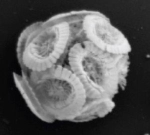
Scanning electron microscope image of E. huxleyi (Image credit. M. Seyedsayamdost)

The team used three dyes to evaluate 13 different features of the deceased cells, such as cell membrane thickness and nucleoid area, comprising TDA’s cytological profile. By comparing to profiles of known drugs, the researchers found a match with a class of compounds called polyethers, which possess anticancer activity.

Given their similar profiles, Seyedsayamdost and coworkers hypothesized that TDA might exhibit anticancer properties as well, and indeed observed its strong anticancer activity in a screen against 60 different cancer cell lines. “The strength of this profiling technique is that it tells you how to repurpose molecules,” Seyedsayamdost said.

The researchers were surprised by the compounds’ shared mode of action because unlike the small sized TDA, polyether compounds are quite large. But through different chemical reactions, they are both able to cause chemical disruptions in the cell membrane that render the bacterium unable to produce the energy needed to perform critical tasks, such as cell division and making proteins.

In addition to TDA’s killing mechanism, the researchers were interested in understanding the mechanism by which a bacterial strain could become resistant to the antibiotic. Particularly, they wondered how the marine roseobacter kept itself safe from the deadly antibiotic weapon that it produced.

The research team approached the task by probing the genes in roseobacter that synthesize TDA as well as the surrounding genes. They identified three nearby genes responsible for transport in and out of the cell, and upon transferring these specific genes to E. coli, were able to produce an elusive TDA resistant bacterial strain.

“We often look at natural products as black boxes,” said Seyedsayamdost, “but these molecules have evolved for millennia to fulfill a certain function. By linking the unusual structural features of TDA to its mode of action, we have begun to explain why TDA looks the way it does.”

Read the abstract:

Wilson, M. Z.; Wang, R.; Gitai, Z.; Seyedsayamdost, M. R. “Tropodithietic Acid: Mode of Action and Mechanism of Resistance.” Proc. Natl. Acad. Sci. 2016, Published online on January 22, 2016.

This work was supported by grants from the National Institutes of Health (GM 098299 and 1DPOD004389).

‘Radiolabeling’ lets scientists track the breakdown of drugs (Nature)

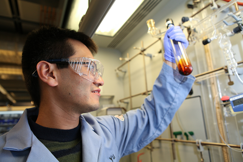
Graduate student Renyuan Pony Yu
Renyuan Pony Yu, a graduate student working with Princeton Professor Paul Chirik, has discovered a new way to radiolabel compounds for use in drug development.

By Tien Nguyen, Department of Chemistry

A new method for labeling molecules with radioactive elements could let chemists more easily track how drugs under development are metabolized in the body.

Chemists consider thousands of compounds in the search for a new drug, and a candidate’s metabolism is a key factor that must be evaluated carefully and quickly. Researchers at Princeton University and pharmaceutical company Merck & Co., Inc. report in the journal Nature that scientists can selectively replace hydrogen atoms in molecules with tritium atoms — a radioactive form of hydrogen that possesses two extra neutrons — to “radiolabel” compounds. This technique can be done in a single step while preserving the biological properties of the parent compound.

While current state-of-the-art techniques are quite reliable, they only work when dissolved in specific solvents, ones that aren’t always capable of dissolving the drug compound of interest. The researchers’ method, however, used an iron-based catalyst that is tolerant to a wider variety of solvents, and it labels the molecules at the opposite positions as compared to existing methods.

“The fact that you can access other positions is what makes this reaction really special,” said corresponding author Paul Chirik, the Edwards S. Sanford Professor of Chemistry at Princeton. Previous methods only incorporate radioactive tritium atoms into the molecule directly next to an atom or a group of atoms called a directing group. The new iron-catalyzed method does not require a directing group, and instead places tritium at whatever positions in the molecules are the least crowded.

“Radiolabeled compounds help medicinal chemists get a better picture of what actually happens to the drug by showing how the drug is metabolized and cleared,” said David Hesk, a collaborator at Merck and co-author on the work. By rapidly assessing the compounds’ metabolism early on, scientists can shorten the time it takes to develop and bring a drug to market. “Having another labeling reaction is very powerful because it gives radiochemists another tool in the toolbox,” he said.

This unique reactivity was actually discovered unexpectedly. Renyuan Pony Yu, a graduate student in the Chirik lab, had originally set out to use their iron catalyst for a different reaction that they were collaborating on with Merck. To study the iron catalyst’s capabilities, Yu subjected it to a technique called proton nuclear magnetic resonance spectroscopy (NMR), which allows chemists to deduce the positions of hydrogen atoms in molecules.

“We started seeing this beautiful, very systematic pattern of signals in the NMR, but we didn’t really know what they were,” said Yu, who is first author on the new study. Particularly puzzling was the fact that the pattern of signals would disappear over time.

The researchers turned to Istvan Pelczer, Director of the NMR Facility at Princeton chemistry and co-author on the work, who developed a special technique that helped them analyze the signals with much greater confidence. Using this method, they realized that the iron catalyst was reacting with the liquid solvent used to dissolve the NMR sample. The solvent’s deuterium atoms, another form of hydrogen that has one extra neutron and is not radioactive, were replacing the hydrogen atoms.

It wasn’t until Yu presented his findings to Matt Tudge, the Princeton authors’ collaborator at Merck, that the catalyst’s potential to introduce tritium atoms into radiolabeled molecules was recognized. “This is a classic example where you really need both partners,” Chirik said. “We were the catalyst experts, but they were the applications experts.”

Though tritium-labeled compounds are used mostly in metabolism studies, they can also be helpful at the very outset of a drug-discovery project to identify a biological target that the potential drugs can be tested against. The biological target could be an enzyme or protein associated with a certain disease. For example, statins are a well-known class of cholesterol-lowering drugs that target a specific enzyme in the body called HMG-CoA reductase.

To explore the scope of the reaction, Yu first optimized the reaction to incorporate deuterium atoms, which is commonly accepted as a model system for tritium. He found that the iron catalyst was surprisingly robust and successfully labeled many different types of compounds, including some from Merck’s library of past drug candidates.

“It was a very exciting project for me because I got to work with real drugs that are fully functionalized and useful,” Yu said. One of their test substrates was Claritin, which Yu bought from a local store; he extracted its active ingredient back in the lab.

Finally, Yu traveled to Merck’s campus in Rahway, where he received radioactivity training — Chirik’s laboratory isn’t equipped to handle radioactivity — and performed the reactions using tritium gas. The reactions were run in a special apparatus that looks like a steel-lined box and releases radioactive tritium gas. The apparatus can capture any unspent gas to limit the amount of radioactive waste produced.

Chemists take care to handle radioactive compounds and waste very carefully, but tritium’s radioactivity is so weak that the particles it emits cannot penetrate simple glassware. For this reason, tritium-labeled compounds can’t be used in any human imaging studies such as PET scans, which require radiolabeled compounds that emit high-energy particles.

This past summer, Yu presented the preliminary results of the iron-catalyzed reaction at the 2015 International Isotope Society Symposia to researchers in the radiolabeling and pharmaceutical community. They were very excited about the research and eager to use the catalyst in their own studies, Yu said.

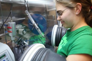
But the major challenge for the researchers is that the iron catalyst is extremely air and moisture sensitive, and it can only be handled inside a glovebox, a special chamber in which oxygen and water vapor have been excluded. The Chirik group is working to develop a more stable catalyst that can be made commercially available, and have recently entered into a partnership with Green Center Canada, a company that helps bring academic research to market.

In the meantime, the Chirik group has found that the iron catalyst can replace hydrogen atoms with other groups besides deuterium and tritium atoms and is extending this chemistry into many other projects in the lab.

“This project is always going to be a special one for me because it’s kind of a pivot point for the type of chemistry that our group can do,” Chirik said, “and there’s this really cool application.”

Read the abstract.

Yu, R. P.; Hesk, D.; Rivera, N.; Pelczer, I.; Chirik, P. J. “Iron-Catalyzed Tritiation of Pharmaceuticals.” Nature, 2016, DOI: 10.1038/nature16464.

This work was supported by Merck & Co. and Princeton University’s Intellectual Property Accelerator Fund.

Scientists predict cool new phase of superionic ice (Nature Communications)

by Tien Nguyen, Department of Chemistry

Uranus as viewed by Voyager 2 in 1986 (NASA/JPL-Caltech)
Uranus (NASA/JPL-Caltech)

Scientists have predicted a new phase of superionic ice, a special form of ice that could exist on Uranus and Neptune, in a theoretical study performed by a team of researchers at Princeton University.

“Superionic ice is this in-between state of matter that we can’t really relate to anything we know of — that’s why it’s interesting,” Salvatore Torquato, a Professor of Chemistry who jointly led the work with Roberto Car, the Ralph W. ‘31 Dornte Professor in Chemistry. Unlike water or regular ice, superionic ice is made up of water molecules that have dissociated into charged atoms called ions, with the oxygen ions locked in a solid lattice and the hydrogen ions moving like the molecules in a liquid.

Published on August 28 in Nature Communications, the research revealed an entirely new type of superionic ice that the investigators call the P21/c-SI phase, which occurs at pressures even higher than those found in the interior of the giant ice planets of our solar system. Two other phases of superionic ice thought to exist on the planets are body-centered cubic superionic ice (BCC-SI) and close-packed superionic ice (CP-SI).

Each phase has a unique arrangement of oxygen ions that gives rise to distinct properties. For example, each of the phases allows hydrogen ions to flow in a characteristic way. The effects of this ionic conductivity may someday be observed by planetary scientists in search of superionic ice. “These unique properties could essentially be used as signatures of superionic ice,” said Torquato. “Now that you know what to look for, you have a better chance of finding it.”

Salvatore Torquato (left) and Roberto Car (right)

Unlike Earth, which has two magnetic poles (north and south), ice giants can have many local magnetic poles, which leading theories suggest may be due to superionic ice and ionic water in the mantle of these planets. In ionic water both oxygen and hydrogen ions show liquid-like behavior. Scientists have proposed that heat emanating outward from the planet’s core may pass through an inner layer of superionic ice, and through convection, create vortices on the outer layer of ionic water that give rise to local magnetic fields.

By using theoretical simulations, the researchers were able to model states of superionic ice that would be difficult to study experimentally. They simulated pressures that were beyond the highest possible pressures attainable in the laboratory with instruments called diamond anvil cells. Extreme pressure can be achieved through shockwave experiments but these rely on creating an explosion and are difficult to interpret, Professor Car explained.

The researchers calculated the ionic conductivity of each phase of superionic ice and found unusual behavior at the transition where the low temperature crystal, in which both oxygen and hydrogen ions are locked together, transforms into superionic ice. In known superionic materials, generally the conductivity can change either abruptly (type I) or gradually (type II), but the type of change will be specific to the material. However, superionic ice breaks from convention, as the conductivity changes abruptly with temperature across the crystal to close-packed superionic transition, and continuously at the crystal to P21/c-SI transition.

As a foundational study, the research team investigated superionic ice treating the ions as if they were classical particles, but in future studies they plan to take quantum effects into account to further understand the properties of the material.

Read the article here:

Sun, J.; Clark, B. K.; Torquato, S.; Car, R. “The phase diagram of high pressure superionic ice.Nature Communications, Published online August 28, 2015.

This work was supported by the National Science Foundation (DMS-1065894) and the US Department of Energy (DE-SC0008626 and DE-SC0005180).


New chemistry makes strong bonds weak (JACS)

By Tien Nguyen, Department of Chemistry

Researchers at Princeton have developed a new chemical reaction that breaks the strongest bond in a molecule instead of the weakest, completely reversing the norm for reactions in which bonds are evenly split to form reactive intermediates.

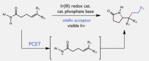
Published on July 13 in the Journal of the American Chemical Society, the non-conventional reaction is a proof of concept that will allow chemists to access compounds that are normally off-limits to this pathway. The team used a two-component catalyst system that works in tandem to selectively activate the strongest bond in the molecule, a nitrogen-hydrogen (N-H) bond through a process known as proton-coupled electron transfer (PCET).

Catalytic alkene carboamination enabled by oxidative proton-coupled electron transfer

“This PCET chemistry was really interesting to us. In particular, the idea that you can use catalysts to modulate an intrinsic property of a molecule allows you to access chemical space that you couldn’t otherwise,” said Robert Knowles, an assistant professor of chemistry who led the research.

Using PCET as a way to break strong bonds is seen in many essential biological systems, including photosynthesis and respiration, he said. Though this phenomenon is known in biological and inorganic chemistry settings, it hasn’t been widely applied to making new molecules—something Knowles hopes to change.

Given the unexplored state of PCET catalysis, Knowles decided to turn to theory instead of the trial and error approach usually taken by synthetic chemists in the initial stages of reaction development. Using a simple mathematical formula, the researchers calculated, for any pair of catalysts, the pair’s combined “effective bond strength,” which is the strength of the strongest bond they could break. Because both molecules independently contribute to this value, the research team had a high degree of flexibility in designing the catalyst system.

When they tested the catalyst pairs in the lab, the researchers observed a striking correlation between the “effective bond strength” and the reaction efficiency. While effective bond strengths that were lower or higher than the target N-H bond strength gave low reaction yields, the researchers found that matching the strengths promoted the reaction in very high yield.

“To see this formula actually working was really inspiring,” said Gilbert Choi, a graduate student in the Knowles lab and lead author on the work. Once he identified a successful catalyst system, he explored the scope of the reaction and its mechanism.

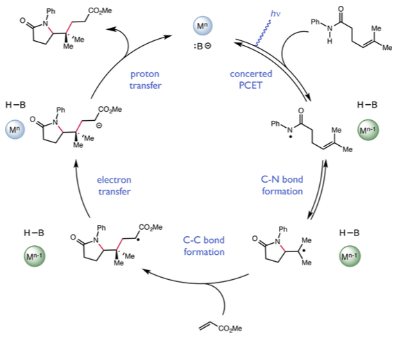
Proposed catalytic cycle

The researchers think that the reaction starts with one of the catalysts, a compound called dibutylphosphate, tugging on a hydrogen atom, which lengthens and weakens the N-H bond. At the same time, the other catalyst, known as a light-activated iridium complex, targets the weakened bond and plucks off one electron from the two-electron bond, slicing it down the middle.

Once the bond is split, the reactive nitrogen intermediate goes on to form a new carbon-nitrogen bond, giving rise to structurally complex products. This finding builds on work the Knowles lab published earlier this year, also in the Journal of the American Chemical Society, on a similar reaction that used a more sensitive catalyst system.

Their research has laid a solid foundation for PCET catalysis as a platform for developing new reactions. “My sincere view is that ideas are a lot more valuable than reactions,” Knowles said. “I’m optimistic that people can use these ideas and do things that we hadn’t even considered.”

Read the abstract: Choi, G. J.; Knowles, R. R. “Catalytic Alkene Carboamination Enabled by Oxidative Proton-Coupled Electron Transfer.2015, J. Am. Chem. Soc., Article ASAP.

This work was supported by Princeton University and the National Institutes of Health (R01 GM113105).

Putting two and two together to make unexplored chemicals (J. American Chemical Society)

Schematic for cobalt-catalyzed [2pi+2pi] reaction.
Schematic for cobalt-catalyzed [2pi+2pi] reaction. Image credit: Chirik group, Princeton University
By Tien Nguyen, Department of Chemistry

Researchers at Princeton have developed a new catalyst that may give unprecedented access to cyclobutanes, four-membered ring-containing molecules that have been relatively unexplored. Held back by the limited scope of previous methods, called [2π+2π] reactions, many cyclobutanes compounds have been out of reach, along with any unique properties that may be of interest to the pharmaceutical or fine chemical industry.

Led by Paul Chirik, the Edwards S. Sanford Professor of Chemistry, the team published the new cobalt-catalyzed [2π+2π] reaction and a thorough investigation of its mechanism in the Journal of the American Chemical Society on June 1.

“Because examples of this reaction are so rare, we wanted to understand why these cobalt complexes were special and how they worked in the reaction,” said Valerie Schmidt, lead author on the article and a postdoctoral researcher in the Chirik lab.

The new cobalt-catalyzed reaction overcame limitations that have plagued other similar methods, such as poor selectivity or requiring very reactive alkenes, which are chemical structures composed of two carbons joined by a double bond, as starting materials. The research team suspected their success came from certain molecules, called bis(imino)pyridine ligands, that are attached to the cobalt center and which are capable of passing electrons to and from the metal.

The Chirik group has used these redox active ligands previously, attached instead to iron to catalyze a [2π+2π] reaction reported in 2006. But the iron catalyst is highly sensitive to air and moisture, an issue that could be mitigated by switching to a less reactive metal like cobalt.

Replacing iron with cobalt presented a unique challenge in analysis because it altered the complex’s overall magnetic state from diamagnetic to paramagnetic. Unlike diamagnetic compounds, paramagnetic compounds can be difficult to identify by nuclear magnetic resonance (NMR) spectroscopy, a technique that uses a strong magnet to pulse atomic nuclei to reveal their environments, and a primary tool for characterizing molecules.

“We really had to be creative in finding ways to confirm our hypotheses about the catalyst,” Schmidt said. One extremely useful tool, analogous to nuclear magnetic resonance but that pulses electrons instead of nuclei, was electron paramagnetic resonance (EPR). This technique allowed the researchers to track the unpaired electrons, called radicals, throughout the reaction.

Additional data gathered from theoretical calculations, kinetic studies and x-ray crystal structure elucidation allowed the research team to sketch out a detailed reaction mechanism. They proposed that the cycle begins with successive coordination of the two tethered alkenes to the metal center.

Coordination of the second alkene was crucial, Schmidt explained, because it changed cobalt’s geometry, from square planar to tetrahedral, and effectively moved the unpaired electron from the ligand to the metal. Only then can the metal based radical promote the carbon-carbon forming event and push the reaction forward.

This action leads to the formation of a metallacycle—a pentagon shaped ring of four carbons and one cobalt atom. Cobalt is then squeezed out of the ring to release the final four-membered cyclobutane product in a process called reductive elimination. After testing a series of catalysts with varying size and electronic properties, the researchers suggested that reductive elimination was the turnover-limiting step, essentially the bottleneck of the reaction.

Armed with a deeper understanding of the cobalt catalyst system, the researchers hope to continually enhance its performance. “We want to make it as easy as possible to access cyclobutane containing molecules, because without this ability, we really have no idea what we are missing out on,” Schmidt said.

Read the abstract.

Schmidt, V. A.; Hoyt, J. M.; Margulieux, G. W.; Chirik, P. J. “Cobalt-Catalyzed [2π+2π] Cycloadditions of Alkenes: Scope, Mechanism and Elucidation of Electronic Structure of Catalytic Intermediates.” Journal of the American Chemical Society 2015, Just Accepted Manuscript.

This work was supported by the National Institutes of Health Ruth L. Kirschstein National Research Service Award (F32 GM109594) and Princeton University Intellectual Property Accelerator Fund.


Solving streptide from structure to biosynthesis (Nature Chemistry)

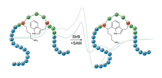
Streptide (Image source: Seyedsayamdost Lab)
(Image source: Seyedsayamdost Lab)

By: Tien Nguyen

Bacteria speak to one another using peptide signals in a soundless language known as quorum sensing. In a step towards translating bacterial communications, researchers at Princeton University have revealed the structure and biosynthesis of streptide, a peptide involved in the quorum sensing system common to many streptococci.

Leah Bushin, Class of 2014

“It’s extremely rare for one research group to do both natural products discovery and mechanistic enzymology,” said Leah Bushin, a member of the Seyedsayamdost lab and co-first author on the article published on April 20 in Nature Chemistry. Bushin worked on elucidating the structure of streptide as part of her undergraduate senior thesis project and will enter Princeton Chemistry’s graduate program in the fall.

To explore how bacteria communicate, first she had to grow them, a challenging process in which oxygen had to be rigorously excluded. Next she isolated the streptide and analyzed it using two-dimensional (2D) nuclear magnetic resonance (NMR) spectroscopy, a technique that allows scientists to deduce the connections between atoms in a molecule by pulsing their nuclei with powerful magnets to pulse atomic nuclei.

The experiments revealed that streptide contained an unprecedented crosslink between two unactivated carbons on lysine and tryptophan, constituting a new class of macrocyclic peptides. “We didn’t think it would be as cool as a carbon-carbon bond between two amino acid side chains, so it was definitely a surprise.” said Bushin.

To figure out how this novel bond was being formed, the researchers took a closer look at the gene cluster that produced streptide. Within the gene cluster, they suspected a radical S-adenosyl methionine (SAM) enzyme, which they dubbed StrB, could be responsible for this unusual modification.

Kelsey Schramma
Kelsey Schramma, a graduate student in the Seyedsayamdost lab

“Radical SAM enzymes catalyze absolutely amazing chemistries,” said Kelsey Schramma, a graduate student in the Seyedsayamdost lab and co-first author on the article. “There are over 48,000 radical SAM enzymes, but only about 50 have been characterized and just a dozen or so studied in detail,” she said.

To probe the enzyme’s role in making streptide, the researchers created a mutated version of the bacteria lacking the strB gene. The mutant failed to produce streptide, confirming that the StrB enzyme was significant and warranted further study.

Schramma determined that in order to function properly, the StrB enzyme required some key components: the pre-crosslinked substrate, which she prepared synthetically, cofactor SAM, reductant, and two iron-sulfur (Fe-S) clusters carefully assembled in the protein interior. The team then showed that one of the FeS clusters reductively activated one molecule of SAM, kicking off a chain of one-electron (radical) reactions that gave rise to the novel carbon-carbon bond.

“The synergy between Leah and Kelsey was great,” said Mohammad Seyedsayamdost, an assistant professor of chemistry at Princeton who led the research team. “They expressed interest in complementary aspects of the project and the whole ended up being greater than the sum of its parts,” he said.

Their efforts included not only chemical and biological approaches, but also theoretical computational studies. While the 2D NMR experiments revealed the flat structure of streptide, its three-dimensional conformation was still unknown.

“Since the crosslink had never been reported, we had to code the modification into the program, which took a bit of creativity,” Bushin said. After corresponding with the software creator, they were able to confidently assign a key residue in the macrocycle with the S-configuration.

Future work will target streptide’s biological function—its meaning in the bacterial language—as well as confirming its production by other streptococcal bacteria strains.

“What we have revealed is a new and unusual mechanism that nature uses to synthesize macrocyclic peptides. There is a lot of novel chemistry to be discovered by interrogating bacterial secondary metabolite biosynthetic pathways,” Seyedsayamdost said.

Read the article here:

Schramma, K. R.; Bushin, L. B.; Seyedsayamdost, M. R. “Structure and biosynthesis of a macrocyclic peptide containing an unprecedented lysine-to-tryptophan crosslink.Nature Chemistry, 2015, 7, 431.

This work was supported by the National Institutes of Health (grant no. GM098299), and by Princeton University start-up funds.

Decoding the Cell’s Genetic Filing System (Nature Chemistry)

By Tien Nguyen, Department of Chemistry

A fully extended strand of human DNA measures about five feet in length. Yet it occupies a space just one-tenth of a cell by wrapping itself around histones—spool-like proteins—to form a dense hub of information called chromatin.

Access to these meticulously packed genes is regulated by post-translational modifications, chemical changes to the structure of histones that act as on-off signals for gene transcription. Mistakes or mutations in histones can cause diseases such as glioblastoma, a devastating pediatric brain cancer.

Source: Nature Chemistry

Researchers at Princeton University have developed a facile method to introduce non-native chromatin into cells to interrogate these signaling pathways. Published on April 6 in the journal Nature Chemistry, this work is the latest chemical contribution from the Muir lab towards understanding nature’s remarkable information indexing system.

Tom Muir, the Van Zandt Williams, Jr. Class of ’65 Professor of Chemistry, began investigating transcriptional pathways in the so-called field of epigenetics almost a decade earlier. Deciphering such a complex and dynamic system posed a formidable challenge, but his research lab was undeterred. “It’s better to fail at something important than to succeed at something trivial,” he said.

Muir recognized the value of introducing chemical approaches to epigenetics to complement early contributions that came mainly from molecular biologists and geneticists. If epigenetics was like a play, he said, molecular biology and genetics could identify the characters but chemistry was needed to understand the subplots.

These subplots, or post-translational modifications of histones, of which there are more than 100, can occur cooperatively and simultaneously. Traditional methods to probe post-translational modifications involved synthesizing modified histones one at a time, which was a very slow process that required large amounts of biological material.

Last year, the Muir group introduced a method that would massively accelerate this process. The researchers generated a library of 54 nucleosomes—single units of chromatin, like pearls on a necklace—encoded with DNA-barcodes, unique genetic tags that can be easily identified. Published in the journal Nature Methods, the high throughput method required only microgram amounts of each nucleosome to run approximately 4,500 biochemical assays.

“The speed and sensitivity of the assay was shocking,” Muir said. Each biochemical assay involved treatment of the DNA-barcoded nucleosome with a writer, reader or nuclear extract, to reveal a particular binding preference of the histone. The products were then isolated using a technique called chromatin immunoprecipitation and characterized by DNA sequencing, essentially an ordered readout of the nucleotides.

“There have been incredible advances in genetic sequencing over the last 10 years that have made this work possible,” said Manuel Müller, a postdoctoral researcher in the Muir lab and co-author on the Nature Methods article.

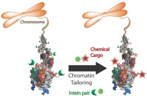
Schematic of approach using split inteins

With this method, researchers could systematically interrogate the signaling system to propose mechanistic pathways. But these mechanistic insights would remain hypotheses unless they could be validated in vivo, meaning inside the cellular environment.

The only method for modifying histones in vivo was extremely complicated and specific, said Yael David, a postdoctoral researcher in the Muir lab and lead author on the recent Nature Chemistry study that demonstrated a new and easily customizable approach.

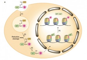
The method relied on using ultra-fast split inteins, protein fragments that have a great affinity for one another. First, one intein fragment was attached to a modified histone, by encoding it into a cell. Then, the other intein fragment was synthetically fused to a label, which could be a small protein tag, fluorophore or even an entire protein like ubiquitin.

Within minutes of being introduced into the cell, the labeled intein fragment bound to the histone intein fragment. Then like efficient and courteous matchmakers, the inteins excised themselves and created a new bond between the label and modified histone. “It’s really a beautiful way to engineer proteins in a cell,” David said.

Regions of the histone may be loosely or tightly packed, depending on signals from the cell indicating whether or not to transcribe a gene. By gradually lowering the amount of labeled intein introduced, the researchers could learn about the structure of chromatin and tease out which areas were more accessible than others.

Future plans in the Muir lab will employ these methods to ask specific biological questions, such as whether disease outcomes can be altered by manipulating signaling pathway. “Ultimately, we’re developing methods at the service of biological questions,” Muir said.

This research was supported by the US National Institutes of Health (grants R37-GM086868 and R01 GM107047).

Read the articles:

Nguyen, U.T.T.; Bittova, L.; Müller, M.; Fierz, B.; David, Y.; Houck-Loomis, B.; Feng, V.; Dann, G.P.; Muir, T.W. “Accelerated chromatin biochemistry using DNA-barcoded nucleosome libraries.” Nature Methods, 2014, 11, 834.

David, Y.; Vila-Perelló, M; Verma, S.; Muir, T.W. “Chemical tagging and customizing of cellular chromatin states using ultrafast trans-splicing inteins.” Nature Chemistry, Advance online publication, April 6, 2015.

Cytomegalovirus hijacks human enzyme for replication (Cell Reports)

DiagramBy: Tien Nguyen, Department of Chemistry
More than 60 percent of the world’s population is infected with a type of herpes virus called human cytomegalovirus. The virus replicates by commandeering the host cell’s metabolism but the details of this maneuver are unclear.

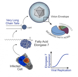
Researchers at Princeton University have discovered that cytomegalovirus manipulates a process called fatty acid elongation, which makes the very-long-chain fatty acids necessary for virus replication. Published in the journal Cell Reports on March 3, the research team identified a specific human enzyme—elongase enzyme 7—that the virus induces to turn on fatty acid elongation.

“Elongase 7 was just screaming, ‘I’m important, study me,’” said John Purdy, a postdoctoral researcher in the Rabinowitz lab and lead author on the study.

He found that once a cell was infected by cytomegalovirus, the level of elongase 7 RNA increased over 150-fold. Purdy then performed a genetic knockdown experiment to silence elongase 7 and established that in its absence the virus was unable to efficiently replicate.

“Elongases are a family of seven related proteins. The particular importance of elongase 7 for cytomegalovirus replication was a pleasant surprise, and enhances its appeal as a drug target,” said Joshua Rabinowitz, a professor of chemistry and the Lewis-Sigler Institute for Integrative Genomics at Princeton and co-author on the paper.

Activation of the elongase enzyme led to an increase in very-long-chain fatty acids, which are used by the virus to build its viral envelope and replicate. The researchers fed infected cells a sugar called heavy isotope-labeled carbon-13 glucose, which is metabolized by the cell to form substrates for fatty acid elongation. The heavy isotope carbon-13 atoms were incorporated into new products that were detected and identified by their mass using a mass spectrometry method. This powerful technique provided insight into the amount of fatty acids produced and how they are constructed.

Cytomegalovirus infection mostly threatens populations with compromised immune systems and developing fetuses, and is the leading cause of hearing loss in children. Current treatments target the DNA replication step of the virus and are not very effective. These findings have advanced the understanding of the virus’s operations and identified fatty acid elongation as a key process that warrants further study.

This work was funded by National Institute of Health grants AI78063, CA82396, and GM71508 and an American Heart Association postdoctoral fellowship to J.G.P. (12POST9190001).

Read the full article here:

Purdy, J. G.; Shenk, T.; Rabinowitz, J. D. “Fatty Acid Elongase 7 Catalyzes the Lipidome Remodeling Essential for Human Cytomegalovirus Replication.” Cell Reports, 2015, 10, 1375.


Computational clues into the structure of a promising energy conversion catalyst (J. Physical Chemistry Letters)

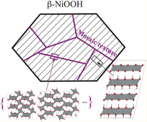
Mosaic structure
Representation of the mosaic texture of β-NiOOH and its possible structures.

By Tien Nguyen, Department of Chemistry

Hydrogen fuel is a promising source of clean energy that can be produced by splitting water into hydrogen and oxygen gas with the help of a catalyst, a material that can speed up the process. Although most known catalysts are inefficient, one called iron-doped nickel oxide is promising but not well understood.

Now researchers at Princeton University have reported new insights into the structure of an active component of the nickel oxide catalyst, known as β-NiOOH, using theoretical calculations. Led by Annabella Selloni, professor of chemistry at Princeton, the findings were published in The Journal of Physical Chemistry Letters on October 28.

“Understanding the structure is the basis for any further study of the material’s properties. If you don’t know the material’s structure you can’t know what it’s doing,” Selloni said. Nickel oxide’s exact structure has been difficult to determine experimentally because it is constantly changing during the reaction.

The research team took a theoretical approach and employed a “genetic algorithm” to search for the structure. Genetic algorithms operate under a set of parameters that draw inspiration from evolution by creating generation after generation of structures to arrive at the most “fit” or most likely candidates.

Taking the results of the genetic algorithm search in combination with computational techniques known as hybrid density functional theory calculations—which estimate a molecule’s electronic structure—co-author Ye-Fei Li, a former postdoctoral researcher at Princeton who is now at Fudan University, and Selloni were able to identify structures of nickel oxide that supported existing observations.

One such observation is the material’s mosaic texture, composed of tiny grain-like microstructures. The researchers propose that these microstructures are stable tunnel structures that relieve stress between layers. Another observed feature is the doubling of the distance between layers made of the same material, referred to as its c axis periodicity, which represents the alternating layers of Ni(OH)2 and NiO2 formed during the reaction.

Armed with a better understanding of the material’s structure, the scientists hope to further map out its activity in the reaction. “I’m interested in the microscopic mechanisms, what are the electrons and atoms doing?” Selloni said.

Read the abstract

Li, Ye-Fei and Annabella Selloni. “Mosaic Texture and Double c-Axis Periodicity of β–NiOOH: Insights from First-Principles and Genetic Algorithm Calculations.” J. Phys. Chem. Lett. 2014, 5, 3981.

This work was supported by the US Department of Energy, Division of Chemical Sciences, Geosciences and Biosciences under award DE-FG0212ER16286.

Quantum mechanical calculations reveal the hidden states of enzyme active sites (Nature Chemistry)

[2Fe–2S] cluster in front of a leaf. (Image by C. Todd Reichart)
Researchers at Princeton University have reported the first direct observation of the electronic states of iron-sulfur clusters, common to many enzyme active sites. Iron-sulfur cluster in front of a leaf. (Image by C. Todd Reichart)
By Tien Nguyen, Department of Chemistry

Enzymes carry out fundamental biological processes such as photosynthesis, nitrogen fixation and respiration, with the help of clusters of metal atoms as “active” sites. But scientists lack basic information about their function because the states thought to be critical to their chemical abilities cannot be experimentally observed.

Now, researchers at Princeton University have reported the first direct observation of the electronic states of iron-sulfur clusters, common to many enzyme active sites. Published on August 31 in the journal Nature Chemistry, the states were revealed by computing the complicated quantum mechanical behavior of the electrons in the clusters.

“These complexes were thought of as impossible to model, due to the complexity of the quantum mechanics,” said Garnet Chan, the A. Barton Hepburn Professor of Chemistry and corresponding author on the paper.

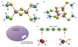
Iron-sulfur clusters
Caption: (a) [2Fe–2S] clusters (b) [4Fe–4S] (c) Area-law entanglement of the physical states can be used to reduce the complexity of quantum calculations (d) Wavefunctions with area-law entanglement can be written compactly as a tensor network where each tensor (represented here by a circle) denotes an active space orbital and the bonds between adjacent orbitals introduce local entanglement between them. (Source: Garnet Chan)
In these systems, the electrons interact strongly with each other, their movements resembling a complicated dance. To reduce the complexity, the researchers drew on a new understanding, gained from fundamental work in quantum information theory, that the motion of the electrons had a special pattern.

“At first glance, the electrons appear to move in a complicated way, but eventually you realize that they only care about what their immediate neighbors are doing, similar to being in a crowded room. This restriction on their behavior leads to important simplifications: the calculations become very difficult rather than impossible — it’s just on the edge of what can be done,” Chan said.

Using their new method, Chan and coworkers found that iron-sulfur clusters possess an order of magnitude more accessible electronic states than previously reported. The researchers suggested that this unusual richness might explain their ubiquity in biological processes.

This finding, that there are many more available electronic states than previously thought, presents many different chemical possibilities. What if these clusters simultaneously used a combination of mechanisms, instead of the accepted chemical idea that there is one distinct electronic pathway, Chan wondered. To test that idea and learn more about the clusters’ behavior, the researchers plan to extend their calculations to observe a chemical transformation in action.

“If you want to understand why iron-sulfur clusters are a ubiquitous biological motif and how we can create even better synthetic analogs, then you need to know what the electrons are doing,” Chan said. “Now we’ve caught a first glimpse as to what they are getting up to.”

Read the abstract.

Sharma, S.; Sivalingam, K.; Neese, F.; Chan, K.-L. G. “Low-energy spectrum of iron sulfur clusters directly from many-particle quantum mechanics.Nat. Chem. 2014, 6, 927.

This work was supported by the US National Science Foundation (CHE-1265277) and used software developed with the support of OCI-1265278. F.N. and K.S acknowledge financial support from the Max Planck Society, the University of Bonn and the SFB 813 “Chemistry at Spin Centers.”