It slices, it dices, and it protects the body from harm (Science)

By Catherine Zandonella, Office of the Dean for Research

RNase L enzyme structure
Researchers at Princeton have deciphered the 3D structure of RNase L, an enzyme that slices through RNA and is a first responder in the innate immune system. The structure contains two subunits, represented in red as two parts of a pair of scissors. Illustration by Sneha Rath, Inset courtesy of Science.

An essential weapon in the body’s fight against infection has come into sharper view. Researchers at Princeton University have discovered the 3D structure of an enzyme that cuts to ribbons the genetic material of viruses and helps defend against bacteria.

The discovery of the structure of this enzyme, a first-responder in the body’s “innate immune system,” could enable new strategies for fighting infectious agents and possibly prostate cancer and obesity. The work was published Feb. 27 in the journal Science.

Until now, the research community has lacked a structural model of the human form of this enzyme, known as RNase L, said Alexei Korennykh, an assistant professor of molecular biology and leader of the team that made the discovery.

“Now that we have the human RNase L structure, we can begin to understand the effects of carcinogenic mutations in the RNase L gene. For example, families with hereditary prostate cancers often carry genetic mutations in the region, or locus, encoding RNase L,” Korennykh said. The connection is so strong that the RNase L locus also goes by the name “hereditary prostate cancer 1.” The newly found structure reveals the positions of these mutations and explains why some of these mutations could be detrimental, perhaps leading to cancer, Korennykh said. RNase L is also essential for insulin function and has been implicated in obesity.

The Princeton team’s work has also led to new insights on the enzyme’s function.

The enzyme is an important player in the innate immune system, a rapid and broad response to invaders that includes the production of a molecule called interferon. Interferon relays distress signals from infected cells to neighboring healthy cells, thereby activating RNase L to turn on its ability to slice through RNA, a type of genetic material that is similar to DNA. The result is new cells armed for destruction of the foreign RNA.

The 3D structure uncovered by Korennykh and his team consists of two nearly identical subunits called protomers. The researchers found that one protomer finds and attaches to the RNA, while the other protomer snips it.

The initial protomer latches onto one of the four “letters” that make up the RNA code, in particular, the “U,” which stands for a component of RNA called uridine. The other protomer “counts” RNA letters starting from the U, skips exactly one letter, then cuts the RNA.

Although the enzyme can slice any RNA, even that of the body’s own cells, it only does so when activated by interferon.

“We were surprised to find that the two protomers were identical but have different roles, one binding and one slicing,” Korennykh said. “Enzymes usually have distinct sites that bind the substrate and catalyze reactions. In the case of RNase L, it appears that the same exact protein surface can do both binding and catalysis. One RNase L subunit randomly adopts a binding role, whereas the other identical subunit has no other choice but to do catalysis.”

To discover the enzyme’s structure, the researchers first created a crystal of the RNase L enzyme. The main challenge was finding the right combination of chemical treatments that would force the enzyme to crystallize without destroying it.

Korennykh group
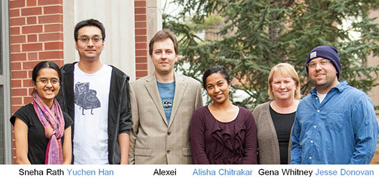
After much trial and error and with the help of an automated system, postdoctoral research associate Jesse Donovan and graduate student Yuchen Han succeeded in making the crystals.

Next, the crystals were bombarded with powerful X-rays, which diffract when they hit the atoms in the crystal and form patterns indicative of the crystal’s structure. The diffraction patterns revealed how the atoms of RNase L are arranged in 3D space.

At the same time Sneha Rath, a graduate student in Korennykh’s laboratory, worked on understanding the RNA cleavage mechanism of RNase L using synthetic RNA fragments. Rath’s results matched the structural findings of Han and Donovan, and the two pieces of data ultimately revealed how RNase L cleaves its RNA targets.

Han, Donovan and Rath contributed equally to the paper and are listed as co-first authors.

Finally, senior research specialist Gena Whitney and graduate student Alisha Chitrakar conducted additional studies of RNase L in human cells, confirming the 3D structure.

Now that the human structure has been solved, researchers can explore ways to either enhance or dampen RNase L activity for medical and therapeutic uses, Korennykh said.

“This work illustrates the wonderful usefulness of doing both crystallography and careful kinetic and enzymatic studies at the same time,” said Peter Walter, professor of biochemistry and biophysics at the University of California-San Francisco School of Medicine. “Crystallography gives a static picture which becomes vastly enhanced by studies of the kinetics.”

Support for the work was provided by Princeton University.

Read the abstract.

Han, Yuchen, Jesse Donovan, Sneha Rath, Gena Whitney, Alisha Chitrakar, and Alexei Korennykh. Structure of Human RNase L Reveals the Basis for Regulated RNA Decay in the IFN Response Science 1249845. Published online 27 February 2014 [DOI:10.1126/science.1249845]

Now in 3D: Video of virus-sized particle trying to enter cell (Nature Nanotechnology)

Video of virus trying to enter cell
3D movie (below) of virus-like nanoparticle trying to gain entry to a cell

By Catherine Zandonella, Office of the Dean for Research

Tiny and swift, viruses are hard to capture on video. Now researchers at Princeton University have achieved an unprecedented look at a virus-like particle as it tries to break into and infect a cell. The technique they developed could help scientists learn more about how to deliver drugs via nanoparticles — which are about the same size as viruses — as well as how to prevent viral infection from occurring.

The video reveals a virus-like particle zipping around in a rapid, erratic manner until it encounters a cell, bounces and skids along the surface, and either lifts off again or, in much less time than it takes to blink an eye, slips into the cell’s interior. The work was published in Nature Nanotechnology.

Video caption: ‘Kiss and run’ on the cell surface. This 3D movie shows actual footage of a virus-like particle (red dot) approaching a cell (green with reddish brown nucleus), as captured by Princeton University researchers Kevin Welcher and Haw Yang. The color of the particle represents its speed, with red indicating rapid movement and blue indicating slower movement. The virus-like particle lands on the surface of the cell, appears to try to enter it, then takes off again. Source: Nature Nanotechnology.

“The challenge in imaging these events is that viruses and nanoparticles are small and fast, while cells are relatively large and immobile,” said Kevin Welsher, a postdoctoral researcher in Princeton’s Department of Chemistry and first author on the study. “That has made it very hard to capture these interactions.”

The problem can be compared to shooting video of a hummingbird as it roams around a vast garden, said Haw Yang, associate professor of chemistry and Welsher’s adviser. Focus the camera on the fast-moving hummingbird, and the background will be blurred. Focus on the background, and the bird will be blurred.

The researchers solved the problem by using two cameras, one that locked onto the virus-like nanoparticle and followed it faithfully, and another that filmed the cell and surrounding environment.

Putting the two images together yielded a level of detail about the movement of nano-sized particles that has never before been achieved, Yang said. Prior to this work, he said, the only way to see small objects at a similar resolution was to use a technique called electron microscopy, which requires killing the cell.

“What Kevin has done that is really different is that he can capture a three-dimensional view of a virus-sized particle attacking a living cell, whereas electron microscopy is in two-dimensions and on dead cells,” Yang said. “This gives us a completely new level of understanding.”

In addition to simply viewing the particle’s antics, the researchers can use the technique to map the contours of the cell surface, which is bumpy with proteins that push up from beneath the surface. By following the particle’s movement along the surface of the cell, the researchers were able to map the protrusions, just as a blind person might use his or her fingers to construct an image of a person’s face.

“Following the motion of the particle allowed us to trace very fine structures with a precision of about 10 nanometers, which typically is only available with an electron microscope,” Welsher said. (A nanometer is one billionth of a meter and roughly 1000 times smaller than the width of a human hair.) He added that measuring changes in the speed of the particle allowed the researchers to infer the viscosity of the extracellular environment just above the cell surface.

The technology has potential benefits for both drug discovery and basic scientific discovery, Yang said.  “We believe this will impact the study of how nanoparticles can deliver medicines to cells, potentially leading to some new lines of defense in antiviral therapies,” he said. “For basic research, there are a number of questions that can now be explored, such as how a cell surface receptor interacts with a viral particle or with a drug.”

Welsher added that such basic research could lead to new strategies for keeping viruses from entering cells in the first place.

“If we understand what is happening to the virus before it gets to your cells,” said Welsher, “then we can think about ways to prevent infection altogether. It is like deflecting missiles before they get there rather than trying to control the damage once you’ve been hit.”

To create the virus-like particle, the researchers coated a miniscule polystyrene ball with quantum dots, which are semiconductor bits that emit light and allow the camera to find the particle. Next, the particle was studded with protein segments known as Tat peptides, derived from the HIV-1 virus, which help the particle find the cell. The width of the final particle was about 100 nanometers.

The researchers then let loose the particles into a dish containing skin cells known as fibroblasts. One camera followed the particle while a second imaging system took pictures of the cell using a technique called laser scanning microscopy, which involves taking multiple images, each in a slightly different focal plane, and combining them to make a three-dimensional picture.

The research was supported by the US Department of Energy (DE-SC0006838) and by the Eric and Wendy Schmidt Transformative Technology Fund at Princeton University.

Read the abstract.

Kevin Welsher and Haw Yang. 2014. Multi-resolution 3D visualization of the early stages of cellular uptake of peptide-coated nanoparticles. Nature nanotechnology. Published online: 23 February 2014 | DOI: 10.1038/NNANO.2014.12

Rife with hype, exoplanet study needs patience and refinement (PNAS)

By Morgan Kelly, Office of Communications

Exoplanet
Exoplanet transiting in front of its star. Princeton’s Adam Burrows argues against drawing too many conclusions about such distant objects with today’s technologies. Photo credit: ESA/C. Carreau

Imagine someone spent months researching new cities to call home using low-resolution images of unidentified skylines. The pictures were taken from several miles away with a camera intended for portraits, and at sunset. From these fuzzy snapshots, that person claims to know the city’s air quality, the appearance of its buildings, and how often it rains.

This technique is similar to how scientists often characterize the atmosphere — including the presence of water and oxygen — of planets outside of Earth’s solar system, known as exoplanets, according to a review of exoplanet research published in the Proceedings of the National Academy of Sciences.

A planet’s atmosphere is the gateway to its identity, including how it was formed, how it developed and whether it can sustain life, stated Adam Burrows, author of the review and a Princeton University professor of astrophysical sciences.

But the dominant methods for studying exoplanet atmospheres are not intended for objects as distant, dim and complex as planets trillions of miles from Earth, Burrows said. They were instead designed to study much closer or brighter objects, such as planets in Earth’s solar system and stars.

Nonetheless, scientific reports and the popular media brim with excited depictions of Earth-like planets ripe for hosting life and other conclusions that are based on vague and incomplete data, Burrows wrote in the first in a planned series of essays that examine the current and future study of exoplanets. Despite many trumpeted results, few “hard facts” about exoplanet atmospheres have been collected since the first planet was detected in 1992, and most of these data are of “marginal utility.”

The good news is that the past 20 years of study have brought a new generation of exoplanet researchers to the fore that is establishing new techniques, technologies and theories. As with any relatively new field of study, fully understanding exoplanets will require a lot of time, resources and patience, Burrows said.

“Exoplanet research is in a period of productive fermentation that implies we’re doing something new that will indeed mature,” Burrows said. “Our observations just aren’t yet of a quality that is good enough to draw the conclusions we want to draw.

“There’s a lot of hype in this subject, a lot of irrational exuberance. Popular media have characterized our understanding as better than it actually is,” he said. “They’ve been able to generate excitement that creates a positive connection between the astrophysics community and the public at large, but it’s important not to hype conclusions too much at this point.”

The majority of data on exoplanet atmospheres come from low-resolution photometry, which captures the variation in light and radiation an object emits, Burrows reported. That information is used to determine a planet’s orbit and radius, but its clouds, surface, and rotation, among other factors, can easily skew the results. Even newer techniques such as capturing planetary transits — which is when a planet passes in front of its star, and was lauded by Burrows as an unforeseen “game changer” when it comes to discovering new planets — can be thrown off by a thick atmosphere and rocky planet core.

All this means that reliable information about a planet can be scarce, so scientists attempt to wring ambitious details out of a few data points. “We have a few hard-won numbers and not the hundreds of numbers that we need,” Burrows said. “We have in our minds that exoplanets are very complex because this is what we know about the planets in our solar system, but the data are not enough to constrain even a fraction of these conceptions.”

Burrows emphasizes that astronomers need to acknowledge that they will never achieve a comprehensive understanding of exoplanets through the direct-observation, stationary methods inherited from the exploration of Earth’s neighbors. He suggests that exoplanet researchers should acknowledge photometric interpretations as inherently flawed and ambiguous. Instead, the future of exoplanet study should focus on the more difficult but comprehensive method of spectrometry, wherein the physical properties of objects are gauged by the interaction of its surface and elemental features with light wavelengths, or spectra. Spectrometry has been used to determine the age and expansion of the universe.

Existing telescopes and satellites are likewise vestiges of pre-exoplanet observation. Burrows calls for a mix of small, medium and large initiatives that will allow the time and flexibility scientists need to develop tools to detect and analyze exoplanet spectra. He sees this as a challenge in a research environment that often puts quick-payback results over deliberate research and observation. Once scientists obtain high-quality spectral data, however, Burrows predicted, “Many conclusions reached recently about exoplanet atmospheres will be overturned.”

“The way we study planets out of the solar system has to be radically different because we can’t ‘go’ to those planets with satellites or probes,” Burrows said. “It’s much more an observational science. We have to be detectives. We’re trying to find clues and the best clues since the mid-19th century have been in spectra. It’s the only means of understanding the atmosphere of these planets.”

A longtime exoplanet researcher, Burrows predicted the existence of “hot-Jupiter” planets — gas planets similar to Jupiter but orbiting very close to the parent star — in a paper in the journal Nature months before the first such planet, 51 Pegasi b, was discovered in 1995.

Read the abstract.

Citation: Burrows, Adam S. 2014. Spectra as windows into exoplanet atmospheres. Proceedings of the National Academy of Sciences. Article first published online: Jan. 13, 2014. DOI: 10.1073/pnas.1304208111