How rabies virus moves through nerve cells, and how it might be stopped

Cells in red against black background

To successfully infect its host, the rabies virus must move from the nerve ending to the nerve cell body where it can replicate. In a study published July 20 in the journal PLoS Pathogens, researchers from Princeton University reveal that the rabies virus moves differently compared to other neuron-invading viruses and that its journey can be blocked by a drug commonly used to treat amoebic dysentery.

Most viruses only infect the nervous system accidentally when the immune system is compromised. But some “neurotropic” viruses have evolved to target neurons as part of their normal infectious cycle. The rabies virus, for example, is transmitted when an infected animal bites into a host’s muscle. It then spreads into the end terminals of motor neurons innervating the muscle and travels along the neurons’ long axon fibers to the neuronal cell bodies. From there, the virus can spread throughout the central nervous system and into the salivary glands, where it can be readily transmitted to other hosts. Though rabies infections in humans are rare in the United States, the virus kills nearly 60,000 people annually.

Alpha herpesviruses, such as herpes simplex viruses, also enter peripheral nerve terminals and move along axons to the neuronal cell body, where they can lie dormant for the life of the host.

“Transport to the neuronal cell body is not a passive process, but an active one relying on the neuron’s own motor proteins and microtubule tracks,” said Lynn Enquist, Princeton’s Henry L. Hillman Professor in Molecular Biology, a professor of molecular biology and the Princeton Neuroscience Institute, and the study’s senior author. “Virus particles must engage this machinery for efficient transport in axons, otherwise infection cannot start.”

Viruses moving in a narrow nerve cell
Twenty-four hours after infecting nerve terminals, the rabies virus (red) reaches the cell bodies (green) of control neurons (left) but not neurons treated with emetine (right). Image credit: MacGibeny et al., 2018.

Enquist and colleagues previously found that alpha herpesviruses engage the neuronal transport machinery by stimulating protein synthesis at infected nerve terminals. Viral transport to the cell body can therefore be blocked by drugs that inhibit protein synthesis, as well as by cellular antiviral proteins called interferons.

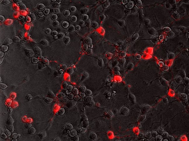
In the current study, Enquist and colleagues investigated how the rabies virus engages the neuronal transport machinery. The researchers infected neurons with a virulent strain of the virus tagged with a red fluorescent protein, allowing the researchers to observe viral transport in real time by live-cell fluorescence microscopy.

The study was led by Margaret MacGibeny, who earned her Ph.D. in 2018, and associate research scholar Orkide Koyuncu, at Princeton, with contributions from research associate Christoph Wirblich and Matthias Schnell, professor and chair of microbiology and immunology at Thomas Jefferson University.

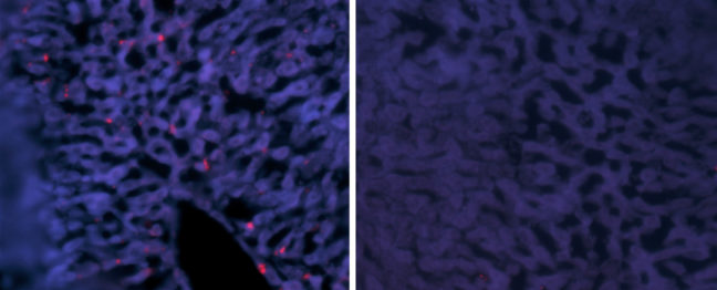
In contrast to alpha herpesvirus infections, the team found that interferons had no effect on rabies virus transport, perhaps because, until it reaches the neuronal cell body, the rabies virus hides out inside cellular structures called endosomes.

“We also couldn’t detect increased protein synthesis in axons upon rabies virus infection,” MacGibeny said. “But, to our surprise, we saw that a protein synthesis inhibitor called emetine efficiently blocked rabies virus transport to the cell body.”

Emetine had no effect on the transport of endosomes devoid of the rabies virus. But endosomes carrying the virus were either completely immobilized, or were only able to move short distances at slower-than-normal speeds.

Other protein synthesis inhibitors did not block rabies virus transport, however, suggesting that emetine works by inhibiting a different process in infected neurons.

“Emetine has been used to treat amoebic dysentery,” Koyuncu said. “In the laboratory it is widely used to inhibit protein synthesis but there are recent reports indicating that emetine has anti-viral effects that are independent of protein synthesis inhibition. Our study shows that this drug can inhibit rabies virus invasion of the nervous system through a novel mechanism that hasn’t been reported before.”

“The manuscript by MacGibeny et al. both advances and complicates our understanding of how neurotropic viruses make their way from the axon terminus to the cell body,” said Professor Glenn Rall, an expert in neurotropic virus infections at Fox Chase Cancer Center, who was not involved in the study. “Revealing variations in the axonal transport of neurotropic viruses, coupled with intriguing insights into new roles for well-known drugs, has both mechanistic and clinical implications for these life-threatening infections.

“Our next step is to figure out how emetine disrupts rabies virus transport in axons,” Enquist says. “Does it inhibit cell signaling pathways after rabies virus entry, or does it directly block the recruitment of motor proteins to virus-carrying endosomes?”

This study was funded by the US National Institutes of Health (grants P40 OD010996, RO1 NS33506, and F30 NS090640).

The study, Retrograde axonal transport of rabies virus is unaffected by interferon treatment but blocked by emetine locally in axons, by Margaret A. MacGibeny, Orkide O. Koyuncu, Christoph Wirblich, Matthias J. Schnell, and Lynn W. Enquist was published in PLoS Pathogens. 2018. DOI: 10.1371/journal.ppat.1007188

Text courtesy of the Department of Molecular Biology

Genetically engineered mice could boost fight against aggressive hepatitis

Article provided by the Department of Molecular Biology

Hepatitis delta virus (HDV) causes the most aggressive form of viral hepatitis in humans, putting at least 20 million people worldwide at risk of developing liver fibrosis, cirrhosis, and liver cancer. Efforts to develop effective treatments against HDV have been hampered by the fact that laboratory mice are not susceptible to the virus. But, in a study published June 27, 2018, in the journal Science Translational Medicine, Alexander Ploss, assistant professor of molecular biology at Princeton University and colleagues describe a genetically engineered mouse that can be persistently infected with HDV.

HDV is a small, RNA-based “satellite” virus that produces just a single protein of its own and therefore requires additional proteins provided by another liver virus, hepatitis B virus (HBV). HDV can infect patients already carrying HBV, or both viruses can infect patients simultaneously. Though infections can be prevented with an anti-HBV vaccine, there are no antiviral therapies available to cure existing HDV infections.

HDV and HBV infect the liver by binding to a protein called NTCP that is present on the surface of liver cells. But the viruses only recognize the version of NTCP present in humans and a few other primates, and therefore can’t infect mice or other small mammals that produce their own versions of NTCP. This has made it difficult to study HBV and HDV infections in the laboratory. Researchers have tried transplanting human liver cells into immunocompromised mice before infecting them with virus, but this approach has produced inconsistent results and is both expensive and time-consuming.

Ploss and colleagues, led by graduate student Benjamin Winer, took a different approach. They generated mice that express the human NTCP protein in their liver cells, allowing these cells to be infected by HBV and HDV.

In these mice, HBV failed to replicate after entering mouse liver cells but HDV was able to establish persistent infection when provided with the HBV proteins it needs to propagate. For example, mice genetically engineered to produce both human NTCP and the entire HBV genome could be infected with HDV for up to 14 days. “To our knowledge, this is the first time the entire HDV life cycle has been recapitulated in a mouse model with inheritable susceptibility to HDV,” Ploss said.

The mice were able to rid themselves of HDV before they developed any liver damage, apparently by mounting an immune response involving antiviral interferon proteins and various white blood cell types, including Natural Killer (NK) cells and T cells. Accordingly, mice expressing human NTCP and the HBV genome, but lacking functional B, T, and NK cells could be infected with HDV for two months or more.

These immunocompromised animals allowed Ploss and colleagues to test the effectiveness of two drugs that are currently being developed as treatments for HDV infection. Both drugs—either alone or in combination—suppressed the levels of HDV in immunocompromised mice after viral infection. But the drugs were not able to completely clear the mice of HDV; viral levels rose again within weeks of stopping treatment.

“This is largely in line with recently reported data from clinical trials, showing the utility of our model for preclinical antiviral drug testing,” Winer said.

“Our model is amenable to genetic manipulations, robust, and can be adopted as a method to rapidly screen for potential treatments,” Ploss added.

Timothy M. Block, president of the Hepatitis B Foundation and its Baruch S. Blumberg Institute who was not involved in the study, said “These systems should be able to provide practical, and presumably economical  tools. Their work is urgently needed, and a desperate community welcomes it. I emphasize that it is often the new methods in science that revolutionize a field such as drug discovery, almost as much as the new drugs themselves.”

The research team included collaborators from Princeton University; Weill Medical College of Cornell University; The Jackson Laboratory; University Medical Center Hamburg-Eppendorf, Hamburg; New York University Medical Center; and North Carolina State University College of Veterinary Medicine.

This study was supported by grants from the National Institutes of Health (R01 AI079031, R01 AI107301, R21AI117213 to Alexander Ploss), a Research Scholar Award from the American Cancer Society (RSG-15-048-01-MPC to Alexander Ploss), a Burroughs Wellcome Fund Award for Investigators in Pathogenesis (to Alexander Ploss) and a Graduate fellowship from the Health Grand Challenge from the Global Health Fund of Princeton University (to Benjamin Y. Winer). The NYU Experimental Pathology Immunohistochemistry Core Laboratory is supported in part by the Laura and Isaac Perlmutter Cancer Center Support Grant; NIH/NCI P30CA016087 and the National Institutes of Health S10 Grants NIH/ORIP S10OD01058 and S10OD018338. Benjamin Y. Winer is a recipient of F31 NIH/NRSA Ruth L. Kirschstein Predoctoral awarded from the NIAID. Julie Sellau and Elham Shirvani-Dastgerdi are both recipients of postdoctoral fellowships from the German Research Foundation. Michael V. Wiles was funded by The Jackson Laboratory.

Benjamin Y. Winer, Elham Shirvani-Dastgerdi, Yaron Bram, Julie Sellau, Benjamin E. Low, Heath Johnson, Tiffany Huang, Gabriela Hrebikova, Brigitte Heller, Yael Sharon, Katja Giersch, Sherif Gerges, Kathleen Seneca, Mihai-Alexandru Pais, Angela S. Frankel, Luis Chiriboga, John Cullen, Ronald G. Nahass, Marc Lutgehetmann, Jared Toettcher, Michael V. Wiles, Robert E. Schwartz, and Alexander Ploss. Preclinical assessment of antiviral combination therapy in a genetically humanized mouse model for persistent hepatitis delta virus infection. Science Translational Medicine. 2018. DOI: 10.1126/scitranslmed.aap9328

Genetic instructions from mom set the pattern for embryonic development

Micrograph of a zebrafish organ called the Kupffer's vesicle

By the Department of Molecular Biology

A new study indicates an essential role for a maternally inherited gene in embryonic development. The study found that zebrafish that failed to inherit specific genetic instructions from mom developed fatal defects earlier in development, even if the fish could make their own version of the gene. The study by researchers at Princeton University was published Nov. 15 in the journal eLife.

When female animals form egg cells inside their ovaries, they deposit messenger RNAs (mRNAs) – a sort of genetic instruction set – in the egg cell cytoplasm. After fertilization, these maternally supplied mRNAs can be translated into proteins required for the early stages of embryonic development, before the embryo is able to produce mRNAs and proteins of its own.

More than thirty years ago, researchers discovered that mRNAs encoding a protein called Vg1 are deposited in the cytoplasm of frog eggs. “vg1 is famous for being one of the first recognized maternal mRNAs,” said Rebecca Burdine, associate professor of molecular biology at Princeton. “Many papers have been written on how this RNA is localized and regulated, but it was never clear what the Vg1 protein actually does in the developing embryo.”

Two zebrafish embryos
Compared to a normal zebrafish embryo (right), an embryo lacking gdf3 (left) inherited from mom shows major defects resulting from its inability to form mesoderm and endoderm cells early in development. Credit: Pelliccia et al., 2017.

In the study, Burdine and two graduate students Jose Pelliccia and Granton Jindal used CRISPR/Cas9 gene editing to remove Vg1, known as Gdf3 in zebrafish. Embryos that couldn’t produce any Gdf3 of their own–but received a healthy portion of the gdf3 mRNA from their mothers–developed perfectly normally. But embryos that didn’t receive maternal gdf3 mRNA showed major defects early on in their development, dying just three days after fertilization.

“If gdf3 is not supplied to the egg by the mother, the fertilized egg cannot produce two of the three major types of cells required for development,” Burdine said. “The embryos lack all [cell types known as] mesoderm and endoderm and are left with skin and some neural tissue, [which derive from the third major cell type, the ectoderm].”

Vg1/Gdf3 is a member of the TGF-beta family of cell-signaling molecules. Two other members of this family, Ndr1 and Ndr2, are required to form the mesoderm and endoderm early in zebrafish development. Embryos lacking maternally supplied gdf3 look very similar to embryos lacking both of these proteins, which are analogous to the Nodal 1 and 2 proteins in mammals.

The researchers found that maternal gdf3 is required for Ndr1 and Ndr2 to signal at the levels necessary to properly induce the formation of mesoderm and endoderm cells in early zebrafish embryos. In the absence of gdf3, Ndr1 and Ndr2 signaling is dramatically reduced and embryonic development goes awry.

Nodal signaling is also required later in zebrafish development when it helps to establish differences between the left and right sides of the developing embryo. It does this, in part, by directing the formation of an organ known as Kupffer’s vesicle, whose asymmetric shape helps determine the embryo’s left and right sides. Subsequently, Nodal signaling induces the expression of a third Nodal protein, called southpaw, in a group of mesoderm cells on the left-hand side of the embryo.

To investigate whether maternally supplied gdf3 mRNA also plays a role in left-right patterning, the researchers used a series of experimental tricks to supply embryos with enough Gdf3 protein to form the mesoderm and endoderm and survive until the later stages of embryonic development.

As predicted, these embryos showed defects in left-right patterning. Their Kupffer’s vesicles were abnormally symmetric in shape, and southpaw expression was greatly reduced, suggesting that gdf3 is also required for optimal Nodal signaling during later stages of embryonic development. At this stage, however, embryonic gdf3 seems to be capable of doing the job if maternally supplied gdf3 is absent.

Nodal and Vg1 proteins are known to bind to each other in other species. “Thus, we hypothesize that Gdf3 combines with Ndr1 and Ndr2 to facilitate Nodal signaling during zebrafish development, acting as an essential factor in embryonic patterning,” said Pelliccia, a graduate student in molecular biology. Co-author Jindal earned his Ph.D. in chemical and biological engineering in 2017.

At the same time as Burdine and colleagues, two other research groups, led by Joe Yost at the University of Utah and Alex Schier at Harvard University, made similar findings on the role of gdf3 during zebrafish development. “All three groups worked together to co-submit and co-publish in eLife, allowing the students involved to all get credit for their hard work,” Burdine said. “It’s a great example of how science should be done.”

###

The research was funded by the Eunice Kennedy Shriver National Institute of Child Health and Human Development (grant R01HD048584) and the National Science Foundation (graduate research fellowship DGE 1148900).

Citation: Pelliccia, J.L., G.A. Jindal, and R.D. Burdine. Gdf3 is required for robust Nodal signaling during germ layer formation and left-right patterning. eLife. 6: e28635 (2017). DOI: 10.7554/eLife.28635

Researchers find an alternative mode of bacterial quorum sensing

By the Department of Molecular Biology

Whether growing in a puddle of dirty water or inside the human body, large groups of bacteria must coordinate their behavior to perform essential tasks that they would not be able to carry out individually. Bacteria achieve this coordination through a process called quorum sensing in which the microorganisms produce and secrete small molecules called autoinducers that can be detected by neighboring bacterial cells. Only when a large number of bacteria are present can the levels of secreted autoinducer build up to the point where the community can detect them and respond as a coordinated group.

In a paper published last month in PLoS Pathogens, a team of researchers led by postdoctoral research associate Sampriti Mukherjee and professor Bonnie Bassler of the Department of Molecular Biology at Princeton University revealed the existence of a new quorum-sensing molecule that increases the virulence of the pathogenic bacterium Pseudomonas aeruginosa. The finding could help researchers develop new antimicrobial drugs to treat the serious infections caused by this bacterium.

P. aeruginosa is an incredibly adaptable organism that can grow in environments ranging from soil and freshwater to the tissues of plants and animals. It thrives on the surfaces of medical equipment and is therefore a major cause of hospital-acquired infections, causing life-threatening conditions such as pneumonia and sepsis in vulnerable patients. The bacterium has become resistant to commonly used antibiotics, making the development of new antimicrobials a priority for both the Centers for Disease Control and Prevention and the World Health Organization.

Quorum sensing is crucial for P. aeruginosa‘s adaptability. The process regulates the development of biofilms, the three-dimensional structures formed by large bacterial communities that promote their ability to establish and maintain infections. “P. aeruginosa strains harboring mutations in the quorum-sensing machinery are attenuated for virulence, and thus, interfering with quorum sensing holds promise for the development of novel anti-microbial therapies,” said Bassler, the Squibb Professor in Molecular Biology at Princeton University and a Howard Hughes Medical Institute Investigator.

P. aeruginosa possesses similar quorum-sensing machinery to other species of bacteria. For example, it produces an enzyme called RhlI that synthesizes an autoinducer molecule known as N-butanoyl-L-homoserine lactone, or C4-HSL. This molecule can then bind and activate a protein called RhlR that regulates the expression of multiple genes that P. aeruginosa needs to form a biofilm and/or infect a host.

Biofilms
Caption: Compared to a typical, or wild-type (WT) colony (left), P. aeruginosa cells lacking RhlR form a much more wrinkled biofilm (middle), while cells lacking RhlI form a biofilm that is abnormally smooth (right). Credit: Mukherjee et al., 2017.

In theory, removing RhlI or RhlR should have the same effect on P. aeruginosa cells, since the latter protein shouldn’t be able to work without the autoinducer produced by the former. But the researchers, led by postdoctoral fellow Sampriti Mukherjee, noticed that bacterial colonies lacking RhlI formed unusually smooth biofilms, whereas strains lacking RhlR formed biofilms that were much more wrinkled than normal.

The researchers went on to show that in biofilms, many genes only depended on RhlR, not on RhlI. “That suggested that RhlR can be activated by an alternative molecule, in addition to C4-HSL,” Bassler said.

The researchers found that bacteria lacking RhlI, which are therefore unable to synthesize the C4-HSL autoinducer, still secrete a molecule capable of activating RhlR. Bassler and colleagues don’t yet know what this molecule is, but it seems to be quite different from C4-HSL. “We are currently working to purify and identify this molecule,” Bassler said.

Researchers at Princeton discovered that a molecule called RhlR, which is important for the ability of P. aeruginosa to infect animals, can be activated by an alternative molecule, in addition to C4-HSL. Image credit: Mukherjee et al., 2017.

Crucially, the activation of RhlR by this unknown molecule may be important for P. aeruginosa‘s ability to infect animals. Mukherjee and the team found that bacteria lacking RhlI were just as effective as wild-type bacteria in infecting both roundworms and mice. But bacteria lacking RhlR were much less virulent and far less able to grow inside these animals. “Targeting RhlR with small-molecule inhibitors could provide an exciting path forward for the development of novel antimicrobial drugs,” Bassler said.

The ability of RhlR to be activated by distinct molecules might also help explain P. aeruginosa‘s adaptability. Bassler and colleagues speculate that different environments could stimulate discrete levels of production of the different autoinducer molecules, each of which could activate RhlR, or a related protein called LasR, to induce expression of the specific genes the bacteria need to thrive in that particular location.

The work was supported by the Howard Hughes Medical Institute, the National Institutes of Health (grant 2R37GM065859), the National Science Foundation (grant MCB-0948112), and a Life Science Research Foundation Postdoctoral Fellowship through the Gordon and Betty Moore Foundation (grant GBMF2550.06).

In addition to Mukherjee and Bassler, the team consisted of postdoctoral fellow Dina Moustafa and professor Joanna Goldberg in the Department of Pediatrics at Emory University School of Medicine, and Chari Smith, a research consultant at Princeton University.

The study, “The RhlR quorum-sensing receptor controls Pseudomonas aeruginosa pathogenesis and biofilm development independently of its canonical homoserine lactone autoinducer,” by Sampriti Mukherjee, Dina Moustafa, Chari D. Smith, Joanna B. Goldberg, and Bonnie L. Bassler, was published in the journal PLoS Pathogens on July 17, 2017. DOI:10.1371/journal.ppat.1006504.

An immune signaling pathway for control of Yellow Fever Virus infection

By the Department of Molecular Biology

Princeton University researchers have uncovered a critical role for a new immune signaling pathway in controlling infection by the flavivirus Yellow Fever Virus (YFV).  The paper describing this discovery was published today in the journal mBio.

Infection with YFV causes a devastating illness with a mortality rate of up to 50%.  Like other members of its viral family—which includes West Nile Virus, Dengue Virus and Zika Virus—YFV is transmitted to humans by mosquitos that are expanding into new areas across the globe, exposing more people to these dangerous viruses. Fortunately, there is an effective vaccine for YFV: a live-attenuated strain of the virus, called YFV-17D, which differs by only a few amino acids from the virulent viral strain YFV-Asibi, but nonetheless provokes a potent and durable protective immune response in humans.

“An improved understanding of the complex mechanisms regulating YFV-17D attenuation will provide insights into key viral-host interactions that regulate host immune responses and infection outcomes, [and] open novel avenues for the development of innovative vaccine strategies,” said Alexander Ploss, assistant professor at Princeton’s Department of Molecular Biology, who led the study with first author Florian Douam, a postdoctoral research associate. However, research efforts have been hampered due to the fact that mice, which are used in the study of viral infections, are resistant to YFV infection. Nonetheless, recent mouse experiments have pointed to an important role for cytokines called interferons (IFN) in controlling the virus.

Mice, like humans, possess three types of interferons, molecules produced by the immune system during infection: type I interferons, which signal through the widely distributed IFN-α/β receptor; type II interferons that act on IFN-γ receptors present in most tissues; and type III interferons, which activate signaling by IFN-λ receptors found on epithelial cells. Mice lacking type I receptors die after infection by YFV-Asibi, but survive YFV-17D infection despite extensive viral replication at early stage of infection.Type II IFN signaling has also been shown to be important for clearing up late stage YFV-Asibi and YFV-17D infection when type I IFN signaling is defective. By contrast, the contribution of type III IFN signaling to control of YFV infection was unknown.

To address this question, Douam and colleagues studied YFV-17D infection in mice lacking the type III receptor. Initial experiments showed that these mice were able to control viral replication and rapidly cleared YFV-17D, indicating that type III signaling alone wasn’t necessary for resistance to YFV-17D. However, mice lacking both type I and type III receptors succumbed after YFV-17D infection, suggesting type III signaling does contribute to the antiviral immune response.

To find out more, the authors examined YFV-17D levels in various tissues. Early in the infection, the virus was present in every tissue of each mouse model examined. However, although viral loads were low in wild-type mice and type III receptor-deficient mice, they were much higher in type I and type I/III receptor-deficient mice. Surprisingly, the viral loads in brains of type I/III receptor-deficient mice increased over time in comparison to type I receptor-deficient mice, showing that loss of type III IFN signaling enhances the susceptibility of type I-receptor deficient animals to brain infection. This is significant because the presence of viruses in the brain can cause brain damage such as spongiosis or encephalitis. The low level of YFV-17FD brain invasion in wild-type mice caused mild spongiosis, whereas type I/III receptor-deficient mice had severe spongiosis—potentially explaining YFV-17D lethality in those animals.  However, this raised the question of why YFV-17 was present at such high levels in the animals’ brains.

Brain tissues
Left panel: Loss of Type I IFN signaling leads to active replication of the attenuated YFV strain (YFV-17D), which is accompanied by viral invasion of the brain and damage to brain tissues (spongiosis). Right panel: The additional loss of Type III IFN signaling in Type I IFN-deficient mice impairs the integrity of the blood-brain-barrier and alters immune cell function, which aggravates spongiosis and is ultimately lethal. Image Credit: Florian Douam and Alexander Ploss

Another study recently showed that type III IFN signaling affects the epithelial cells that make up the blood brain barrier (BBB), and modulates BBB integrity during infection by another flavivirus, West Nile Virus. Consistent with this, Ploss’s group observed that the BBB of type I/III receptor-deficient mice was especially leaky to a blue dye. But this wasn’t the only way that loss of type III IFN signaling impaired the body’s response to YFV; the researchers also found evidence that type III receptor deficiency provokes strong imbalances in several different kinds of immune cells during YFV-17D infection. In particular, type I/III receptor-deficient mice were defective in the activation of T cells, critical immune cells that control YFV-17D infection.

“We uncovered a critical role of type III IFN-mediated signaling in preserving the integrity of the blood brain barrier and preventing viral brain invasion,” Ploss said. More work is needed to explore how type III IFN signaling affects YFV infection in primates, but this study already provides important new insights about a poorly understood immune signaling pathway.

The study was supported by grants from National Institutes of Health (R01 AI107301, R21AI117213 to Alexander Ploss and R01 AI104669 to Sergei Kotenko). Additional funding included a Research Scholar Award from the American Cancer Society (RSG-15-048-01-MPC), the Princeton Environmental Institute‘s Grand Health Challenge program from Princeton University, and an Investigator in Pathogenesis Award by the Burroughs Wellcome Fund (all to Alexander Ploss).

The study, “Type III Interferon-mediated signaling is critical for controlling live attenuated Yellow Fever Virus infection in vivo.,” by Florian Douam, Yentil E. Soto-Albrecht, Gabriela Hrebikova, Evita Sadimin, Christian Davidson, Sergei V. Kotenko and Alexander Ploss was published in the journal mBio.  (2017). doi:10.1128/mBio.00819-17

‘Acidic patch’ regulates access to genetic information

Histone image

By Pooja Makhijani for the Department of Chemistry

Chromatin remodelers — protein machines that pack and unpack chromatin, the tightly wound DNA-protein complex in cell nuclei — are essential and powerful regulators for critical cellular processes, such as replication, recombination and gene transcription and repression. In a new study published Aug. 2 in the journal Nature, a team led by researchers from Princeton University unravels more details on how a class of ATP-dependent chromatin remodelers, called ISWI, regulate access to genetic information.

The researchers reported that ISWI remodelers use a structural feature of the nucleosome, known as the “acidic patch,” to remodel chromatin. The nucleosome is the fundamental structural subunit of chromatin, and is often compared to thread wrapped around a spool.

Geoffrey Dann
Geoffrey Dann. Photo by C. Todd Reichart

“The acidic patch is a negatively charged surface, presented on each face of the nucleosome disc, that is formed by amino acids contributed by two different histone proteins, H2A and H2B,” said Geoffrey Dann, a graduate student in the Department of Molecular Biology at Princeton and the study’s lead author. “Histone proteins are overall very positively charged, which makes the negatively charged acidic patch region of the nucleosome very unique. Recognition of the acidic patch has never before been implicated in chromatin remodeling.”

The research was conducted in the laboratory of Tom Muir, the Van Zandt Williams Jr. Class of 1965 Professor of Chemistry and chair of the Department of Chemistry. Research in the Muir group centers on elucidating the physiochemical basis of protein functions in biomedically relevant systems.

Because ISWI remodelers are known to interact extensively with nucleosomes, the researchers hypothesized that signals, in the form of chemical modifications on histone proteins embedded within nucleosomes, communicate to the remodelers on which nucleosome to act. Using high throughput screening technology, an assay process often used in drug discovery, allowed the researchers to quickly conduct tens of thousands of biochemical measurements to test their assumptions. “The number of chromatin modifications known to exist in vivo is astronomical,” Dann said.

Not only did the experiments reveal that ISWI remodelers use the “acidic patch” to remodel chromatin, but also determined that remodeling enzymes outside the family of ISWI remodelers also use this structural feature, “suggesting that this feature may be a general requirement for chromatin remodeling to occur,” Dann said.

Certain chemical modifications that act on histone proteins that are adjacent to the acidic patch also have the ability to enhance or inhibit ISWI remodeling activity, he explained. “A handful of other proteins are known to engage the acidic patch in their interaction with chromatin as well, and we also found that the biochemistry of several of these proteins was affected by such modifications. Interestingly, each protein tested had its own signature response to this collection of modifications.”

The high throughput screening technology method also generated a vast library of data to drive the design of future studies geared toward further understanding ISWI regulation. “This study generated an immense amount of data pointing to many other novel regulatory inputs, in the form of chromatin modifications, into ISWI remodeling activity,” Dann said. “A long-term goal in our lab is to use this data resource as a launch pad for additional studies investigating how chromatin modifications affect ISWI remodeling, and how this plays into the various roles ISWI remodelers assume in the cell.”

histone diagram
Diagram depicting all histone modifications, mutants, and variants present in the 115-member nucleosome library used in this study. Residues modified or mutated were mapped on to the nucleosome in black. H2A (light yellow), H2B (light red), H3 (light blue), and H4 (light green) modification and mutation locations are indicated by boxes and lines. For clarity, connections are only shown to a single copy of each histone protein.

Their findings may also identify a new instrument in cells’ molecular repertoire of chromatin-remodeling tools and spur investigations into potential cancer therapeutic targets. “Mutations in the acidic patch are known to occur in certain types of human cancers, which underscores the emerging importance of the acidic patch in chromatin biology,” Dann said.

The study, “ISWI chromatin remodellers sense nucleosome modifications to determine substrate preference,” was published Aug. 2 by Nature. doi:10.1038/nature23671

The authors at Princeton University were Geoffrey P. Dann, Glen Liszczak, John D. Bagert, Manuel M. Müller, Uyen T. T. Nguyen, Felix Wojcik, Zachary Z. Brown, Jeffrey Bos, Rasmus Pihl, Samuel B. Pollock, Katharine L. Diehl and Tom W. Muir. Also contributing to the study were Tatyana Panchenko & C. David Allis at The Rockefeller University.

The research was funded in part by the German Research Foundation and the National Institutes of Health (GM112365, R01 GM107047).

Read the full article here: https://www.nature.com/nature/journal/vaap/ncurrent/full/nature23671.html


Princeton researchers report new system to study chronic hepatitis B

A co-culture of human hepatocytes
A co-culture of human hepatocytes and non-parenchymal stromal cells self-assembles into liver-like structures that can be infected for extended periods with the hepatitis B virus. Image courtesy of Benjamin Winer and Alexander Ploss, Princeton University Department of Molecular Biology.

By the Department of Molecular Biology

Scientists from Princeton University‘s Department of Molecular Biology have successfully tested a cell-culture system that will allow researchers to perform laboratory-based studies of long-term hepatitis B virus (HBV) infections. The technique, which is described in a paper published July 25 in the journal Nature Communications, will aid the study of viral persistence and accelerate the development of antiviral drugs to cure chronic hepatitis B, a condition that affects over 250 million people worldwide and can cause severe liver disease, including liver cancer.

HBV specifically infects the liver by binding to a protein called sodium-taurocholate co-transporting polypeptide (NTCP) that is only present on the surface of liver cells. Once inside the cell, HBV hijacks its host’s cellular machinery to convert the virus’s DNA into a stable “mini-chromosome.” This allows the virus to establish persistent, long-term infections that can ultimately cause liver fibrosis, cirrhosis and hepatocellular carcinoma. The World Health Organization estimates that 600,000 people die every year as a result of HBV infection.

Researchers have so far failed to develop drugs that can cure chronic HBV infections, partly because they have not been able to study the long-term infection of liver cells grown in the laboratory. Liver cells—also known as hepatocytes—lose their function within days of being isolated from donor livers, preventing researchers from studying anything other than the acute stage of HBV infection. Hepatocytes can be maintained for longer when they are co-cultured with other, supportive cells.

“In previous studies using hepatocytes and cells known as fibroblasts grown on micro-patterned surfaces, HBV infections worked with only a few donors, and infection lasted for no longer than 14-19 days and required the suppression of antiviral cell signaling pathways, which poses problems for studying host-cell responses to HBV and for antiviral drug testing,” said Alexander Ploss, an assistant professor of molecular biology at Princeton University.

Dr. Ploss and colleagues at Princeton and the Hurel Corporation, led by graduate student Benjamin Winer, tested a different system, in which primary human hepatocytes are co-cultured with non-parenchymal stromal cells, which are cells that support the function of the parenchymal hepatocytes in the liver. When plated in collagen-coated labware, the co-cultures self-assemble into liver-like structures. These self-assembling liver-like cultures could be persistently infected with HBV for over 30 days, without the aid of antiviral signaling inhibitors. Moreover, the system worked with hepatocytes grown from a variety of donors and with viruses isolated from chronically-infected patients, which are harder to work with than lab-grown strains of HBV.

“The establishment of a co-culturing system of human primary hepatocytes and non-parenchymal stromal cells for extended HBV infection is a valuable addition to the armamentarium of cell culture model systems for the study of HBV biology and therapeutic development, which has been hampered by a relative lack of efficient infectious cell culture systems,” said T. Jake Liang, a senior investigator at the National Institute of Diabetes and Digestive and Kidney Diseases, who was not involved in the research.

Ploss and colleagues were able to scale down their co-culture infections to volumes as small as a few hundred microliters. This will be important for future high-throughput screens for anti-HBV drug candidates. As a proof-of-principle for these screens, the researchers found that they could block HBV infections in their co-culture system using drugs that either prevent the virus’ entry into hepatocytes or inhibit a viral enzyme that is essential for the virus’ replication. “The platform presented here may aid the identification and testing of novel therapeutic regimens,” Ploss said.

This study is supported in part by grants from the National Institutes of Health (R21AI117213 to Alexander Ploss and R37GM086868 to Tom W. Muir), a Burroughs Wellcome Fund Award for Investigators in Pathogenesis (to Alexander Ploss) and funds from Princeton University (to Alexander Ploss). Benjamin Y. Winer is a recipient of F31 NIH/NRSA Ruth L. Kirschstein Predoctoral awarded from the National Institute of Allergy and Infectious Diseases. Felix Wojcik is supported by a German Research Foundation (DFG) postdoctoral fellowship.

The study, “Long-term hepatitis B infection in a scalable hepatic co-culture system,” by Benjamin Y. Winer, Tiffany S. Huang, Eitan Pludwinski, Brigitte Heller, Felix Wojcik, Gabriel E. Lipkowitz, Amit Parekh, Cheul Cho, Anil Shrirao, Tom W. Muir, Eric Novik, Alexander Ploss, was published in Nature Communications on July 25, 2017. DOI: 10.1038/s41467-017-00200-8.

Read more in this commentary in Nature Microbiology.

How TPX2 helps microtubules branch out

By Staff, Department of Molecular Biology

Branching microtubules, which are structures involved in cell division, form in response to a protein known as TPX2, according to a study conducted at Princeton University in the laboratory of Sabine Petry, assistant professor of molecular biology. The image was featured on the cover of the Journal of Cell Biology. Image credit: Alfaro-Aco et al.

A new study has revealed insights into how new microtubules branch from the sides of existing ones. Researchers at Princeton University investigated proteins that control the formation of the thin, hollow tubes, which play an essential role in cellular structure and cell division. In a study published in the Journal of Cell Biology in March, the team found that one of these microtubule regulators—a protein called TPX2—controls the formation of new microtubule branches.

“TPX2 is often overexpressed in various cancers, and, in many cases, serves as a prognostic indicator,” said Raymundo Alfaro-Aco, a graduate student in the Department of Molecular Biology. Aco conducted the study with graduate student Akanksha Thawani in the Department of Chemical and Biological Engineering in the laboratory of Sabine Petry, assistant professor of molecular biology. “Therefore, elucidating the role of TPX2 in cell division in general can have important implications in our understanding of human diseases,” Alfaro-Aco said.

Microtubules are formed by the polymerization of two proteins, α- and ß-tubulin, but a third form of tubulin—γ-tubulin—helps to initiate (or “nucleate”) microtubule polymerization inside cells. γ-Tubulin combines with several other proteins to form γ-tubulin ring complexes (γ-TuRCs) that localize, for example, to the cell’s centrosomes, which nucleate and organize most of the microtubules that assemble into the mitotic spindle, the cellular structure that segregates chromosomes into newly forming daughter cells during cell division.

While a postdoc at the University of California-San Francisco, Petry demonstrated that spindles also contain microtubules that are nucleated from the sides of other microtubules (Petry et al., Cell. 152: 768-777, 2013). This “branching nucleation” process depends, in part, on a microtubule-binding protein called TPX2. Petry and Alfaro-Aco decided to investigate exactly how this protein stimulates branching microtubule nucleation.

To explore this question, the researchers used cell-free extracts prepared from frog eggs, which are capable of forming functional spindles in vitro, Alfaro-Aco said. “This powerful system allows us to easily add or remove factors, such as proteins or small molecules, to probe different aspects of spindle assembly,” he said. “Combining this extract system with a powerful imaging method — known as total internal reflection fluorescence microscopy — allows us to observe and measure microtubule events, such as nucleation, at the level of single microtubules.”

By adding different fragments of TPX2 to egg extracts and observing their effects on microtubules, Alfaro-Aco found that a fragment containing three of the protein’s seven alpha-helical domains was the smallest piece capable of stimulating branching microtubule nucleation.

This minimal fragment contained three short stretches of amino acids that are similar to sequences found in proteins that bind and activate γ-TuRC. The researchers found that deleting or mutating these sequences eliminated the TPX2 fragment’s capacity to stimulate microtubule branching, without affecting the protein’s ability to bind to microtubules.

The team also found that this region of TPX2 binds to γ-TuRC. Mutating the three sequences found in other γ-TuRC-binding proteins didn’t inhibit this interaction but, because these mutants no longer stimulate branching microtubule nucleation, Alfaro-Aco and colleagues think that the sequences are required to activate γ-TuRC. TPX2 may therefore bind to existing spindle microtubules and then bind and activate γ-TuRC to initiate the formation of a new microtubule branch. This process is crucial for spindle assembly and the accurate segregation of chromosomes.

This work was supported by the National Institutes of Health/National Institute of General Medical Sciences (grant # 4R00GM100013), the Pew Scholars Program in the Biomedical Sciences, the Sidney Kimmel Foundation, and the David and Lucile Packard Foundation. In addition, Alfaro-Aco received support from the Howard Hughes Medical Institute and the National Science Foundation.

Alfaro-Aco, R., A. Thawani, and S. Petry. Structural analysis of the role of TPX2 in branching microtubule nucleation. Journal of Cell Biology, 216: 983-997, 2017. DOI: 10.1083/jcb.201607060 | Published March 6, 2017.

Study reveals the multitasking secrets of an RNA-binding protein

RNA-binding domains
Two views of one of Glo’s RNA-binding domains highlight the amino acids required for binding G-tract RNA (left) and U-A stem structures (right). Courtesy of Cell Reports.

By Staff, Department of Molecular Biology

Researchers from Princeton University and the National Institute of Environmental Health Sciences have discovered how a fruit fly protein binds and regulates two different types of RNA target sequence. The study, published April 4 in the journal Cell Reports, may help explain how various RNA-binding proteins, many of which are implicated in cancer and neurodegenerative disease, perform so many different functions in the cell.

There are hundreds of RNA-binding proteins in the human genome that together regulate the processing, turnover and localization of the many thousands of RNA molecules expressed in cells. These proteins also control the translation of RNA into proteins. RNA-binding proteins are crucial for maintaining normal cellular function, and defects in this family of proteins can lead to disease. For example, RNA-binding proteins are overexpressed in many human cancers, and mutations in some of these proteins have been linked to neurological and neurodegenerative disorders such as amyotrophic lateral sclerosis. “Understanding the fundamental properties of this class of proteins is very relevant,” said Elizabeth Gavis, the Damon B. Pfeiffer Professor in the Life Sciences and a professor of molecular biology.

Gavis and colleagues are particularly interested in a protein called Glorund (Glo), a type of RNA-binding protein that performs several functions in fruit fly development. This protein was originally identified due to its ability to repress the translation of an RNA molecule called nanos to protein in fly eggs. By binding to a stem structure formed by uracil and adenine nucleotides in the nanos RNA, Glo prevents the production of Nanos protein at the front of the embryo, a step that enables the fly’s head to form properly.

Like many other RNA-binding proteins, however, Glo is multifunctional. It regulates several other steps in fly development, apparently by binding to RNAs other than nanos. The mammalian counterparts of Glo, known as heterogeneous nuclear ribonucleoprotein (hnRNP) F/H proteins, bind to RNAs containing stretches of guanine nucleotides known as G-tracts, and, rather than repressing translation, mammalian hnRNP F/H proteins regulate processes such as RNA splicing, in which RNAs are rearranged to produce alternative versions of the proteins they encode.

To understand how Glo might bind to diverse RNAs and regulate them in different ways, Gavis and graduate student Joel Tamayo collaborated with Traci Tanaka Hall and Takamasa Teramoto from the National Institute of Environmental Health Sciences to generate X-ray crystallographic structures of Glo’s three RNA-binding domains. As expected, the three domains were almost identical to the corresponding domains of mammalian hnRNP F/H proteins. They retained, for example, the amino acid residues that bind to G-tract RNA, and the researchers confirmed that, like their mammalian counterparts, each RNA-binding domain of Glo can bind to this type of RNA sequence.

However, the researchers also saw something new. “When we looked at the structures, we realized that there were also some basic amino acids that projected from a different part of the RNA-binding domains that could be involved in contacting RNA,” Gavis explained.

The researchers found that these basic amino acids mediate binding to uracil-adenine (U-A) stem structures like the one found in nanos RNA. Each of Glo’s RNA-binding domains therefore contains two distinct binding surfaces that interact with different types of RNA target sequence. “While there have been examples previously of RNA-binding proteins that carry more than one binding domain, each with a different specificity, this represents the first example of a single domain harboring two different specificities,” said Howard Lipshitz, a professor of molecular genetics at the University of Toronto who was not involved in the study.

To investigate which of Glo’s two RNA-binding modes was required for its different functions in flies, Gavis and colleagues generated insects carrying mutant versions of the RNA-binding protein. Glo’s ability to repress nanos translation during egg development required both of the protein’s RNA-binding modes. The researchers discovered that, as well as binding the U-A stem in the nanos RNA, Glo also recognized a nearby G-tract sequence. But Glo’s ability to regulate other RNAs at different developmental stages only depended on the protein’s capacity to bind G-tracts.

“We think that the binding mode may correlate with Glo’s activity towards a particular RNA,” said Gavis. “If it binds to a G-tract, Glo might promote RNA splicing. If it simultaneously binds to both a G-tract and a U-A stem, Glo acts as a translational repressor.”

The RNA-binding domains of mammalian hnRNP F/H proteins probably have a similar ability to bind two different types of RNA, allowing them to regulate diverse target RNAs within the cell. “This paper represents an exciting advance in a field that has become increasingly important with the discovery that defects in RNA-binding proteins contribute to human diseases such as metabolic disorders, cancer and neurodegeneration,” Lipshitz said. “Since these proteins are evolutionarily conserved from fruit flies to humans, experiments of this type tell us a lot about how their human versions normally work or can go wrong.”

The research was supported in part by a National Science Foundation Graduate Research Fellowship (DGE 1148900), a Japan Society for the Promotion of Science fellowship, the National Institutes of Health (R01 GM061107) and the Intramural Research Program of the National Institute of Environmental Health Sciences. The Advanced Photon Source used for this study is supported by the US Department of Energy, Office of Science, Office of Basic Energy Sciences, under contract W-31-109-Eng-38.

The study, “The Drosophila hnRNP F/H Homolog Glorund Uses Two Distinct RNA-binding Modes to Diversify Target Recognition,” by Joel Tamayo, Takamasa Teramoto, Seema Chatterjee, Traci Tanaka Hall, and Elizabeth Gavis, was published in the journal Cell Reports on April 4, 2017.  http://dx.doi.org/10.1016/j.celrep.2017.03.022

Researchers develop technique to track yellow fever virus replication

Infection with a strain of yellow fever virus
Infection with a strain of yellow fever virus (YFD-17D) in mouse liver. The liver of a mouse whose immune cells lack the immune signaling component known as STAT1 shows severe lymphocyte infiltration and inflammation, as well as necrosis, after infection with YFV-17D. Credit: Florian Douam and Alexander Ploss

By Staff, Department of Molecular Biology

Researchers from Princeton University‘s Department of Molecular Biology have developed a new method that can precisely track the replication of yellow fever virus in individual host immune cells. The technique, which is described in a paper published March 14 in the journal Nature Communications, could aid the development of new vaccines against a range of viruses, including Dengue and Zika.

Yellow fever virus (YFV) is a member of the flavivirus family that also includes Dengue and Zika virus. The virus, which is thought to infect a variety of cell types in the body, causes up to 200,000 cases of yellow fever every year, despite the widespread use of a highly effective vaccine. The vaccine consists of a live, attenuated form of the virus called YFV-17D, whose RNA genome is more than 99 percent identical to the virulent strain. This one percent difference in the attenuated virus’ genome may subtly alter interactions with the host immune system so that it induces a protective immune response without causing disease.

To explore how viruses interact with their hosts, and how these processes lead to virulence and disease, Alexander Ploss, assistant professor of molecular biology, and colleagues at Princeton University adapted a technique — called RNA Prime flow — that can detect RNA molecules within individual cells. They used the technique to track the presence of replicating viral particles in various immune cells circulating in the blood of infected mice. Mice are usually resistant to YFV, but Ploss and colleagues found that even the attenuated YFV-17D strain was lethal if the transcription factor STAT1, part of the antiviral interferon signaling pathway, was removed from mouse immune cells. The finding suggests that interferon signaling within immune cells protects mice from YFV, and that species-specific differences in this pathway allow the virus to replicate in humans and certain other primates but not mice.

Accordingly, YFV-17D was able to replicate efficiently in mice whose immune systems had been replaced with human immune cells capable of activating interferon signaling. However, just like humans immunized with the attenuated YFV vaccine, these “humanized” mice didn’t develop disease symptoms when infected with YFV-17D, allowing Ploss and colleagues to study how the attenuated virus interacts with the human immune system. Using their viral RNA flow technique, the researchers determined that the virus can replicate inside certain human immune cell types, including B lymphocytes and natural killer cells, in which the virus has not been detected previously. The researchers found that the panel of human cell types targeted by the virus changes over the course of infection in both the blood and the spleen of the animals, highlighting the distinct dynamics of YFV-17D replication in the human immune system.

The next step, said Florian Douam, a postdoctoral research associate in the Department of Molecular Biology and first author on the study, is to confirm YFV replication in these subsets of immune cells in YFV-infected patients and in recipients of the YFV-17D vaccine. Viral RNA flow now provides the means to perform such analyses, Douam said.

The researchers also plan to study whether the virulent and attenuated strains of yellow fever virus infect different host immune cells. The approach may help explain why some people infected with the virus die while others develop only the mildest of symptoms, as well as which changes in the YFV-17D genome weaken the virus’ ability to cause disease. “This could guide the rational design of vaccines against related pathogens, such as Zika and Dengue virus,” Ploss said.

This work was supported by a grant from the Health Grand Challenge program from Princeton University, the New Jersey Commission on Cancer Research (Grant No. DHFS16PPC007), the Genentech Foundation and Princeton University’s Anthony Evnin ’62 Senior Thesis Fund.

Florian Douam, Gabriela Hrebikova, Yentli E. Soto Albrecht, Julie Sellau, Yael Sharon, Qiang Ding and Alexander Ploss. Single-cell tracking of flavivirus RNA uncovers species-specific interactions with the immune system dictating disease outcome. Nature Communications. 8: 14781. (2017). doi: 10.1038/ncomms14781