It slices, it dices, and it protects the body from harm (Science)

By Catherine Zandonella, Office of the Dean for Research
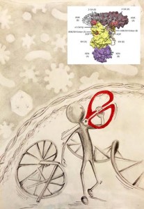
RNase L enzyme structure
Researchers at Princeton have deciphered the 3D structure of RNase L, an enzyme that slices through RNA and is a first responder in the innate immune system. The structure contains two subunits, represented in red as two parts of a pair of scissors. Illustration by Sneha Rath, Inset courtesy of Science.

An essential weapon in the body’s fight against infection has come into sharper view. Researchers at Princeton University have discovered the 3D structure of an enzyme that cuts to ribbons the genetic material of viruses and helps defend against bacteria.

The discovery of the structure of this enzyme, a first-responder in the body’s “innate immune system,” could enable new strategies for fighting infectious agents and possibly prostate cancer and obesity. The work was published Feb. 27 in the journal Science.

Until now, the research community has lacked a structural model of the human form of this enzyme, known as RNase L, said Alexei Korennykh, an assistant professor of molecular biology and leader of the team that made the discovery.

“Now that we have the human RNase L structure, we can begin to understand the effects of carcinogenic mutations in the RNase L gene. For example, families with hereditary prostate cancers often carry genetic mutations in the region, or locus, encoding RNase L,” Korennykh said. The connection is so strong that the RNase L locus also goes by the name “hereditary prostate cancer 1.” The newly found structure reveals the positions of these mutations and explains why some of these mutations could be detrimental, perhaps leading to cancer, Korennykh said. RNase L is also essential for insulin function and has been implicated in obesity.

The Princeton team’s work has also led to new insights on the enzyme’s function.

The enzyme is an important player in the innate immune system, a rapid and broad response to invaders that includes the production of a molecule called interferon. Interferon relays distress signals from infected cells to neighboring healthy cells, thereby activating RNase L to turn on its ability to slice through RNA, a type of genetic material that is similar to DNA. The result is new cells armed for destruction of the foreign RNA.

The 3D structure uncovered by Korennykh and his team consists of two nearly identical subunits called protomers. The researchers found that one protomer finds and attaches to the RNA, while the other protomer snips it.

The initial protomer latches onto one of the four “letters” that make up the RNA code, in particular, the “U,” which stands for a component of RNA called uridine. The other protomer “counts” RNA letters starting from the U, skips exactly one letter, then cuts the RNA.

Although the enzyme can slice any RNA, even that of the body’s own cells, it only does so when activated by interferon.

“We were surprised to find that the two protomers were identical but have different roles, one binding and one slicing,” Korennykh said. “Enzymes usually have distinct sites that bind the substrate and catalyze reactions. In the case of RNase L, it appears that the same exact protein surface can do both binding and catalysis. One RNase L subunit randomly adopts a binding role, whereas the other identical subunit has no other choice but to do catalysis.”

To discover the enzyme’s structure, the researchers first created a crystal of the RNase L enzyme. The main challenge was finding the right combination of chemical treatments that would force the enzyme to crystallize without destroying it.
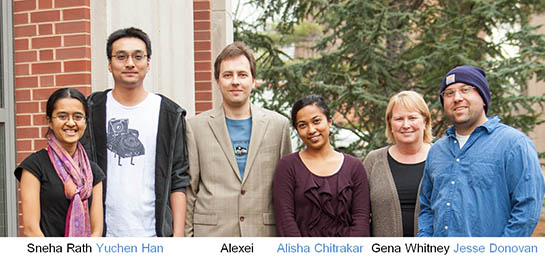
Korennykh groupAfter much trial and error and with the help of an automated system, postdoctoral research associate Jesse Donovan and graduate student Yuchen Han succeeded in making the crystals.

Next, the crystals were bombarded with powerful X-rays, which diffract when they hit the atoms in the crystal and form patterns indicative of the crystal’s structure. The diffraction patterns revealed how the atoms of RNase L are arranged in 3D space.

At the same time Sneha Rath, a graduate student in Korennykh’s laboratory, worked on understanding the RNA cleavage mechanism of RNase L using synthetic RNA fragments. Rath’s results matched the structural findings of Han and Donovan, and the two pieces of data ultimately revealed how RNase L cleaves its RNA targets.

Han, Donovan and Rath contributed equally to the paper and are listed as co-first authors.

Finally, senior research specialist Gena Whitney and graduate student Alisha Chitrakar conducted additional studies of RNase L in human cells, confirming the 3D structure.

Now that the human structure has been solved, researchers can explore ways to either enhance or dampen RNase L activity for medical and therapeutic uses, Korennykh said.

“This work illustrates the wonderful usefulness of doing both crystallography and careful kinetic and enzymatic studies at the same time,” said Peter Walter, professor of biochemistry and biophysics at the University of California-San Francisco School of Medicine. “Crystallography gives a static picture which becomes vastly enhanced by studies of the kinetics.”

Support for the work was provided by Princeton University.

Read the abstract.

Han, Yuchen, Jesse Donovan, Sneha Rath, Gena Whitney, Alisha Chitrakar, and Alexei Korennykh. Structure of Human RNase L Reveals the Basis for Regulated RNA Decay in the IFN Response Science 1249845. Published online 27 February 2014 [DOI:10.1126/science.1249845]

DNA Gridlock – Cells undo glitches to prevent mutations (Nature)

By Catherine Zandonella, Office of the Dean for Research

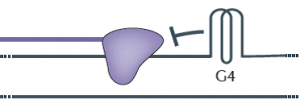
G4 Quadruplex
The diagram shows a G-quadruplex (G4) on the upper of the two strands that make up DNA. The purple shape represents DNA polymerase, which is blocked by the G4 in its attempt to copy DNA. Regions of the genome that are especially susceptible to forming G-quadruplexes are ones rich in guanine, which is one of the four nucleotides, designated by the letters G, A, C, and T, in DNA. Adapted from Nature Genetics, 2012.

Roughly six feet of DNA are packed into every human cell, so it is not surprising that our genetic material occasionally folds into odd shapes such as hairpins, crosses and clover leafs. But these structures can block the copying of DNA during cell division, leading to gene mutations that could have implications in cancer and aging.

Now researchers based at Princeton University have uncovered evidence that cells contain a built-in system for eliminating one of the worst of these roadblocks, a structure known as a G-quadruplex. In a paper published earlier this month in Nature, a group of researchers led by Princeton’s Virginia Zakian reported that an enzyme known as the Pif1 helicase can unfold these structures both in test tubes and in cells, bringing DNA replication back on track.

Given that Pif1 mutations have been associated with an increased risk of breast cancer, Zakian said, the study of how Pif1 ensures proper DNA replication could be relevant to human health. Zakian is Princeton’s Harry C. Wiess Professor in the Life Sciences.

Most DNA is made of up of two strands twisted about each other in a way that resembles a spiral staircase. Every time a cell divides, each DNA molecule must be duplicated, a process that involves unwinding the staircase so that an enzyme known as DNA polymerase can work down each strand, copying each letter in the DNA code. During this exposed period, regions of the unwound single strands can fold into G-quadruplexes (see diagram).

Like a car that encounters a pile-up on an Interstate, the DNA polymerase halts when it encounters a G-quadruplex, explained Matthew Bochman, a postdoctoral researcher who was a co-first author with Katrin Paeschke, now an independent investigator at University of Würzburg in Germany. The work also included Princeton graduate student Daniela Garcia.

“The DNA that is folded into a G-quadruplex cannot be replicated, so essentially it is skipped,” Bochman said. “Failure to copy specific areas of DNA that you really need is a serious problem, especially in regions that control genes that either suppress or contribute to cancer,” Bochman said.

Last year, the Zakian group in collaboration with human geneticists at the University of Washington reported that a mutation in human Pif1 is associated with an increased risk of breast cancer, suggesting that the ability to unwind G-quadruplexes could be important for protecting against cancer. The finding was published in the journal PLoS One.

G-quadruplexes could also be implicated in the process of aging, according to the researchers. The structures are thought to form at the ends of chromosomes in regions called telomeres, said Zakian, an expert on telomere biology.  Damaged or shortened telomeres are associated with premature aging and cancer.

To explore the role of Pif1 helicases in tackling G-quadruplexes, Bochman and Paeschke purified Pif1 helicases from yeast and bacteria and found that in test tubes, all of the Pif1 helicases unwind G-quadruplex structures extremely fast and very efficiently, much better than other helicases tested in the same way.

Next, these investigators set up an experiment to determine if Pif1 acts on G-quadruplexes inside cells. Using a system that could precisely evaluate the effects of G-quadruplex structures on the integrity of chromosomes, the researchers found that normal cells had no problem with the addition of a G-quadruplex structure, but when cells lack Pif1 helicases, the G-quadruplex induced a high amount of genome instability.

“To me, the most remarkable aspect of the study was the demonstration that Pif1-like helicases taken from species ranging from bacteria to humans and placed in yeast cells can suppress G-quadruplex-induced DNA damage,” Zakian said. “This finding suggests that resolving G-quadruplexes is an evolutionarily conserved function of Pif1 helicases.”

The Zakian lab also found that replicating through G-quadruplexes in the absence of Pif1 helicases results not only in mutations of the DNA at the site of the G-quadruplex but also in intriguing “epigenetic” effects on expression of nearby genes that were totally unexpected. Epigenetic events cause changes in gene expression that are inherited, yet they do not involve loss or mutation of DNA. Graduate student Daniela Garcia has proposed that the epigenetic silencing of gene expression that occurs near G-quadruplexes in the absence of Pif1 helicases is a result of the addition or removal of molecular tags on histones, which are proteins that bind DNA and regulate gene expression. This hypothesis is currently being studied.

The study involved contributions from Petr Cejka and Stephen C. Kowalczykowski of the University of California-Davis, and Katherine Friedman of Vanderbilt University.

Read the abstract.

Paeschke, Katrin, Matthew L. Bochman, P. Daniela Garcia, Petr Cejka, Katherine L. Friedman, Stephen C. Kowalczykowski & Virginia A. Zakian. Pif1 family helicases suppress genome instability at G-quadruplex motifs. Nature. 2013. doi:10.1038/nature12149.

This research was supported by the National Institutes of Health (V.A.Z., GM026938-34; S.C.K.GM041347), the National Science Foundation (K.L.F., MCB-0721595), the German Research Foundation (DFG), the New Jersey Commission on Cancer Research (K.P.) and the American Cancer Society (M.L.B., PF-10-145-02-01).


Cancer cells exchange leaders during invasion (PNAS)

By Catherine Zandonella, Office of the Dean for Research

A new study has found that cancer cells appear to exchange leading roles as they migrate out of a tumor in the early stages of invasion, or metastasis, of other sites in the body. Metastatic cancer accounts for more than 90% of cancer-related deaths.

A team led by Robert Austin, professor of physics at Princeton University, found that individual cancer cells take turns as trailblazers when they carve their way through the dense wall — known as the extracellular matrix — that stands between a tumor and the blood vessels which can carry the cells to other parts of the body.

The researchers also found that the cells leave the tumor in search of food, since cells that had plenty of available nutrients did not migrate. The finding reinforces the hypothesis that metastasis occurs when tumors become so densely packed that blood vessels cannot penetrate the interior and cancer cells must migrate to survive.

The researchers included first author Liyu Liu of the Chinese Academy of Sciences; Guillaume Duclos of the National Center for Scientific Research in Paris; Bo Sun, Jeongseog Lee, Amy Wu, Howard Stone and James Sturm of Princeton University; Yoonseok Kam and Robert Gatenby of H. Lee Moffitt Cancer Center in Tampa; and Eduardo Sontag of Rutgers University. The article appeared in the Proceedings of the National Academy of Sciences.

To study cancer cell behavior, the researchers constructed a small chamber with three compartments arranged like floors in an apartment building. On the bottom floor was a well of glucose, the preferred food for metastatic cells. The middle floor contained a dense layer of collagen, a protein that makes up the extracellular matrix that surrounds tumors. On the top floor they placed metastatic cancer cells, which were labeled with fluorescent dye for visibility. They trained a microscope and camera on the chamber.

Through the microscope, the researchers filmed the cancer cells as they moved down through the chamber toward the glucose. The researchers found that a single cell would become the leader for some time, then drop back as another cell took the lead in what the authors term a “collective invasion strategy.” They also found that the collagen was pushed aside, leaving a wake in which cells behind the leader could travel.

Because the collagen is very dense, the cells must expend a lot of energy to reach the glucose, and indeed the researchers found that cells without a need for glucose did not bother to burrow down into the collagen. The researchers used collagen with a density similar to that of human breast tissue.

The study adds to the growing understanding of metastasis and could serve to assist researchers in developing strategies for its prevention.

Liyu Liu, Guillaume Duclos, Bo Sun, Jeongseog Lee, Amy Wu, Yoonseok Kam, Eduardo D. Sontag, Howard A. Stone, James C. Sturm, Robert A. Gatenby, and Robert H. Austin. Minimization of thermodynamic costs in cancer cell invasion. PNAS January 14, 2013 201221147.

Read the paper (open access).

This work was supported by the National Science Foundation and the National Cancer Institute.

The role of breast structure in tumor development (PNAS)

Why do some breast tumors grow aggressively while others grow slowly? In this study, researchers found that the stiffness of the cells in the area around an emerging tumor influences its ability to grow and invade the breast. Using a 3-D fabrication process, the researchers created artificial breast ducts containing normal breast cells and a single tumor cell. They found that regions characterized by stiffness among the normal cells were more likely to give rise to tumors that are aggressive and invasive, while regions that were less stiff gave rise to tumors that are less invasive.

Eline Boghaert, Jason P. Gleghorn, KangAe Lee, Nikolce Gjorevski, Derek C. Radisky, and Celeste M. Nelson. Host epithelial geometry regulates breast cancer
cell invasiveness. Published online before print November 12, 2012, doi: 10.1073/pnas.1118872109 PNAS November 12, 2012

Read the abstract.