Genetically engineered mice could boost fight against aggressive hepatitis

Article provided by the Department of Molecular Biology

Hepatitis delta virus (HDV) causes the most aggressive form of viral hepatitis in humans, putting at least 20 million people worldwide at risk of developing liver fibrosis, cirrhosis, and liver cancer. Efforts to develop effective treatments against HDV have been hampered by the fact that laboratory mice are not susceptible to the virus. But, in a study published June 27, 2018, in the journal Science Translational Medicine, Alexander Ploss, assistant professor of molecular biology at Princeton University and colleagues describe a genetically engineered mouse that can be persistently infected with HDV.

HDV is a small, RNA-based “satellite” virus that produces just a single protein of its own and therefore requires additional proteins provided by another liver virus, hepatitis B virus (HBV). HDV can infect patients already carrying HBV, or both viruses can infect patients simultaneously. Though infections can be prevented with an anti-HBV vaccine, there are no antiviral therapies available to cure existing HDV infections.

HDV and HBV infect the liver by binding to a protein called NTCP that is present on the surface of liver cells. But the viruses only recognize the version of NTCP present in humans and a few other primates, and therefore can’t infect mice or other small mammals that produce their own versions of NTCP. This has made it difficult to study HBV and HDV infections in the laboratory. Researchers have tried transplanting human liver cells into immunocompromised mice before infecting them with virus, but this approach has produced inconsistent results and is both expensive and time-consuming.

Ploss and colleagues, led by graduate student Benjamin Winer, took a different approach. They generated mice that express the human NTCP protein in their liver cells, allowing these cells to be infected by HBV and HDV.

In these mice, HBV failed to replicate after entering mouse liver cells but HDV was able to establish persistent infection when provided with the HBV proteins it needs to propagate. For example, mice genetically engineered to produce both human NTCP and the entire HBV genome could be infected with HDV for up to 14 days. “To our knowledge, this is the first time the entire HDV life cycle has been recapitulated in a mouse model with inheritable susceptibility to HDV,” Ploss said.

The mice were able to rid themselves of HDV before they developed any liver damage, apparently by mounting an immune response involving antiviral interferon proteins and various white blood cell types, including Natural Killer (NK) cells and T cells. Accordingly, mice expressing human NTCP and the HBV genome, but lacking functional B, T, and NK cells could be infected with HDV for two months or more.

These immunocompromised animals allowed Ploss and colleagues to test the effectiveness of two drugs that are currently being developed as treatments for HDV infection. Both drugs—either alone or in combination—suppressed the levels of HDV in immunocompromised mice after viral infection. But the drugs were not able to completely clear the mice of HDV; viral levels rose again within weeks of stopping treatment.

“This is largely in line with recently reported data from clinical trials, showing the utility of our model for preclinical antiviral drug testing,” Winer said.

“Our model is amenable to genetic manipulations, robust, and can be adopted as a method to rapidly screen for potential treatments,” Ploss added.

Timothy M. Block, president of the Hepatitis B Foundation and its Baruch S. Blumberg Institute who was not involved in the study, said “These systems should be able to provide practical, and presumably economical  tools. Their work is urgently needed, and a desperate community welcomes it. I emphasize that it is often the new methods in science that revolutionize a field such as drug discovery, almost as much as the new drugs themselves.”

The research team included collaborators from Princeton University; Weill Medical College of Cornell University; The Jackson Laboratory; University Medical Center Hamburg-Eppendorf, Hamburg; New York University Medical Center; and North Carolina State University College of Veterinary Medicine.

This study was supported by grants from the National Institutes of Health (R01 AI079031, R01 AI107301, R21AI117213 to Alexander Ploss), a Research Scholar Award from the American Cancer Society (RSG-15-048-01-MPC to Alexander Ploss), a Burroughs Wellcome Fund Award for Investigators in Pathogenesis (to Alexander Ploss) and a Graduate fellowship from the Health Grand Challenge from the Global Health Fund of Princeton University (to Benjamin Y. Winer). The NYU Experimental Pathology Immunohistochemistry Core Laboratory is supported in part by the Laura and Isaac Perlmutter Cancer Center Support Grant; NIH/NCI P30CA016087 and the National Institutes of Health S10 Grants NIH/ORIP S10OD01058 and S10OD018338. Benjamin Y. Winer is a recipient of F31 NIH/NRSA Ruth L. Kirschstein Predoctoral awarded from the NIAID. Julie Sellau and Elham Shirvani-Dastgerdi are both recipients of postdoctoral fellowships from the German Research Foundation. Michael V. Wiles was funded by The Jackson Laboratory.

Benjamin Y. Winer, Elham Shirvani-Dastgerdi, Yaron Bram, Julie Sellau, Benjamin E. Low, Heath Johnson, Tiffany Huang, Gabriela Hrebikova, Brigitte Heller, Yael Sharon, Katja Giersch, Sherif Gerges, Kathleen Seneca, Mihai-Alexandru Pais, Angela S. Frankel, Luis Chiriboga, John Cullen, Ronald G. Nahass, Marc Lutgehetmann, Jared Toettcher, Michael V. Wiles, Robert E. Schwartz, and Alexander Ploss. Preclinical assessment of antiviral combination therapy in a genetically humanized mouse model for persistent hepatitis delta virus infection. Science Translational Medicine. 2018. DOI: 10.1126/scitranslmed.aap9328

Researchers develop technique to track yellow fever virus replication

Infection with a strain of yellow fever virus
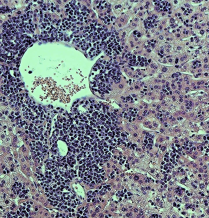
Infection with a strain of yellow fever virus (YFD-17D) in mouse liver. The liver of a mouse whose immune cells lack the immune signaling component known as STAT1 shows severe lymphocyte infiltration and inflammation, as well as necrosis, after infection with YFV-17D. Credit: Florian Douam and Alexander Ploss

By Staff, Department of Molecular Biology

Researchers from Princeton University‘s Department of Molecular Biology have developed a new method that can precisely track the replication of yellow fever virus in individual host immune cells. The technique, which is described in a paper published March 14 in the journal Nature Communications, could aid the development of new vaccines against a range of viruses, including Dengue and Zika.

Yellow fever virus (YFV) is a member of the flavivirus family that also includes Dengue and Zika virus. The virus, which is thought to infect a variety of cell types in the body, causes up to 200,000 cases of yellow fever every year, despite the widespread use of a highly effective vaccine. The vaccine consists of a live, attenuated form of the virus called YFV-17D, whose RNA genome is more than 99 percent identical to the virulent strain. This one percent difference in the attenuated virus’ genome may subtly alter interactions with the host immune system so that it induces a protective immune response without causing disease.

To explore how viruses interact with their hosts, and how these processes lead to virulence and disease, Alexander Ploss, assistant professor of molecular biology, and colleagues at Princeton University adapted a technique — called RNA Prime flow — that can detect RNA molecules within individual cells. They used the technique to track the presence of replicating viral particles in various immune cells circulating in the blood of infected mice. Mice are usually resistant to YFV, but Ploss and colleagues found that even the attenuated YFV-17D strain was lethal if the transcription factor STAT1, part of the antiviral interferon signaling pathway, was removed from mouse immune cells. The finding suggests that interferon signaling within immune cells protects mice from YFV, and that species-specific differences in this pathway allow the virus to replicate in humans and certain other primates but not mice.

Accordingly, YFV-17D was able to replicate efficiently in mice whose immune systems had been replaced with human immune cells capable of activating interferon signaling. However, just like humans immunized with the attenuated YFV vaccine, these “humanized” mice didn’t develop disease symptoms when infected with YFV-17D, allowing Ploss and colleagues to study how the attenuated virus interacts with the human immune system. Using their viral RNA flow technique, the researchers determined that the virus can replicate inside certain human immune cell types, including B lymphocytes and natural killer cells, in which the virus has not been detected previously. The researchers found that the panel of human cell types targeted by the virus changes over the course of infection in both the blood and the spleen of the animals, highlighting the distinct dynamics of YFV-17D replication in the human immune system.

The next step, said Florian Douam, a postdoctoral research associate in the Department of Molecular Biology and first author on the study, is to confirm YFV replication in these subsets of immune cells in YFV-infected patients and in recipients of the YFV-17D vaccine. Viral RNA flow now provides the means to perform such analyses, Douam said.

The researchers also plan to study whether the virulent and attenuated strains of yellow fever virus infect different host immune cells. The approach may help explain why some people infected with the virus die while others develop only the mildest of symptoms, as well as which changes in the YFV-17D genome weaken the virus’ ability to cause disease. “This could guide the rational design of vaccines against related pathogens, such as Zika and Dengue virus,” Ploss said.

This work was supported by a grant from the Health Grand Challenge program from Princeton University, the New Jersey Commission on Cancer Research (Grant No. DHFS16PPC007), the Genentech Foundation and Princeton University’s Anthony Evnin ’62 Senior Thesis Fund.

Florian Douam, Gabriela Hrebikova, Yentli E. Soto Albrecht, Julie Sellau, Yael Sharon, Qiang Ding and Alexander Ploss. Single-cell tracking of flavivirus RNA uncovers species-specific interactions with the immune system dictating disease outcome. Nature Communications. 8: 14781. (2017). doi: 10.1038/ncomms14781

Now in 3D: Video of virus-sized particle trying to enter cell (Nature Nanotechnology)

Video of virus trying to enter cell
3D movie (below) of virus-like nanoparticle trying to gain entry to a cell

By Catherine Zandonella, Office of the Dean for Research

Tiny and swift, viruses are hard to capture on video. Now researchers at Princeton University have achieved an unprecedented look at a virus-like particle as it tries to break into and infect a cell. The technique they developed could help scientists learn more about how to deliver drugs via nanoparticles — which are about the same size as viruses — as well as how to prevent viral infection from occurring.

The video reveals a virus-like particle zipping around in a rapid, erratic manner until it encounters a cell, bounces and skids along the surface, and either lifts off again or, in much less time than it takes to blink an eye, slips into the cell’s interior. The work was published in Nature Nanotechnology.

Video caption: ‘Kiss and run’ on the cell surface. This 3D movie shows actual footage of a virus-like particle (red dot) approaching a cell (green with reddish brown nucleus), as captured by Princeton University researchers Kevin Welcher and Haw Yang. The color of the particle represents its speed, with red indicating rapid movement and blue indicating slower movement. The virus-like particle lands on the surface of the cell, appears to try to enter it, then takes off again. Source: Nature Nanotechnology.

“The challenge in imaging these events is that viruses and nanoparticles are small and fast, while cells are relatively large and immobile,” said Kevin Welsher, a postdoctoral researcher in Princeton’s Department of Chemistry and first author on the study. “That has made it very hard to capture these interactions.”

The problem can be compared to shooting video of a hummingbird as it roams around a vast garden, said Haw Yang, associate professor of chemistry and Welsher’s adviser. Focus the camera on the fast-moving hummingbird, and the background will be blurred. Focus on the background, and the bird will be blurred.

The researchers solved the problem by using two cameras, one that locked onto the virus-like nanoparticle and followed it faithfully, and another that filmed the cell and surrounding environment.

Putting the two images together yielded a level of detail about the movement of nano-sized particles that has never before been achieved, Yang said. Prior to this work, he said, the only way to see small objects at a similar resolution was to use a technique called electron microscopy, which requires killing the cell.

“What Kevin has done that is really different is that he can capture a three-dimensional view of a virus-sized particle attacking a living cell, whereas electron microscopy is in two-dimensions and on dead cells,” Yang said. “This gives us a completely new level of understanding.”

In addition to simply viewing the particle’s antics, the researchers can use the technique to map the contours of the cell surface, which is bumpy with proteins that push up from beneath the surface. By following the particle’s movement along the surface of the cell, the researchers were able to map the protrusions, just as a blind person might use his or her fingers to construct an image of a person’s face.

“Following the motion of the particle allowed us to trace very fine structures with a precision of about 10 nanometers, which typically is only available with an electron microscope,” Welsher said. (A nanometer is one billionth of a meter and roughly 1000 times smaller than the width of a human hair.) He added that measuring changes in the speed of the particle allowed the researchers to infer the viscosity of the extracellular environment just above the cell surface.

The technology has potential benefits for both drug discovery and basic scientific discovery, Yang said.  “We believe this will impact the study of how nanoparticles can deliver medicines to cells, potentially leading to some new lines of defense in antiviral therapies,” he said. “For basic research, there are a number of questions that can now be explored, such as how a cell surface receptor interacts with a viral particle or with a drug.”

Welsher added that such basic research could lead to new strategies for keeping viruses from entering cells in the first place.

“If we understand what is happening to the virus before it gets to your cells,” said Welsher, “then we can think about ways to prevent infection altogether. It is like deflecting missiles before they get there rather than trying to control the damage once you’ve been hit.”

To create the virus-like particle, the researchers coated a miniscule polystyrene ball with quantum dots, which are semiconductor bits that emit light and allow the camera to find the particle. Next, the particle was studded with protein segments known as Tat peptides, derived from the HIV-1 virus, which help the particle find the cell. The width of the final particle was about 100 nanometers.

The researchers then let loose the particles into a dish containing skin cells known as fibroblasts. One camera followed the particle while a second imaging system took pictures of the cell using a technique called laser scanning microscopy, which involves taking multiple images, each in a slightly different focal plane, and combining them to make a three-dimensional picture.

The research was supported by the US Department of Energy (DE-SC0006838) and by the Eric and Wendy Schmidt Transformative Technology Fund at Princeton University.

Read the abstract.

Kevin Welsher and Haw Yang. 2014. Multi-resolution 3D visualization of the early stages of cellular uptake of peptide-coated nanoparticles. Nature nanotechnology. Published online: 23 February 2014 | DOI: 10.1038/NNANO.2014.12

New mouse model for hepatitis C (Nature)

By Catherine Zandonella, Office of the Dean for Research

Hepatitis C affects about three million people in the U.S. and is a leading cause of chronic liver disease, so creating a vaccine and new treatments is an important public health goal. Most research to date has been done in chimpanzees because they are one of a handful of species that become infected and spread the virus.

Now researchers led by Alexander Ploss of Princeton University and Charles Rice of the Rockefeller University have generated a mouse that can become infected with hepatitis C virus (HCV).  They reported the advance in the Sept 12 issue of the journal Nature. “The entire life cycle of the virus — from infection of liver cells to viral replication, assembly of new particles, and release from the infected cell — occurs in these mice,” said Ploss, who joined the Princeton faculty in July 2013 as assistant professor of molecular biology.

Ploss and his colleagues have been working for some time on the challenge of creating a small animal model for studying the disease. Four years ago, while at the Rockefeller University in New York, Ploss and Rice identified two human proteins, known as CD81 and occludin, that enable mouse cells to become infected with HCV (Nature 2009). In a follow up study Ploss and colleagues showed that a mouse engineered to express these human proteins could become infected with HCV, although the animals could not spread the virus (Nature 2011).

In the present study, which included colleagues at Osaka University and the Scripps Research Institute, the researchers bred the human-protein-containing mice with another strain that had a defective immune system – one that could not easily rid the body of viruses. The resulting mice not only become infected, but could potentially pass the virus to other susceptible mice.

The availability of this new way to study HCV could help researchers discover new vaccines and treatments, although Ploss cautioned that more work needs to be done to refine the model.

The study was supported in part by award number RC1DK087193 from the National Institute of Diabetes and Digestive and Kidney Diseases; R01AI072613, R01AI099284, and R01AI079031 from the National Institute for Allergy and Infectious Disease; R01CA057973 from the National Cancer Institute; and several foundations and contributors, as well as the Infectious Disease Society of America and the American Liver Foundation.

Read the abstract

Marcus Dorner, Joshua A. Horwitz, Bridget M. Donovan, Rachael N. Labitt, William C. Budell, Tamar Friling, Alexander Vogt, Maria Teresa Catanese, Takashi Satoh, Taro Kawai, Shizuo Akira, Mansun Law, Charles Rice & Alexander Ploss. 2013. Completion of the entire hepatitis C virus life cycle in genetically humanized mice. Nature 501, 237–241 (First published online on 31 July 2013)  doi:10.1038/nature12427.


Shingles symptoms may be caused by neuronal short circuit (Proceedings of the National Academy of Sciences)

By Catherine Zandonella, Office of the Dean for Research

Neurons firing in synchrony could be responsible for pain, itch in shingles and herpes infection. Click to view movie. (Source: PNAS)

The pain and itching associated with shingles and herpes may be due to the virus causing a “short circuit” in the nerve cells that reach the skin, Princeton researchers have found.

This short circuit appears to cause repetitive, synchronized firing of nerve cells, the researchers reported in the journal Proceedings of the National Academy of Sciences. This cyclical firing may be the cause of the persistent itching and pain that are symptoms of oral and genital herpes as well as shingles and chicken pox, according to the researchers.

These diseases are all caused by viruses of the herpes family. Understanding how these viruses cause discomfort could lead to better strategies for treating symptoms.

The team studied what happens when a herpes virus infects neurons. For research purposes the investigators used a member of the herpes family called pseudorabies virus. Previous research indicated that these viruses can drill tiny holes in neurons, which pass messages in the form of electrical signals along long conduits known as axons.

The researchers’ findings indicate that electrical current can leak through these holes, or fusion pores, and spread to nearby neurons that were similarly damaged, causing the neurons to fire all at once rather than as needed. The pores were likely created for the purpose of infecting new cells, the researchers said.

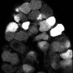
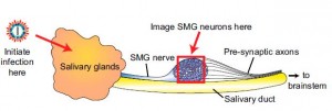
Researchers at Princeton University imaged the synchronized, repetitive firing of herpes-infected neurons in a region known as the submandibular ganglia (SMG) between the salivary glands and the brain in mice. Image source: PNAS.
Researchers at Princeton University imaged the synchronized, repetitive firing of herpes-infected neurons in a region known as the submandibular ganglia (SMG) between the salivary glands and the brain in mice. (Source: PNAS)

The investigators observed the cyclical firing of neurons in a region called the submandibular ganglia between the salivary glands and the brain in mice using a technique called 2-photon microscopy and dyes that flash brightly when neurons fire. (Movie of synchronized firing of herpes-infected neurons.)

The team found that two viral proteins appear to work together to cause the simultaneous firing, according to Andréa Granstedt, who received her Ph.D. in molecular biology at Princeton in 2013 and is the first author on the article.  The team was led by Lynn Enquist, Princeton’s Henry L. Hillman Professor in Molecular Biology and a member of the Princeton Neuroscience Institute.

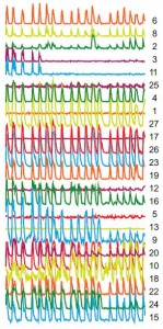
Each colored line and number on the right represents an individual neuron. The overlapping peaks indicate synchronized firing of neurons, which occurs when electrical current is able to leak from one neuron to the next. (Source: PNAS)

The first of these two proteins is called glycoprotein B, a fusion protein that drills the holes in the axon wall. A second protein, called Us9, acts as a shuttle that sends glycoprotein B into axons, according to the researchers. “The localization of glycoprotein B is crucial,” Granstedt said. “If glycoprotein B is present but not in the axons, the synchronized flashing won’t happen.”

The researchers succeeded in stopping the short circuit from occurring in engineered viruses that lacked the gene for either glycoprotein B or Us9. Such genetically altered viruses are important as research tools, Enquist said.

Finding a way to block the activity of the proteins could be a useful strategy for treating the pain and itching associated with herpes viral diseases, Enquist said. “If you could block fusion pore formation, you could stop the generation of the signal that is causing pain and discomfort,” he said.

Granstedt conducted the experiments with Jens-Bernhard Bosse, a postdoctoral research associate in molecular biology. Assistance with 2-photon microscopy was provided by Stephan Thiberge, director of the Bezos Center for Neural Circuit Dynamics at the Princeton Neuroscience Institute.

The team previously observed the synchronized firing in laboratory-grown neurons (PLoS Pathogens, 2009), but the new study expands on the previous work by observing the process in live mice and including the contribution of Us9, Granstedt said.

Shingles, which is caused by the virus herpes zoster and results in a painful rash, will afflict almost one out of three people in the United States over their lifetime. Genital herpes, which is caused by herpes simplex virus-2, affects about one out of six people ages 14 to 49 years in the United States, according the Centers for Disease Control and Prevention.

This research was funded by National Institutes of Health (NIH) Grants NS033506 and NS060699. The Imaging Core Facility at the Lewis-Sigler Institute is funded by NIH National Institute of General Medical Sciences Center Grant PM50 GM071508.

Read the abstract

Granstedt, Andréa E., Jens B. Bosse, Stephan Y. Thiberge, and Lynn W. Enquist. 2013. In vivo imaging of alphaherpesvirus infection reveals synchronized activity dependent on axonal sorting of viral proteins. PNAS 2013 ; published ahead of print August 26, 2013, doi:10.1073/pnas.1311062110

Herpes viruses commandeer protein production to storm neurons (Cell Host and Microbe)

By Morgan Kelly, Office of Communications

Viruses in the herpes family most commonly found in humans infect nervous system cells by “turning on” and then seizing control of the internal system these cells rely on to sense injury, among other signaling functions.

Princeton University researchers report in the journal Cell Host and Microbe that the pseudorabies virus (PRV) — a model herpes virus that infects animals — initiates and commandeers protein production in axons, the long offshoots of the cell body that connect neurons to other neurons and to tissue. After entering the neuron at the axon, the virus particles — which deliver the viral DNA that infects host cells — use the newly made proteins to travel to and infect the cell nucleus. Once there, the infection can spread to other neurons.

The research is the latest from the laboratory of senior researcher Lynn Enquist, the Henry L. Hillman Professor in Molecular Biology, to unravel the puzzling efficiency with which PRV and related herpes viruses invade the nervous system. PRV is an alpha-herpes virus, a prolific herpes subfamily that includes herpes simplex virus 1 (HSV-1), an extremely common human virus that causes cold sores and other lesions.

In the current paper, the researchers write that PRV “cleverly exploited” a natural cell process to speed up infection, a theme that resonates in past work from the Enquist lab on alpha-herpes viruses. In 2012, another researcher in the lab reported in Cell Host and Microbe that PRV and HSV-1 infections affect movement of neuronal mitochondria, the mobile organelles that regulate a cell’s energy supply, communication, and self-destruction response to infection.

For this newest research, Enquist worked with lead author Orkide Koyuncu, a postdoctoral research associate in molecular biology, and David Perlman, head of the molecular biology department’s mass spectrometry facility. They suggest that PRV particles first replicate in non-neuronal (such as skin and other tissue) cells at the site of body entry. The particles then enter axon terminals as the axon carries out its regular status-reports with those cells. The process of viral-particle entry is sensed by the neuron as a damage signal, which begins the protein production that will carry the virus particles to the nucleus.

Interestingly, the researchers discovered that the movement of incoming virus particles was disrupted by a genuine damage signal initiated before PRV infection. They hypothesized that the immediate response spurred by injury, infection or inflammation slows down other processes within the axon, which the researchers call “competitive inhibition.” When the molecular details of this crosstalk are fully understood, these signals could be used clinically to prevent the spread of alpha-herpes viruses.

Read the paper.

Citation: Koyuncu, Orkide O., David H. Perlman, Lynn W. Enquist. 2013. Efficient Retrograde Transport of Pseudorabies Virus within Neurons Requires Local Protein Synthesis in Axons. Cell Host & Microbe Vol. 13, no. 1, pp. 54-66.

This work was supported by U.S. National Institutes of Health grant R01NS033506-18.