An immune signaling pathway for control of Yellow Fever Virus infection

By the Department of Molecular Biology

Princeton University researchers have uncovered a critical role for a new immune signaling pathway in controlling infection by the flavivirus Yellow Fever Virus (YFV).  The paper describing this discovery was published today in the journal mBio.

Infection with YFV causes a devastating illness with a mortality rate of up to 50%.  Like other members of its viral family—which includes West Nile Virus, Dengue Virus and Zika Virus—YFV is transmitted to humans by mosquitos that are expanding into new areas across the globe, exposing more people to these dangerous viruses. Fortunately, there is an effective vaccine for YFV: a live-attenuated strain of the virus, called YFV-17D, which differs by only a few amino acids from the virulent viral strain YFV-Asibi, but nonetheless provokes a potent and durable protective immune response in humans.

“An improved understanding of the complex mechanisms regulating YFV-17D attenuation will provide insights into key viral-host interactions that regulate host immune responses and infection outcomes, [and] open novel avenues for the development of innovative vaccine strategies,” said Alexander Ploss, assistant professor at Princeton’s Department of Molecular Biology, who led the study with first author Florian Douam, a postdoctoral research associate. However, research efforts have been hampered due to the fact that mice, which are used in the study of viral infections, are resistant to YFV infection. Nonetheless, recent mouse experiments have pointed to an important role for cytokines called interferons (IFN) in controlling the virus.

Mice, like humans, possess three types of interferons, molecules produced by the immune system during infection: type I interferons, which signal through the widely distributed IFN-α/β receptor; type II interferons that act on IFN-γ receptors present in most tissues; and type III interferons, which activate signaling by IFN-λ receptors found on epithelial cells. Mice lacking type I receptors die after infection by YFV-Asibi, but survive YFV-17D infection despite extensive viral replication at early stage of infection.Type II IFN signaling has also been shown to be important for clearing up late stage YFV-Asibi and YFV-17D infection when type I IFN signaling is defective. By contrast, the contribution of type III IFN signaling to control of YFV infection was unknown.

To address this question, Douam and colleagues studied YFV-17D infection in mice lacking the type III receptor. Initial experiments showed that these mice were able to control viral replication and rapidly cleared YFV-17D, indicating that type III signaling alone wasn’t necessary for resistance to YFV-17D. However, mice lacking both type I and type III receptors succumbed after YFV-17D infection, suggesting type III signaling does contribute to the antiviral immune response.

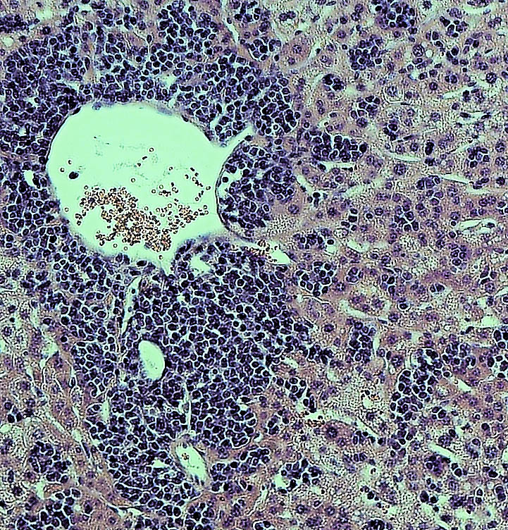
To find out more, the authors examined YFV-17D levels in various tissues. Early in the infection, the virus was present in every tissue of each mouse model examined. However, although viral loads were low in wild-type mice and type III receptor-deficient mice, they were much higher in type I and type I/III receptor-deficient mice. Surprisingly, the viral loads in brains of type I/III receptor-deficient mice increased over time in comparison to type I receptor-deficient mice, showing that loss of type III IFN signaling enhances the susceptibility of type I-receptor deficient animals to brain infection. This is significant because the presence of viruses in the brain can cause brain damage such as spongiosis or encephalitis. The low level of YFV-17FD brain invasion in wild-type mice caused mild spongiosis, whereas type I/III receptor-deficient mice had severe spongiosis—potentially explaining YFV-17D lethality in those animals.  However, this raised the question of why YFV-17 was present at such high levels in the animals’ brains.

Brain tissues
Left panel: Loss of Type I IFN signaling leads to active replication of the attenuated YFV strain (YFV-17D), which is accompanied by viral invasion of the brain and damage to brain tissues (spongiosis). Right panel: The additional loss of Type III IFN signaling in Type I IFN-deficient mice impairs the integrity of the blood-brain-barrier and alters immune cell function, which aggravates spongiosis and is ultimately lethal. Image Credit: Florian Douam and Alexander Ploss

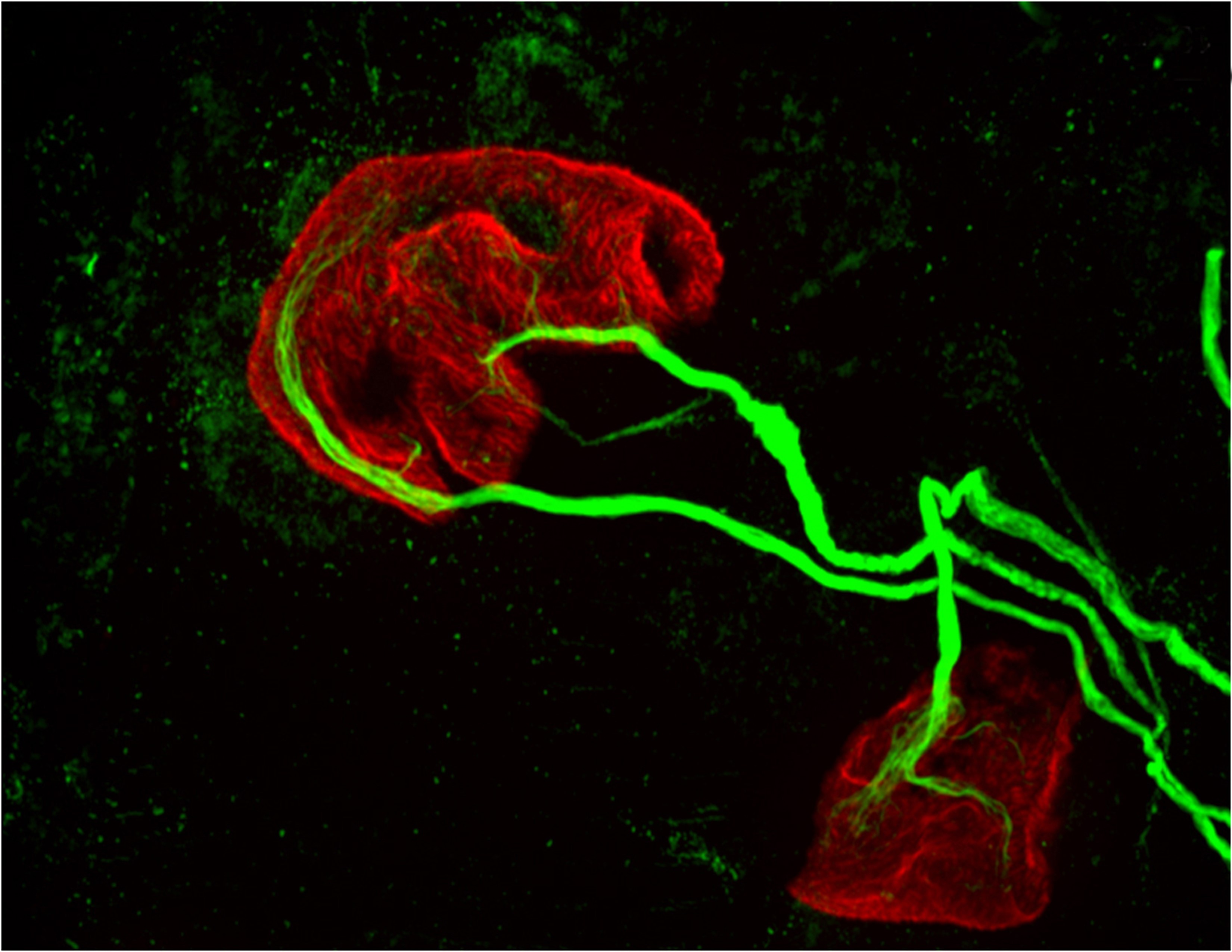
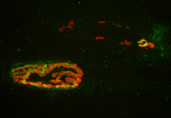
Another study recently showed that type III IFN signaling affects the epithelial cells that make up the blood brain barrier (BBB), and modulates BBB integrity during infection by another flavivirus, West Nile Virus. Consistent with this, Ploss’s group observed that the BBB of type I/III receptor-deficient mice was especially leaky to a blue dye. But this wasn’t the only way that loss of type III IFN signaling impaired the body’s response to YFV; the researchers also found evidence that type III receptor deficiency provokes strong imbalances in several different kinds of immune cells during YFV-17D infection. In particular, type I/III receptor-deficient mice were defective in the activation of T cells, critical immune cells that control YFV-17D infection.

“We uncovered a critical role of type III IFN-mediated signaling in preserving the integrity of the blood brain barrier and preventing viral brain invasion,” Ploss said. More work is needed to explore how type III IFN signaling affects YFV infection in primates, but this study already provides important new insights about a poorly understood immune signaling pathway.

The study was supported by grants from National Institutes of Health (R01 AI107301, R21AI117213 to Alexander Ploss and R01 AI104669 to Sergei Kotenko). Additional funding included a Research Scholar Award from the American Cancer Society (RSG-15-048-01-MPC), the Princeton Environmental Institute‘s Grand Health Challenge program from Princeton University, and an Investigator in Pathogenesis Award by the Burroughs Wellcome Fund (all to Alexander Ploss).

The study, “Type III Interferon-mediated signaling is critical for controlling live attenuated Yellow Fever Virus infection in vivo.,” by Florian Douam, Yentil E. Soto-Albrecht, Gabriela Hrebikova, Evita Sadimin, Christian Davidson, Sergei V. Kotenko and Alexander Ploss was published in the journal mBio.  (2017). doi:10.1128/mBio.00819-17

Researchers develop technique to track yellow fever virus replication

Infection with a strain of yellow fever virus
Infection with a strain of yellow fever virus (YFD-17D) in mouse liver. The liver of a mouse whose immune cells lack the immune signaling component known as STAT1 shows severe lymphocyte infiltration and inflammation, as well as necrosis, after infection with YFV-17D. Credit: Florian Douam and Alexander Ploss

By Staff, Department of Molecular Biology

Researchers from Princeton University‘s Department of Molecular Biology have developed a new method that can precisely track the replication of yellow fever virus in individual host immune cells. The technique, which is described in a paper published March 14 in the journal Nature Communications, could aid the development of new vaccines against a range of viruses, including Dengue and Zika.

Yellow fever virus (YFV) is a member of the flavivirus family that also includes Dengue and Zika virus. The virus, which is thought to infect a variety of cell types in the body, causes up to 200,000 cases of yellow fever every year, despite the widespread use of a highly effective vaccine. The vaccine consists of a live, attenuated form of the virus called YFV-17D, whose RNA genome is more than 99 percent identical to the virulent strain. This one percent difference in the attenuated virus’ genome may subtly alter interactions with the host immune system so that it induces a protective immune response without causing disease.

To explore how viruses interact with their hosts, and how these processes lead to virulence and disease, Alexander Ploss, assistant professor of molecular biology, and colleagues at Princeton University adapted a technique — called RNA Prime flow — that can detect RNA molecules within individual cells. They used the technique to track the presence of replicating viral particles in various immune cells circulating in the blood of infected mice. Mice are usually resistant to YFV, but Ploss and colleagues found that even the attenuated YFV-17D strain was lethal if the transcription factor STAT1, part of the antiviral interferon signaling pathway, was removed from mouse immune cells. The finding suggests that interferon signaling within immune cells protects mice from YFV, and that species-specific differences in this pathway allow the virus to replicate in humans and certain other primates but not mice.

Accordingly, YFV-17D was able to replicate efficiently in mice whose immune systems had been replaced with human immune cells capable of activating interferon signaling. However, just like humans immunized with the attenuated YFV vaccine, these “humanized” mice didn’t develop disease symptoms when infected with YFV-17D, allowing Ploss and colleagues to study how the attenuated virus interacts with the human immune system. Using their viral RNA flow technique, the researchers determined that the virus can replicate inside certain human immune cell types, including B lymphocytes and natural killer cells, in which the virus has not been detected previously. The researchers found that the panel of human cell types targeted by the virus changes over the course of infection in both the blood and the spleen of the animals, highlighting the distinct dynamics of YFV-17D replication in the human immune system.

The next step, said Florian Douam, a postdoctoral research associate in the Department of Molecular Biology and first author on the study, is to confirm YFV replication in these subsets of immune cells in YFV-infected patients and in recipients of the YFV-17D vaccine. Viral RNA flow now provides the means to perform such analyses, Douam said.

The researchers also plan to study whether the virulent and attenuated strains of yellow fever virus infect different host immune cells. The approach may help explain why some people infected with the virus die while others develop only the mildest of symptoms, as well as which changes in the YFV-17D genome weaken the virus’ ability to cause disease. “This could guide the rational design of vaccines against related pathogens, such as Zika and Dengue virus,” Ploss said.

This work was supported by a grant from the Health Grand Challenge program from Princeton University, the New Jersey Commission on Cancer Research (Grant No. DHFS16PPC007), the Genentech Foundation and Princeton University’s Anthony Evnin ’62 Senior Thesis Fund.

Florian Douam, Gabriela Hrebikova, Yentli E. Soto Albrecht, Julie Sellau, Yael Sharon, Qiang Ding and Alexander Ploss. Single-cell tracking of flavivirus RNA uncovers species-specific interactions with the immune system dictating disease outcome. Nature Communications. 8: 14781. (2017). doi: 10.1038/ncomms14781

Same immune-system proteins may first giveth, then taketh away motor control (Brain, Behavior, and Immunity)

two motor neurons (green) connected to a single muscle fiber (red)
Princeton University researchers found that proteins in the MHCI, or major histocompatibility complex class I, family can “prune” the connections, or synapses, between motor neurons and muscle fibers, which is necessary during early development. But the researchers also found that MHCI levels can rise again in old age, and that the proteins may resume pruning nerve-muscle synapses. This image from a mouse bred to express less MHCI shows two motor neurons (green) connected to a single muscle fiber (red) at an age when only one connection should remain. (Image by Lisa Boulanger, Princeton Neuroscience Institute, and Mazell Tetruashvily, Department of Molecular Biology)

By Morgan Kelly, Office of Communications

Princeton University researchers have found that a family of proteins with important roles in the immune system may be responsible for fine-tuning a person’s motor control as they grow — and for their gradual loss of muscle function as they age. The research potentially reveals a biological cause of weakness and instability in older people, as well as a possible future treatment that would target the proteins specifically.

The researchers reported in the journal Brain, Behavior, and Immunity that proteins in the family MHCI, or major histocompatibility complex class I, “prune” the connections, or synapses, between motor neurons and muscle fibers. Pruning is necessary during early development because at birth each muscle fiber in humans, mice and other vertebrates receives signals from dozens of neural connections. Proper motor control, however, requires that each muscle fiber receive signals from only a single motor neuron, so without the pruning carried out by MHCI proteins, fine motor control would never emerge.

But the researchers also found that MHCI levels can rise again in old age, and that the proteins may resume pruning nerve-muscle synapses — except that in a mature organism there are no extra synapses. The result is that individual muscle fibers become completely “denervated,” or detached from nervous system control. Denervated muscle fibers cannot be recruited during muscle contraction, which can leave older people weaker and more susceptible to devastating falls, making independent living difficult.

However, the Princeton researchers discovered that when MHCI levels were reduced in mice, denervation during aging was largely prevented. These findings could help scientists identify and treat the neurological causes of denervation and muscle weakness in the elderly.

Corresponding author Lisa Boulanger, an assistant professor in the Princeton Neuroscience Institute, explained that in infants, motor neurons initially make far too many connections to muscle fibers, which is part of why infants lack fine motor control.  Synapse overproduction followed by pruning occurs in many different regions of the vertebrate nervous system, and the neuromuscular junction has often been used as a model for studying this process.

It is not known why more synapses are made during development than are needed.

One possibility is that it allows the wiring diagram of the nervous system to be precisely tuned based on the way the circuit is used, Boulanger said. MHCI proteins help limit the final number of connections so that communication between neurons and muscles is more precise and efficient than would be possible using just a molecular code that produced a set number of connections.

“Molecules might get you to the right zip code, but pruning can make sure you arrive at the right house,” Boulanger said. “During development, it’s essential to get rid of extra synapses. But when you up-regulate MHCI when you’re older and start pruning synapses again, but you don’t have any extras to replace them.”

Boulanger worked with first author Mazell Tetruashvily, who received her doctorate in molecular biology from Princeton in 2015 and is now completing her M.D. training at University of Medicine and Dentistry of New Jersey; Marin McDonald, who received her doctorate in neuroscience from the University of California-San Diego (UCSD) in 2010, and is now a radiology resident at UCSD; and Karla Frietze, a doctoral student in Princeton’s Department of Molecular Biology. Boulanger was on the UCSD faculty before moving her lab to Princeton in 2009.

In the immune system, MHCI proteins present protein fragments, or peptides, to T cells, which are white blood cells with a central role in the body’s response to infection. This peptide presentation allows T cells to recognize and kill infected and cancerous cells, which present abnormal or foreign peptides on their MHCI proteins. It is unknown if the proteins’ ability to help recognize and destroy infected or cancerous cells is mechanistically related to the proteins’ ability to help eliminate excess synapses that the Princeton researchers discovered.

In the nervous system, MHCI proteins stop pruning synapses early in life. Why they may resume their synapse-eliminating activity later in life is unknown, Boulanger said. As immune-system proteins, MHCI levels increase with inflammation, she said. Aging is associated with chronic inflammation, which could explain the observed increase in MHCI levels and the reactivation of its former role.

Neuron image
The researchers found that MHCI the proteins may resume pruning nerve-muscle synapses in older organisms, an age when there are no extra synapses. Instead, muscle fibers become completely “denervated,” or detached from nervous system control, which could lead to weakness and instability in older people. This image from a 2-year-old (elderly) normal mouse shows denervation in the upper-right synapse, as noted by the lack of overlap of the red and green fluorescent markers used to indicate cells where the neuron and muscle fiber meet. At lower left, a healthy-looking synapse displays good overlap. (Image by Lisa Boulanger, Department of Molecular Biology)

The Princeton researchers found that mice bred to express less MHCI proteins had “more youthful” patterns of muscle innervation, since they were protected from denervation as they aged, Boulanger said. The mice actually lacked a protein known as beta-2 microglobulin, which forms a complex with MHCI and is necessary for MHCI expression on the surface of cells. This could be beneficial from a clinical perspective because beta-2 microglobulin is a soluble protein and can be removed from the blood, Boulanger said.

“If a rise in MHCI is the problem, having less beta-2 microglobulin might be protective,” Boulanger said. Recent results from a lab at Stanford University showed that reducing beta-2 microglobulin also helped with cognitive aging because of its effects on MHCI proteins. “Our studies raise the possibility that targeting one protein could help with both motor and cognitive aspects of aging,” Boulanger said.

Because MHCI proteins are important in the immune system, however, such an approach could result in compromised immunity, Boulanger said. The mice bred to not express beta-2 microglobulin had weakened immune systems, as a result of their lower levels of MHCI proteins. Future work will include exploring the effectiveness of other approaches to reducing the proteins’ synapse-eliminating activity in older nervous systems, ideally while leaving their immune functions intact, she said.

The research was supported by the Princeton Department of Molecular Biology and the Princeton Neuroscience Institute (grant no. 1F30AG046044-01A1), the UCSD School of Medicine, the Alfred P. Sloan Foundation, the Whitehall Foundation, and the Princeton Neuroscience Institute Innovation Fund.

Read more.

Mazell M. Tetruashvily, Marin A. McDonald, Karla K. Frietze and Lisa M. Boulanger. “MHCI promotes developmental synapse elimination and aging-related synapse loss at the vertebrate neuromuscular junction.” Brain, Behavior, and Immunity, in press. DOI: 10.1016/j.bbi.2016.01.008epartment of Molecular Biology)