Genetic instructions from mom set the pattern for embryonic development

Micrograph of a zebrafish organ called the Kupffer's vesicle

By the Department of Molecular Biology

A new study indicates an essential role for a maternally inherited gene in embryonic development. The study found that zebrafish that failed to inherit specific genetic instructions from mom developed fatal defects earlier in development, even if the fish could make their own version of the gene. The study by researchers at Princeton University was published Nov. 15 in the journal eLife.

When female animals form egg cells inside their ovaries, they deposit messenger RNAs (mRNAs) – a sort of genetic instruction set – in the egg cell cytoplasm. After fertilization, these maternally supplied mRNAs can be translated into proteins required for the early stages of embryonic development, before the embryo is able to produce mRNAs and proteins of its own.

More than thirty years ago, researchers discovered that mRNAs encoding a protein called Vg1 are deposited in the cytoplasm of frog eggs. “vg1 is famous for being one of the first recognized maternal mRNAs,” said Rebecca Burdine, associate professor of molecular biology at Princeton. “Many papers have been written on how this RNA is localized and regulated, but it was never clear what the Vg1 protein actually does in the developing embryo.”

Two zebrafish embryos
Compared to a normal zebrafish embryo (right), an embryo lacking gdf3 (left) inherited from mom shows major defects resulting from its inability to form mesoderm and endoderm cells early in development. Credit: Pelliccia et al., 2017.

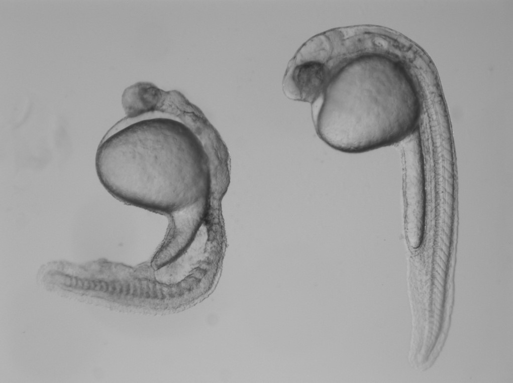
In the study, Burdine and two graduate students Jose Pelliccia and Granton Jindal used CRISPR/Cas9 gene editing to remove Vg1, known as Gdf3 in zebrafish. Embryos that couldn’t produce any Gdf3 of their own–but received a healthy portion of the gdf3 mRNA from their mothers–developed perfectly normally. But embryos that didn’t receive maternal gdf3 mRNA showed major defects early on in their development, dying just three days after fertilization.

“If gdf3 is not supplied to the egg by the mother, the fertilized egg cannot produce two of the three major types of cells required for development,” Burdine said. “The embryos lack all [cell types known as] mesoderm and endoderm and are left with skin and some neural tissue, [which derive from the third major cell type, the ectoderm].”

Vg1/Gdf3 is a member of the TGF-beta family of cell-signaling molecules. Two other members of this family, Ndr1 and Ndr2, are required to form the mesoderm and endoderm early in zebrafish development. Embryos lacking maternally supplied gdf3 look very similar to embryos lacking both of these proteins, which are analogous to the Nodal 1 and 2 proteins in mammals.

The researchers found that maternal gdf3 is required for Ndr1 and Ndr2 to signal at the levels necessary to properly induce the formation of mesoderm and endoderm cells in early zebrafish embryos. In the absence of gdf3, Ndr1 and Ndr2 signaling is dramatically reduced and embryonic development goes awry.

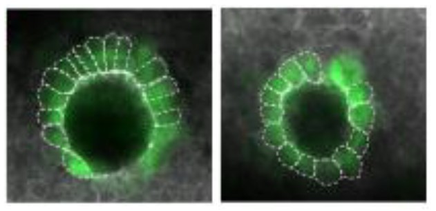
Nodal signaling is also required later in zebrafish development when it helps to establish differences between the left and right sides of the developing embryo. It does this, in part, by directing the formation of an organ known as Kupffer’s vesicle, whose asymmetric shape helps determine the embryo’s left and right sides. Subsequently, Nodal signaling induces the expression of a third Nodal protein, called southpaw, in a group of mesoderm cells on the left-hand side of the embryo.

To investigate whether maternally supplied gdf3 mRNA also plays a role in left-right patterning, the researchers used a series of experimental tricks to supply embryos with enough Gdf3 protein to form the mesoderm and endoderm and survive until the later stages of embryonic development.

As predicted, these embryos showed defects in left-right patterning. Their Kupffer’s vesicles were abnormally symmetric in shape, and southpaw expression was greatly reduced, suggesting that gdf3 is also required for optimal Nodal signaling during later stages of embryonic development. At this stage, however, embryonic gdf3 seems to be capable of doing the job if maternally supplied gdf3 is absent.

Nodal and Vg1 proteins are known to bind to each other in other species. “Thus, we hypothesize that Gdf3 combines with Ndr1 and Ndr2 to facilitate Nodal signaling during zebrafish development, acting as an essential factor in embryonic patterning,” said Pelliccia, a graduate student in molecular biology. Co-author Jindal earned his Ph.D. in chemical and biological engineering in 2017.

At the same time as Burdine and colleagues, two other research groups, led by Joe Yost at the University of Utah and Alex Schier at Harvard University, made similar findings on the role of gdf3 during zebrafish development. “All three groups worked together to co-submit and co-publish in eLife, allowing the students involved to all get credit for their hard work,” Burdine said. “It’s a great example of how science should be done.”

###

The research was funded by the Eunice Kennedy Shriver National Institute of Child Health and Human Development (grant R01HD048584) and the National Science Foundation (graduate research fellowship DGE 1148900).

Citation: Pelliccia, J.L., G.A. Jindal, and R.D. Burdine. Gdf3 is required for robust Nodal signaling during germ layer formation and left-right patterning. eLife. 6: e28635 (2017). DOI: 10.7554/eLife.28635

‘Acidic patch’ regulates access to genetic information

Histone image

By Pooja Makhijani for the Department of Chemistry

Chromatin remodelers — protein machines that pack and unpack chromatin, the tightly wound DNA-protein complex in cell nuclei — are essential and powerful regulators for critical cellular processes, such as replication, recombination and gene transcription and repression. In a new study published Aug. 2 in the journal Nature, a team led by researchers from Princeton University unravels more details on how a class of ATP-dependent chromatin remodelers, called ISWI, regulate access to genetic information.

The researchers reported that ISWI remodelers use a structural feature of the nucleosome, known as the “acidic patch,” to remodel chromatin. The nucleosome is the fundamental structural subunit of chromatin, and is often compared to thread wrapped around a spool.

Geoffrey Dann
Geoffrey Dann. Photo by C. Todd Reichart

“The acidic patch is a negatively charged surface, presented on each face of the nucleosome disc, that is formed by amino acids contributed by two different histone proteins, H2A and H2B,” said Geoffrey Dann, a graduate student in the Department of Molecular Biology at Princeton and the study’s lead author. “Histone proteins are overall very positively charged, which makes the negatively charged acidic patch region of the nucleosome very unique. Recognition of the acidic patch has never before been implicated in chromatin remodeling.”

The research was conducted in the laboratory of Tom Muir, the Van Zandt Williams Jr. Class of 1965 Professor of Chemistry and chair of the Department of Chemistry. Research in the Muir group centers on elucidating the physiochemical basis of protein functions in biomedically relevant systems.

Because ISWI remodelers are known to interact extensively with nucleosomes, the researchers hypothesized that signals, in the form of chemical modifications on histone proteins embedded within nucleosomes, communicate to the remodelers on which nucleosome to act. Using high throughput screening technology, an assay process often used in drug discovery, allowed the researchers to quickly conduct tens of thousands of biochemical measurements to test their assumptions. “The number of chromatin modifications known to exist in vivo is astronomical,” Dann said.

Not only did the experiments reveal that ISWI remodelers use the “acidic patch” to remodel chromatin, but also determined that remodeling enzymes outside the family of ISWI remodelers also use this structural feature, “suggesting that this feature may be a general requirement for chromatin remodeling to occur,” Dann said.

Certain chemical modifications that act on histone proteins that are adjacent to the acidic patch also have the ability to enhance or inhibit ISWI remodeling activity, he explained. “A handful of other proteins are known to engage the acidic patch in their interaction with chromatin as well, and we also found that the biochemistry of several of these proteins was affected by such modifications. Interestingly, each protein tested had its own signature response to this collection of modifications.”

The high throughput screening technology method also generated a vast library of data to drive the design of future studies geared toward further understanding ISWI regulation. “This study generated an immense amount of data pointing to many other novel regulatory inputs, in the form of chromatin modifications, into ISWI remodeling activity,” Dann said. “A long-term goal in our lab is to use this data resource as a launch pad for additional studies investigating how chromatin modifications affect ISWI remodeling, and how this plays into the various roles ISWI remodelers assume in the cell.”

histone diagram
Diagram depicting all histone modifications, mutants, and variants present in the 115-member nucleosome library used in this study. Residues modified or mutated were mapped on to the nucleosome in black. H2A (light yellow), H2B (light red), H3 (light blue), and H4 (light green) modification and mutation locations are indicated by boxes and lines. For clarity, connections are only shown to a single copy of each histone protein.

Their findings may also identify a new instrument in cells’ molecular repertoire of chromatin-remodeling tools and spur investigations into potential cancer therapeutic targets. “Mutations in the acidic patch are known to occur in certain types of human cancers, which underscores the emerging importance of the acidic patch in chromatin biology,” Dann said.

The study, “ISWI chromatin remodellers sense nucleosome modifications to determine substrate preference,” was published Aug. 2 by Nature. doi:10.1038/nature23671

The authors at Princeton University were Geoffrey P. Dann, Glen Liszczak, John D. Bagert, Manuel M. Müller, Uyen T. T. Nguyen, Felix Wojcik, Zachary Z. Brown, Jeffrey Bos, Rasmus Pihl, Samuel B. Pollock, Katharine L. Diehl and Tom W. Muir. Also contributing to the study were Tatyana Panchenko & C. David Allis at The Rockefeller University.

The research was funded in part by the German Research Foundation and the National Institutes of Health (GM112365, R01 GM107047).

Read the full article here: https://www.nature.com/nature/journal/vaap/ncurrent/full/nature23671.html


“Kurly” protein keeps cilia moving, oriented in the right direction (Cell Reports)

Cilia with a mutant form of the Kurly protein are wild and disorganized.

By Catherine Zandonella, Office of the Dean for Research

A new study of a protein found in cilia – the hair-like projections on the cell surface – may help explain how genetic defects in cilia play a role in developmental abnormalities, kidney disease and a number of other disorders.

The researchers at Princeton University and Northwestern University found that the protein, which goes by the name C21orf59 or “Kurly,” is needed for cilia to undulate to keep fluid moving over the surface of cells. They also found that the protein is needed during development to properly orient the cilia so that they are facing the right direction to move the fluid.

“It’s extremely exciting that we’ve found a single protein that is responsible for these two functions – orientation and motility – in cilia,” said Rebecca Burdine, an associate professor of molecular biology at Princeton University. “Despite their importance in human disease, very little is known about how cilia motility and orientation are coordinated, so this protein will provide an important gateway into looking at this process.” The finding is published online and in the March 1 issue of the journal Cell Reports.

Kurly panels
Caption: Staining of cilia (hair-like projections in green and nuclei in blue) in zebrafish kidney tubules show cilia are disorganized and oriented incorrectly in fish with mutated Kurly protein (bottom panel) versus normal Kurly (top panel). Image courtesy of the Burdine lab.

The studies were conducted in zebrafish at Princeton and in African clawed frogs (Xenopus laevis) at Northwestern. In the zebrafish kidney, the researchers found that the Kurly protein enabled cilia to orient themselves in a uniform direction, and most importantly, in the proper direction to facilitate the flow of fluid along the narrow channels in the kidney. In frogs, the cilia on skin cells help move fluid along the surface of the animal during its larval stage. In both cases, knocking out the gene for Kurly caused the cilia to orient incorrectly thereby losing their ability to move in the waving fashion that helps push fluid along.

The discovery of Kurly’s role in cilia movement and orientation stemmed from work in the Burdine lab on fetal organ development, specifically an investigation of mutations that alter the left-right asymmetric orientation of the heart. Such mutations can result in an organ that is working properly but is an exact mirror image of a normal heart. During a search for genes involved in this left-right patterning, the Burdine team discovered that mutations in a gene they called kur, which codes for the Kurly protein, were linked to errors in left-right orientation in zebrafish heart.

When the kurly protein is mutated, the cilia cannot orient and move properly.
Image credit: Burdine lab

As the team investigated kur, they noted that the mutation also affected the function of cilia. It has been known for some time that cilia are important for a number of jobs, from sensing the environment to facilitating fluid flow, to ensuring that the lungs excrete inhaled contaminants. Cilia genetic defects are linked to a number of human diseases, including polycystic kidney disease, respiratory distress, hearing loss, infertility, and left-right patterning disorders such as the one Burdine studies.

Researchers in Burdine’s laboratory found that Kurly’s role in cilia movement stems from its ability to ensure proteins called dynein arms are correctly located in the cilia. When the researchers knocked out the kur gene, the dynein proteins failed to form in the proper location.

The finding that a single protein is involved in both movement and orientation is surprising, said co-first author Daniel Grimes, a postdoctoral research associate in the Burdine lab. “These are two aspects that are both required to generate fluid flow, and we’d like to know how they are linked molecularly. This work adds a new gene that aids this discovery.”

The gene for Kurly has also been detected in relation to human cilia disorders, so the work may have an impact on understanding the mechanisms of human disease, Grimes added. The researchers also found that the mutation they discovered rendered the Kurly protein sensitive to temperature, and used this trait to find that the Kurly protein may be involved in initiating movement rather than keeping the cilia moving once they’ve started.

The team also explored proteins that interact with Kurly. The Northwestern team showed that when the kur gene was inactivated using a gene-editing technique called CRISPR-Cas9, the lack of a functioning Kurly protein led to the mis-positioning of a second protein on the cell surface called Prickle2, which helps cells know which direction they face. Without proper Prickle2 positioning, the cilia pushed fluid in the wrong direction.

The study of the Kurly protein involved Grimes as well as two additional co-authors, Kimberly Jaffe and Jodi Schottenfeld-Roames, a former postdoctoral researcher and graduate student respectively, in the Burdine lab. The initial studies on the Kurly protein were conducted as part of an undergraduate research project by Tse-shuen (Jade) Ku, Class of 2007. Additional work was contributed by Nicholas Morante and José Pelliccia, graduate students in the Burdine lab.

The work at Northwestern University was performed in the laboratory of Brian Mitchell with the assistance of Michael Werner and Sun Kim.

The research was supported by a National Institutes of Health (NIH) Ruth L. Kirschstein Institutional National Research Service Award grant to K. Jaffe (#1F32HD060396-01A1), an NIH National Institute of General Medical Sciences grant to B. Mitchell (#2R01GM089970), and an NIH Eunice Kennedy Shriver National Institute of Child Health and Human Development grant to R. Burdine (#2R01HD048584).

Read the article.

Kimberly M. Jaffe, Daniel T. Grimes, Jodi Schottenfeld-Roames, Michael E. Werner, Tse-Shuen J. Ku, Sun K. Kim, Jose L. Pelliccia, Nicholas F.C. Morante, Brian J. Mitchell, Rebecca D. Burdine.c21orf59/kurly controls both cilia motility and polarization. Cell Reports (2016), http://dx.doi.org/10.1016/j.celrep.2016.01.069. In Press Corrected Proof.