Study of individual neurons in flies reveals memory-related changes in gene activity (Cell Reports)

Image of the Drosophila brain (magenta) with a subset of mushroom body neurons expressing green fluorescent protein (GFP) via a genetic marker. This marker was used to harvest these neurons following the learning and memory assay. (Credit: Crocker, et al.)
Image of the Drosophila brain (magenta) with a subset of mushroom body neurons expressing green fluorescent protein (GFP) via a genetic marker. (Credit: Janelia Farm/HHMI – FlyLight)

By Kristin Qian for the Office of the Dean for Research

Researchers at Princeton University have developed a highly sensitive and precise method to explore genes important for memory formation within single neurons of the Drosophila fly brain. With this method, the researchers found an unexpected result: certain genes involved in creating long-term memories in the brain are the same ones that the eye uses for sensing light.

The study, published in the May 17 issue of the journal Cell Reports, demonstrated the utility of the new method and also identified new patterns of gene expression that drive long-term memory formation.

“Ultimately, to understand the brain, we want to know what individual neurons are doing,” said Mala Murthy, assistant professor in the Princeton Neuroscience Institute and the Department of Molecular Biology. “We found that single neurons can be defined by their pattern of their gene expression, even if they’re all in the same brain network.”

To their surprise, the researchers found that many of the active genes in these neurons produce proteins that are best known for their roles in detecting light in the fly’s eye or sensing odor in the fly’s nose. “It is possible that these sensory proteins have been repurposed by the brain for a different function,” Murthy said.

“Even though the paper is focused on the methodology, which I think will be impactful for the field, there is this new science here—a whole new class of molecules we found that is in the central brain and seems to be involved in memory formation,” Murthy said.

Researchers have known that genes “turn on,” or start making proteins, during the formation of long-term memories in Drosophila, a widely used organism in studies of neurobiology, but they didn’t know exactly which genes in which neurons were involved.

To investigate this question, the researchers first trained flies to form long-term memories. Then they extracted single neurons from the fly brains and evaluated all of the gene readouts, or transcripts, which encode proteins. By comparing the transcripts of the memory-trained flies to those of non-trained flies, researchers were able to identify genes involved in long-term memory formation.

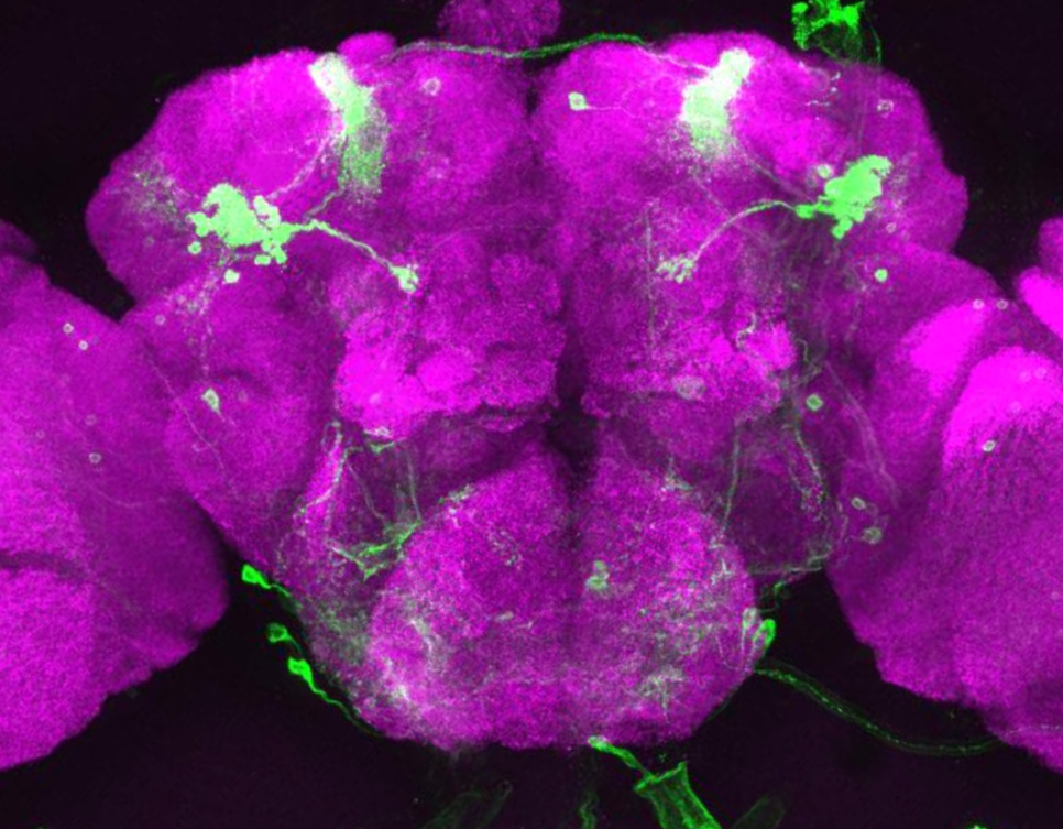
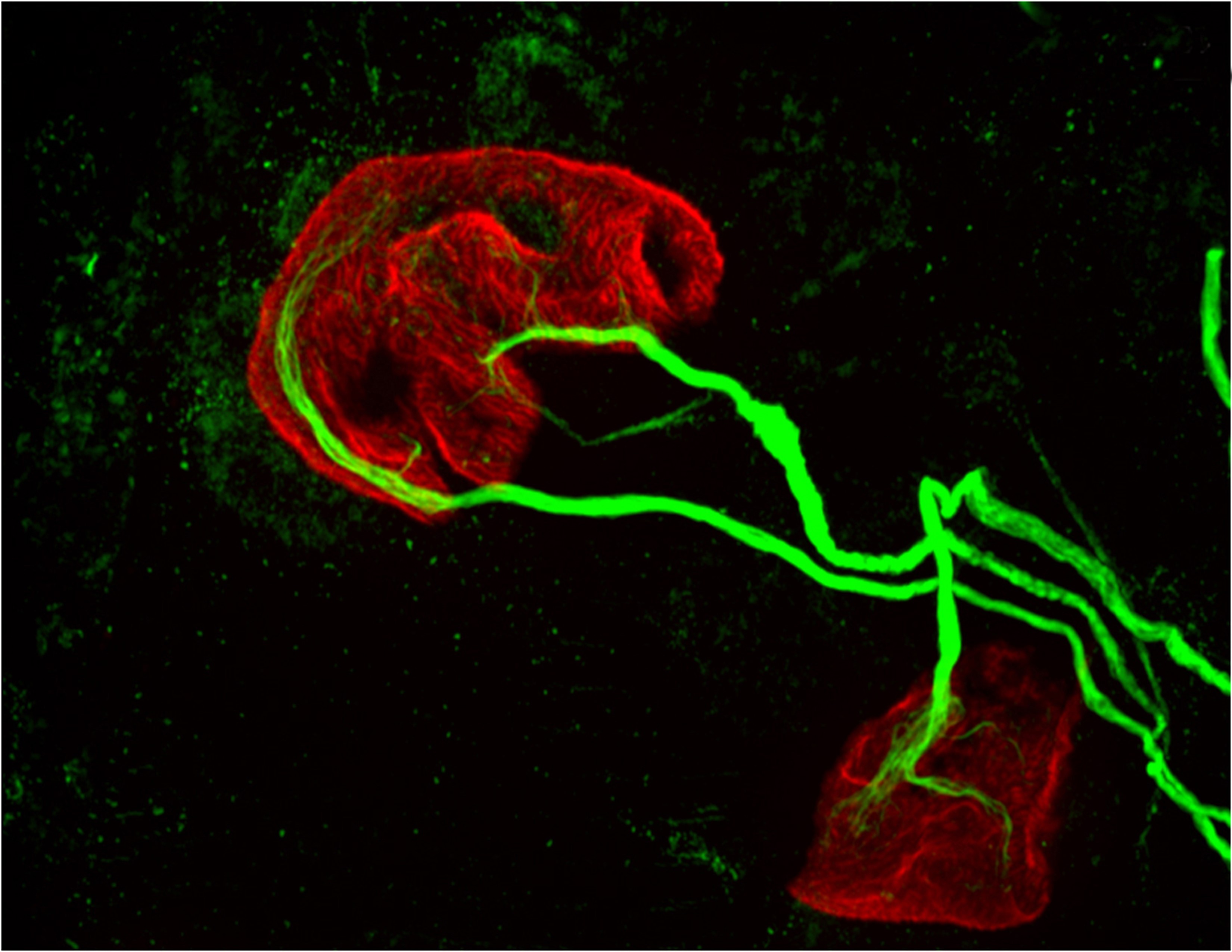
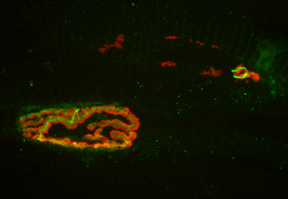
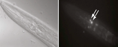
The task was complicated by the tiny size of the fly’s head, which is just one millimeter across, and contains fewer than 100,000 neurons. Murthy’s team focused on neuron types in one part of the brain, the mushroom body, named for its distinctive shape.

First author Amanda Crocker, a former postdoctoral fellow in Murthy’s lab and now an assistant professor of neuroscience at Middlebury College, conducted the experiments in collaboration with co-authors Xiao-Juan Guan, a senior research specialist in the Princeton Neuroscience Institute; Coleen Murphy, professor of molecular biology and the Lewis-Sigler Institute for Integrative Genomics; and Murthy.

“Our work opens up the ability to use Drosophila as a way to study how gene expression in single neurons relates to brain function,” Crocker said. “This has been a challenge because the fly brain is very small and contains fewer neurons than other organisms that neuroscientists study. The advantage of using flies is that they have significantly less redundancy in the neurons that they do have. We can look at specific neurons and gene expression, and ask what the genes are doing in that cell to cause the behavior.”

The researchers trained the flies to form long-term memories by exposing them to an odor – either an earthy, mushroom-like smell (3-octanol) or a menthol-like smell (4-methylcyclohexanol) – while simultaneously delivering a negative stimulus in the form of an electric shock.

Flies experience two odor spaces in each tube. If neither odor has been paired with electric shock, flies spend an equal amount of time on both sides of the tube (control). If one of the odors is paired with electric shock, flies avoid that side of the tube - for example, flies trained to associate the odor 3-OCT with electric shock, avoid the red side (containing 3-OCT) of the tube. (Credit: Crocker, et al.)
Flies experience two odor spaces in each tube. If neither odor has been paired with electric shock, flies spend an equal amount of time on both sides of the tube (control). If one of the odors is paired with electric shock, flies avoid that side of the tube. For example, flies trained to associate the odor 3-OCT with electric shock avoided the red side (containing 3-OCT) of the tube. (Credit: Murthy lab, Princeton University)

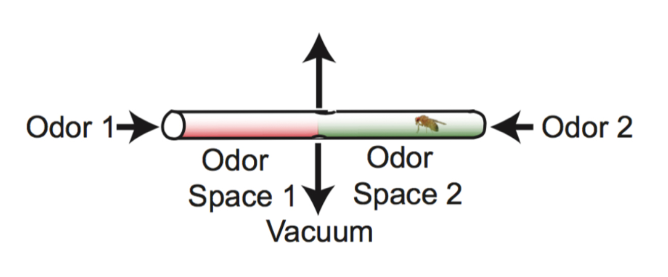
The training took place in a tube containing the two odors, one at each end of the tube. Researchers paired one of the odors with the electric shock, and as a result the fly avoided that end of the tube. The assay was conducted in the dark, so that the flies could use only their sense of smell, not their vision, to navigate the tube.

A second group of flies received the electric shock and the odor, but not at the same time, so they did not form the memory that linked odor to shock.

The researchers then isolated single neurons from the fly brains using tiny glass tubes to suction out the cells. Harvesting neurons using this technique is not common, Murthy said, and it had not been combined with a complete analysis of gene activity in fly neurons before. With this novel method, they were able to use only 10 to 90 femtograms – a quintillionth of a kilogram – of genetic material.

They evaluated gene activity by looking at the production of messenger ribonucleic acid (mRNA), an intermediary between DNA and proteins. The result is a “transcriptome,” or readout of all of the genetic messages that the cell uses to produce proteins. The researchers then read the transcriptome to see which genes produced proteins in the memory-trained flies versus the non-trained flies, and found that some of the active genes in memory-trained flies were the same as ones used in the sensory organs to detect light, odors and taste.

To follow-up, the researchers bred mutant flies that lacked genes for some of the light-sensing proteins and thus could not see. The same memory experiments as before were carried out, and the researchers confirmed that the flies lacking light-sensing proteins were both unable to see and unable to form long-term memories.

The discovery of the expression of genes for classical ‘light-sensing’ proteins, such as rhodopsin, as well as other sensory-related proteins for odor and taste detection, was unexpected because these proteins were not known to be utilized in mushroom bodies, Murthy said. Although studies in other organisms, including humans, have detected sensory genes in areas of the brain unrelated to the sensory organ itself, this may be the first study to link these genes to memory formation.

The study was funded by a National Institutes of Health Ruth L. Kirschstein Institutional National Research Service Award, the Alfred P. Sloan Foundation, the Human Frontier Science Program, a National Science Foundation (NSF) CAREER award, the McKnight Endowment Fund for Neuroscience, the Klingenstein Foundation, a National Institutes of Health New Innovator award, and an NSF BRAIN Initiative EAGER award. The study was also funded in part through Princeton’s Glenn Center for Quantitative Aging Research, directed by Coleen Murphy.

The paper, “Cell-Type-Specific Transcriptome Analysis in the Drosophila Mushroom Body Reveals Memory-Related Changes in Gene Expression,” was published in the May 17 issue of Cell Reports.

Read the journal article.

Dopamine neurons have a role in movement, new study finds (Nature Neuroscience)

Dopamine neurons. Image credit: Witten et al., Nature Neuroscience

By Catherine Zandonella, Office of the Dean for Research

Princeton University researchers have found that dopamine – a brain chemical involved in learning, motivation and many other functions – also has a direct role in representing or encoding movement. The finding could help researchers better understand dopamine’s role in movement-related disorders such as Parkinson’s disease.

The researchers used a new, more precise technique to record the activity of dopamine neurons at two regions within a part of the brain known as the striatum, which oversees action planning, motivation and reward perception. The researchers found that while all of the neurons carried signals needed to learn and plan movement, one of the nerve bundles, the one that went to the region called the dorsomedial striatum, also carried a signal that could be used to control movements.

The work was published online in the journal Nature Neuroscience this week.

“What we learned from this study is that dopamine neurons that go to one part of the brain act differently than dopamine neurons that go to another part of the brain,” said Ilana Witten, assistant professor of psychology and the Princeton Neuroscience Institute. “This is contrary to what has been the mainstream view of dopamine neurons.”

The research may shed light on how Parkinson’s disease, which involves the destruction of dopamine neurons in the dorsomedial striatum, deprives patients of the ability to move. Previous studies have failed to find a direct link between dopamine neuron activity and the control of movement or actions. Instead, the mainstream view suggested an indirect role for dopamine: the neurons make it possible for us to learn which actions are likely to lead to a rewarding experience, which in turn enables us to plan to take that action. When dopamine neurons are destroyed, the individual cannot learn to plan actions and thus cannot move.

The new study affirmed the role of dopamine in reward-based learning, but also found that in the dorsomedial striatum, dopamine neurons can play a direct role in movement. The researchers used a method for measuring neuron activity at very precise locations in the brain. They measured the activity at the ends of neurons – the terminals where dopamine is released into the junction, or synapse, between two cells – in two locations in the striatum: the nucleus accumbens, known to be involved in processing reward, and the dorsomedial striatum, known for evaluating and generating actions.

Until recently, it has been difficult to measure dopamine neuron activity in these regions due to the small size of the regions and the fact that there are many other neurons present that are delivering other brain chemicals, or neurotransmitters, to the same areas of the brain.

To restrict their measurements to only dopamine-carrying neurons, the researchers used mice whose brains carry genetically altered cells that glow green when active. The mice also contain a second gene that ensured that the glowing could only occur when dopamine was present.

The researchers then recorded neuron activity from either the nucleus accumbens or the dorsomedial striatum by inserting a very thin optical fiber into each region to record the fluorescing dopamine cells in only the desired regions.

Once the ability to measure neuron activity was in place, the researchers gave the mice a task that involved both reward-based learning as well as movement.

The task involved presenting the mice with two levers, one of which, when pressed, gave a drink of sweetened water. Through trial and error, the mice learned which lever would give the reward. During the task, the researchers recorded their brain activity.

The task is analogous to playing slot machines at a casino. Picture yourself at a casino with two slot machines in front of you. You pull the lever on the machine to your left and it spits out some coins. Your brain learns that the left lever leads to a reward, so you plan and execute an action: you pull the left lever again. After a few more pulls on the left lever without a reward, you switch to the machine on the right.

When an action is rewarding, you are likely to remember it, an important step in learning. The difference between how much reward you expect, and how much you get, is also important, because it tells you whether or not something is new and how much you should pay attention to it. Researchers call this gap between your predicted reward and the reward you actually get the “reward-prediction error” and consider it an important teaching signal.

By matching the mice’s actions to the dopamine activity in their brains during these tasks, the researchers could determine which parts of the brain were active during reward-based learning, and which parts were active when choosing to press a lever. Assistance with computational modeling of the mice’s behaviors was provided by Nathaniel Daw, a professor of the Princeton Neuroscience Institute and Psychology.

The researchers found that the dopamine neurons that innervate the nucleus accumbens and the dorsomedial striatum did indeed encode reward-prediction cues, which is consistent with previous findings. But they also found that in the dorsomedial striatum, the dopamine neurons carried information about what actions the animal is going to take.

“This idea was that dopamine neurons carry this reward-prediction error signal, and that could indirectly affect movement or actions, because if you don’t have this, you won’t correctly learn which actions to perform,” Witten said. “We show that while this is true, it is certainly not the whole story. There is also a layer where dopamine is directly coding movement or actions.”

Nathan Parker, a graduate student in the Witten lab who designed and conducted the experiments and is first author on the paper, added that new findings were made possible both by the improvements in recording of neurons and by the experimental design, which gave researchers a detailed evaluation of neuron activity during a relatively complex task.

Additional research assistance was provided by Princeton postdoctoral research associates Courtney Cameron and Junuk Lee, and graduate student Jung Yoon Choi. Research Specialist Joshua Taliaferro, Class of 2015, begin working on the project as part of his senior thesis. The study also involved contributions from Thomas Davidson, a postdoctoral researcher at Stanford University.

The study also addresses the general question of how dopamine can be involved in so many functions in the brain, Witten said. “We think that some of the way that dopaminergic neurons achieve such diverse functions in the brain is by having specific roles based on their anatomical target.”

Naoshige Uchida, a professor of molecular and cellular biology at Harvard University who was not involved in the study, said the results challenge long-held views and open up new directions of research. “This study by the Witten lab elegantly shows that the activity of some dopamine neurons is modulated by the direction of motion,” Uchida said. “More importantly, they found some of the clearest evidence indicating the heterogeneity of dopamine neurons: A specific population of dopamine neurons projecting to the dorsomedial striatum encodes movement direction more so compared to another population projecting to the ventral striatum.”

Uchida continued, “A similar phenomenon has also been reported in an independent study in non-human primates (Kim, et al., Cell, 2015), suggesting that the Witten lab finding is more universal and not specific to mice. This is particularly important because dopamine has been implicated in Parkinson’s disease but how dopamine regulates movement remains a large mystery.”

Funding for the study was provided by the Pew Charitable Trusts, the McKnight Foundation, the Brain & Behavior Research (NARSAD) Foundation, the Alfred P. Sloan Foundation, the National Institutes of Health, the National Science Foundation, and Princeton’s Stuart M. Essig ’83 and Erin S. Enright ’82 Fund for Innovation in Engineering and Neuroscience.

Read the abstract.

The study, “Reward and choice encoding in terminals of midbrain dopamine neurons depends on striatal target,” by Nathan Parker, Courtney Cameron, Joshua Taliaferro, Junuk Lee, Jung Yoon Choi, Thomas Davidson, Nathaniel Daw and Ilana Witten, was published online in the journal Nature Neuroscience (Advance Online Publication, http://dx.doi.org/10.1038/nn.4287).

Same immune-system proteins may first giveth, then taketh away motor control (Brain, Behavior, and Immunity)

two motor neurons (green) connected to a single muscle fiber (red)
Princeton University researchers found that proteins in the MHCI, or major histocompatibility complex class I, family can “prune” the connections, or synapses, between motor neurons and muscle fibers, which is necessary during early development. But the researchers also found that MHCI levels can rise again in old age, and that the proteins may resume pruning nerve-muscle synapses. This image from a mouse bred to express less MHCI shows two motor neurons (green) connected to a single muscle fiber (red) at an age when only one connection should remain. (Image by Lisa Boulanger, Princeton Neuroscience Institute, and Mazell Tetruashvily, Department of Molecular Biology)

By Morgan Kelly, Office of Communications

Princeton University researchers have found that a family of proteins with important roles in the immune system may be responsible for fine-tuning a person’s motor control as they grow — and for their gradual loss of muscle function as they age. The research potentially reveals a biological cause of weakness and instability in older people, as well as a possible future treatment that would target the proteins specifically.

The researchers reported in the journal Brain, Behavior, and Immunity that proteins in the family MHCI, or major histocompatibility complex class I, “prune” the connections, or synapses, between motor neurons and muscle fibers. Pruning is necessary during early development because at birth each muscle fiber in humans, mice and other vertebrates receives signals from dozens of neural connections. Proper motor control, however, requires that each muscle fiber receive signals from only a single motor neuron, so without the pruning carried out by MHCI proteins, fine motor control would never emerge.

But the researchers also found that MHCI levels can rise again in old age, and that the proteins may resume pruning nerve-muscle synapses — except that in a mature organism there are no extra synapses. The result is that individual muscle fibers become completely “denervated,” or detached from nervous system control. Denervated muscle fibers cannot be recruited during muscle contraction, which can leave older people weaker and more susceptible to devastating falls, making independent living difficult.

However, the Princeton researchers discovered that when MHCI levels were reduced in mice, denervation during aging was largely prevented. These findings could help scientists identify and treat the neurological causes of denervation and muscle weakness in the elderly.

Corresponding author Lisa Boulanger, an assistant professor in the Princeton Neuroscience Institute, explained that in infants, motor neurons initially make far too many connections to muscle fibers, which is part of why infants lack fine motor control.  Synapse overproduction followed by pruning occurs in many different regions of the vertebrate nervous system, and the neuromuscular junction has often been used as a model for studying this process.

It is not known why more synapses are made during development than are needed.

One possibility is that it allows the wiring diagram of the nervous system to be precisely tuned based on the way the circuit is used, Boulanger said. MHCI proteins help limit the final number of connections so that communication between neurons and muscles is more precise and efficient than would be possible using just a molecular code that produced a set number of connections.

“Molecules might get you to the right zip code, but pruning can make sure you arrive at the right house,” Boulanger said. “During development, it’s essential to get rid of extra synapses. But when you up-regulate MHCI when you’re older and start pruning synapses again, but you don’t have any extras to replace them.”

Boulanger worked with first author Mazell Tetruashvily, who received her doctorate in molecular biology from Princeton in 2015 and is now completing her M.D. training at University of Medicine and Dentistry of New Jersey; Marin McDonald, who received her doctorate in neuroscience from the University of California-San Diego (UCSD) in 2010, and is now a radiology resident at UCSD; and Karla Frietze, a doctoral student in Princeton’s Department of Molecular Biology. Boulanger was on the UCSD faculty before moving her lab to Princeton in 2009.

In the immune system, MHCI proteins present protein fragments, or peptides, to T cells, which are white blood cells with a central role in the body’s response to infection. This peptide presentation allows T cells to recognize and kill infected and cancerous cells, which present abnormal or foreign peptides on their MHCI proteins. It is unknown if the proteins’ ability to help recognize and destroy infected or cancerous cells is mechanistically related to the proteins’ ability to help eliminate excess synapses that the Princeton researchers discovered.

In the nervous system, MHCI proteins stop pruning synapses early in life. Why they may resume their synapse-eliminating activity later in life is unknown, Boulanger said. As immune-system proteins, MHCI levels increase with inflammation, she said. Aging is associated with chronic inflammation, which could explain the observed increase in MHCI levels and the reactivation of its former role.

Neuron image
The researchers found that MHCI the proteins may resume pruning nerve-muscle synapses in older organisms, an age when there are no extra synapses. Instead, muscle fibers become completely “denervated,” or detached from nervous system control, which could lead to weakness and instability in older people. This image from a 2-year-old (elderly) normal mouse shows denervation in the upper-right synapse, as noted by the lack of overlap of the red and green fluorescent markers used to indicate cells where the neuron and muscle fiber meet. At lower left, a healthy-looking synapse displays good overlap. (Image by Lisa Boulanger, Department of Molecular Biology)

The Princeton researchers found that mice bred to express less MHCI proteins had “more youthful” patterns of muscle innervation, since they were protected from denervation as they aged, Boulanger said. The mice actually lacked a protein known as beta-2 microglobulin, which forms a complex with MHCI and is necessary for MHCI expression on the surface of cells. This could be beneficial from a clinical perspective because beta-2 microglobulin is a soluble protein and can be removed from the blood, Boulanger said.

“If a rise in MHCI is the problem, having less beta-2 microglobulin might be protective,” Boulanger said. Recent results from a lab at Stanford University showed that reducing beta-2 microglobulin also helped with cognitive aging because of its effects on MHCI proteins. “Our studies raise the possibility that targeting one protein could help with both motor and cognitive aspects of aging,” Boulanger said.

Because MHCI proteins are important in the immune system, however, such an approach could result in compromised immunity, Boulanger said. The mice bred to not express beta-2 microglobulin had weakened immune systems, as a result of their lower levels of MHCI proteins. Future work will include exploring the effectiveness of other approaches to reducing the proteins’ synapse-eliminating activity in older nervous systems, ideally while leaving their immune functions intact, she said.

The research was supported by the Princeton Department of Molecular Biology and the Princeton Neuroscience Institute (grant no. 1F30AG046044-01A1), the UCSD School of Medicine, the Alfred P. Sloan Foundation, the Whitehall Foundation, and the Princeton Neuroscience Institute Innovation Fund.

Read more.

Mazell M. Tetruashvily, Marin A. McDonald, Karla K. Frietze and Lisa M. Boulanger. “MHCI promotes developmental synapse elimination and aging-related synapse loss at the vertebrate neuromuscular junction.” Brain, Behavior, and Immunity, in press. DOI: 10.1016/j.bbi.2016.01.008epartment of Molecular Biology)

Fruit flies adjust their courtship song based on distance (Neuron)

A fly runs on an air-supported ball. The audio traces of the fly’s courtship song are shown.

Article courtesy of Joseph Caputo, Cell Press

Outside of humans, the ability to adjust the intensity of acoustic signals with distance has only been identified in songbirds. Research published February 3 in Neuron now demonstrates that the male fruit fly also displays this complex behavior during courtship, adjusting the amplitude of his song depending on how far away he is from a female. Studying this process in the fruit fly can help shed light on the building blocks for social interactions across the animal kingdom.

Mala Murthy, of Princeton University, and her colleagues have revealed an unanticipated level of control in insect acoustic communication by analyzing each stage of the neuronal pathway underlying male fruit flies’ ability to adjust their courtship song—from the visual cues that help estimate distance to the signals that pass through nervous system and cause changes in muscle activity that drive softer or louder song. The complexity is remarkable considering that the fruit fly has only 100,000 neurons, one-millionth that of a human brain.

During courtship, males chase females, extending and vibrating one wing at a time to produce a courtship song. Songs, which consist primarily of two modes: sine and pulse, are extremely quiet and must be recorded on sensitive microphones, then amplified more than 1 million times in order to be heard by humans. When amplified, the sine song sounds like the whine of an approaching mosquito, while the pulse song is more akin to a cat’s purr.

“Females listen to many minutes of male song before deciding whether to accept him,” says Murthy, of the Princeton Neuroscience Institute and Department of Molecular Biology. “There is thus enormous evolutionary pressure for males to optimize their song to match the female’s preference while simultaneously minimizing the energetic cost of singing for long periods of time.” Adjusting the amplitude of song to compensate for female distance allows males to conserve energy and thereby court for longer periods of time and better compete with other males.

“While the precise neural mechanisms underlying the generation and patterning of fly song may be distinct from humans or even songbirds, the fundamental problem is the same: how can a neural network produce such a complex and dynamic signal?” Murthy says. “For this reason, we anticipate that similar neural mechanisms will be employed in all systems, and the genetic model system of the fruit fly is an ideal starting point from which to dissect them.”

The researchers were funded by the Howard Hughes Medical Institute, the DAAD (German Academic Exchange Foundation), the Alfred P. Sloan Foundation, the Human Frontiers Science Program, a National Science Foundation CAREER award, a NIH New Innovator Award, the NSF BRAIN Initiative, an EAGER award, the McKnight Foundation, and the Klingenstein-Simons Foundation.

Read the abstract

Philip Coen, Marjorie Xie, Jan Clemens and Mala Murthy. Sensorimotor Transformations Underlying Variability in Song Intensity during Drosophila Courtship. Neuron. Vol. 89, Issue 3, p629–644, 3 February 2016.


Revisiting the mechanics of the action potential (Nature Communications)

By Staff

AW_Pic
The action potential (AP) and the accompanying action wave (AW) constitute an electromechanical pulse traveling along the axon.

The action potential is widely understood as an electrical phenomenon. However, a long experimental history has documented the existence of co-propagating mechanical signatures.

In a new paper in the journal Nature Communications, two Princeton University researchers have proposed a theoretical model to explain these mechanical signatures, which they term “action waves.” The research was conducted by Ahmed El Hady, a visiting postdoctoral research associate at the Princeton Neuroscience Institute and a postdoctoral fellow at the Howard Hughes Medical Institute, and Benjamin Machta, an associate research scholar at the Lewis-Sigler Institute for Integrative Genomics and a lecturer in physics and the Lewis-Sigler Institute for Integrative Genomics.

In the model, the co-propagating waves are driven by changes in charge separation across the axonal membrane, just as a speaker uses charge separation to drive sound waves through the air. The researchers argue that these forces drive surface waves involving both the axonal membrane and cytoskeleton as well as its surrounding fluid. Their model may help shed light on the functional role of the surprisingly structured axonal cytoskeleton that recent super-resolution techniques have uncovered, and suggests a wider role for mechanics in neuronal function.

Read the paper.

Ahmed El Hady & Benjamin B. Machta. Mechanical surface waves accompany action potential propagation. Nature Communications 6, No. 6697 doi:10.1038/ncomms7697

When attention is a deficit: How the brain switches strategies to find better solutions (Neuron)

By Catherine Zandonella, Office of the Dean for Research

2015_03_26_JW_Schuck_NYC3Sometimes being too focused on a task is not a good thing.

During tasks that require our attention, we might become so engrossed in what we are doing that we fail to notice there is a better way to get the job done.

For example, let’s say you are coming out of a New York City subway one late afternoon and you want to find out which way is west. You might begin to scan street signs and then suddenly realize that you could just look for the setting sun.

A new study explored the question of how the brain switches from an ongoing strategy to a new and perhaps more efficient one. The study, conducted by researchers at Princeton University, Humboldt University of Berlin, the Bernstein Center for Computational Neuroscience in Berlin, and the University of Milan-Bicocca, found that activity in a region of the brain known as the medial prefrontal cortex was involved in monitoring what is happening outside one’s current focus of attention and shifting focus from a successful strategy to one that is even better. They published the finding in the journal Neuron.

“The human brain at any moment in time has to process quite a wealth of information,” said Nicolas Schuck, a postdoctoral research associate in the Princeton Neuroscience Institute and first author on the study. “The brain has evolved mechanisms that filter that information in a way that is useful for the task that you are doing. But the filter has a disadvantage: you might miss out on important information that is outside your current focus.”

Schuck and his colleagues wanted to study what happens at the moment when people realize there is a different and potentially better way of doing things. They asked volunteers to play a game while their brains were scanned with magnetic resonance imaging (MRI). The volunteers were instructed to press one of two buttons depending on the location of colored squares on a screen. However, the game contained a hidden pattern that the researchers did not tell the participants about, namely, that if the squares were green, they always appeared in one part of the screen and if the squares were red, they always appeared in another part. The researchers refrained from telling players that they could improve their performance by paying attention to the color instead of the location of the squares.

Volunteers played a game where they had to press one button or another depending on the location of squares on a screen. Participants that switched to a strategy based on the color of the square were able to improve their performance on the game. (Image source: Schuck, et al.)
Volunteers played a game where they had to press one button or another depending on the location of squares on a screen. Participants that switched to a strategy based on the color of the squares were able to improve their performance on the game. (Image source: Schuck, et al.)

Not all of the players figured out that there was a more efficient way to play the game. However, among those that did, their brain images revealed specific signals in the medial prefrontal cortex that corresponded to the color of the squares. These signals arose minutes before the participants switched their strategies. This signal was so reliable that the researchers could use it to predict spontaneous strategy shifts ahead of time, Schuck said.

“These findings are important to better understand the role of the medial prefrontal cortex in the cascade of processes leading to the final behavioral change, and more generally, to understand the role of the medial prefrontal cortex in human cognition,” said Carlo Reverberi, a researcher at the University of Milan-Bicocca and senior author on the study. “Our findings suggest that the medial prefrontal cortex is ‘simulating’ in the background an alternative strategy, while the overt behavior is still shaped by the old strategy.”

The study design – specifically, not telling the participants that there was a more effective strategy – enabled the researchers to show that the brain can monitor background information while focused on a task, and choose to act on that background information.

“What was quite special about the study was that the behavior was completely without instruction,” Schuck said. “When the behavior changed, this reflected a spontaneous internal process.”

Before this study, he said, most researchers had focused on the question of switching strategies because you made a mistake or you realized that your current approach isn’t working. “But what we were able to explore,” he said, “is what happens when people switch to a new way of doing things based on information from their surroundings.” In this way, the study sheds light on how learning and attention can interact, he said.

The study has relevance for the question of how the brain balances the need to maintain attention with the need to incorporate new information about the environment, and may eventually help our understanding of disorders that involve attention deficits.

Schuck designed and conducted the experiments while a graduate student at Humboldt University and the International Max Planck Research School on the Life Course (LIFE) together with the other authors, and conducted the analysis at Princeton University in the laboratory of Yael Niv, assistant professor of psychology and the Princeton Neuroscience Institute in close collaboration with Reverberi.

The research was supported through a grant from the U.S. National Institutes of Health, the International Max Planck Research School LIFE, the Italian Ministry of University, the German Federal Ministry of Education and Research, and the German Research Foundation.

Read the abstract.

Nicolas W. Schuck, Robert Gaschler, Dorit Wenke, Jakob Heinzle, Peter A. Frensch, John-Dylan Haynes, and Carlo Reverberi. Medial Prefrontal Cortex Predicts Internally Driven Strategy Shifts, Neuron (2015) http://dx.doi.org/10.1016/j.neuron.2015.03.015.


Genome-wide search reveals new genes involved in long-term memory (Neuron)

By Catherine Zandonella, Office of the Dean for Research

Whole genome expression data reveals new genes involved in long-term memory formation in worms. (Image source: Murphy lab)

A new study has identified genes involved in long-term memory in the worm as part of research aimed at finding ways to retain cognitive abilities during aging.

The study, which was published in the journal Neuron, identified more than 750 genes involved in long-term memory, including many that had not been found previously and that could serve as targets for future research, said senior author Coleen Murphy, an associate professor of molecular biology and the Lewis-Sigler Institute for Integrative Genomics at Princeton University.

“We want to know, are there ways to extend memory?” Murphy said. “And eventually, we would like to ask, are there compounds that could maintain memory with age?”

Long-term memory training in worms (left) led to induction of the transcription factor CREB in AIM neurons (shown by arrows in right). CREB-induced genes were shown to be involved in forming long-term memories in worm neurons. (Image source: Murphy lab)

The newly pinpointed genes are “turned on” by a molecule known as CREB (cAMP-response element-binding protein), a factor known to be required for long-term memory in many organisms, including worms and mice.

“There is a pretty direct relationship between CREB and long-term memory,” Murphy said, “and many organisms lose CREB as they age.” By studying the CREB-activated genes involved in long-term memory, the researchers hope to better understand why some organisms lose their long-term memories as they age.

To identify the genes, the researchers first instilled long-term memories in the worms by training them to associate meal-time with a butterscotch smell. Trained worms were able to remember that the butterscotch smell means dinner for about 16 hours, a significant amount of time for the worm.

The researchers then scanned the genomes of both trained worms and non-trained worms, looking for genes turned on by CREB.

The researchers detected 757 CREB-activated genes in the long-term memory-trained worms, and showed that these genes were turned on primarily in worm cells called the AIM interneurons.

They also found CREB-activated genes in non-trained worms, but the genes were not turned on in AIM interneurons and were not involved in long-term memory. CREB turns on genes involved in other biological functions such as growth, immune response, and metabolism. Throughout the worm, the researchers noted distinct non-memory (or “basal”) genes in addition to the memory-related genes.

The next step, said Murphy, is to find out what these newly recognized long-term memory genes do when they are activated by CREB. For example, the activated genes may strengthen connections between neurons.

Worms are a perfect system in which to explore that question, Murphy said. The worm Caenorhabditis elegans has only 302 neurons, whereas a typical mammalian brain contains billions of the cells.

“Worms use the same molecular machinery that higher organisms, including mammals, use to carry out long-term memory,” said Murphy. “We hope that other researchers will take our list and look at the genes to see whether they are important in more complex organisms.”

Murphy said that future work will involve exploring CREB’s role in long-term memory as well as reproduction in worms as they age.

The team included co-first-authors Postdoctoral Research Associate Vanisha Lakhina, Postdoctoral Research Associate Rachel Arey, and Associate Research Scholar Rachel Kaletsky of the Lewis-Sigler Institute for Integrative Genomics. Additional research was performed by Amanda Kauffman, who earned her Ph.D. in Molecular Biology in 2010; Geneva Stein, who earned her Ph.D. in Molecular Biology in 2014; William Keyes, a laboratory assistant in the Department of Molecular Biology; and Daniel Xu, who earned his B.A. in Molecular Biology in 2014.

Funding for the research was provided by the National Institutes of Health and the Paul F. Glenn Laboratory for Aging Research at Princeton University.

Read the abstract

Citation: Vanisha Lakhina, Rachel N. Arey, Rachel Kaletsky, Amanda Kauffman, Geneva Stein, William Keyes, Daniel Xu, and Coleen T. Murphy. Genome-wide Functional Analysis of CREB/Long-Term Memory-Dependent Transcription Reveals Distinct Basal and Memory Gene Expression Programs, Neuron (2015), http://dx.doi.org/10.1016/j.neuron.2014.12.029.

Pupil study reveals learning styles, brain activity (Nature Neuroscience)

Test of learning styles experiment
To test people’s learning styles, participants were presented with a choice between two images (objects or words) and rewarded according to their choices. In this exercise, to maximize reward, participants had to learn by trial and error that office-related images provide a higher reward than food-related images (semantic, or related to meaning), and that grayscale images provide a higher reward than color images (visual features). Image credit: Nature Neuroscience.

By Catherine Zandonella, Office of the Dean for Research

People are often said to have “learning styles” – for example, some people pay attention to visual details while others grab onto abstract concepts and meanings. A new study from Princeton University researchers found that changes in pupil size can reveal whether people are learning using their dominant learning style, or whether they are learning in modes outside of that style.

The researchers found that pupil dilation was smaller when people learned using their usual style and larger when people diverged from their normal style. The study was published in the journal Nature Neuroscience.

The study compared brain activity in individuals with two different learning styles – those who learn best by absorbing concrete visual details and those who are better at learning abstract concepts or meanings.

“We showed that changes in pupil dilation are associated with the degree to which learners use the style for which they have a predisposition,” said Eldar Eran, a graduate student in the Princeton Neuroscience Institute, who led the study.

The researchers used changes in pupil size as an indicator of variations in “neural gain,” which can be thought of as an amplifier of neural communication: when gain is increased, excited neurons become even more active and inhibited neurons become even less active. Smaller pupil dilation indicates more neural gain and larger pupil dilation indicates less neural gain.

The team showed that neural gain was correlated with different modes of communication between parts of the brain. In studies of human volunteers undergoing brain scans, when neural gain was high, communication tended to be tightly concentrated in certain regions of the brain that govern specific tasks, whereas low neural gain is associated with communication across wider regions of the brain.

“We showed that the brain has different modes of communication,” said Yael Niv, assistant professor of psychology and the Princeton Neuroscience Institute, “one mode where everything talks to everything else, and another mode where communication is more segregated into areas that don’t talk to each other.” The study also involved Jonathan Cohen, Princeton’s Robert Bendheim and Lynn Bendheim Thoman Professor in Neuroscience.

These modes are linked to the level of neural gain and to learning style, Niv said. Neural gain can be thought of as a contrast amplifier that increases intensity of both activation and inhibition of communication among brain areas. “If one area is trying to activate another, or trying to inhibit another – both effects are stronger, everything is more potent,” she said. “This is correlated with communication being segregated into clusters of activation in the brain, so each network is talking to itself loudly, but connections across networks are inhibited. In situations of lower gain, however, the areas can talk to each other across networks, so information flows more globally.”

“These two modes [of communication in the brain] seem to be associated with different constraints on learning,” she said. “According to our study, in the mode where everything talks to everything, learning is very flexible. In contrast, in the mode where communication is stronger and more focused, but also more segregated between brain areas, subjects were more true to their personal learning style. Neither of these modes are better than the other – in both cases participants were equally successful in the task, but in different ways.”

“We interpreted these results to mean that although we tend to have a dominant learning style, we are not a slave to that style, and when operating in the proper mode, we can overcome dominant styles to learn in other ways,” she said.

This research was funded by NIH grants R03 DA029073 and R01 MH098861, a Howard Hughes Medical Institute International Student Research fellowship, and a Sloan Research Fellowship. The authors also wish to thank the generous support of the Regina and John Scully Center for the Neuroscience of Mind and Behavior within the Princeton Neuroscience Institute.

Read the article

Eldar, Eran, Jonathan D Cohen & Yael Niv. 2013. The effects of neural gain on attention and learning.  Nature Neuroscience. Published online June 16, 2013 doi:10.1038/nn.3428

Researchers discover workings of brain’s ‘GPS system’ (Nature)

By Catherine Zandonella, Office of the Dean for Research

Just as a global positioning system (GPS) helps find your location, the brain has an internal system for helping determine the body’s location as it moves through its surroundings.

A new study from researchers at Princeton University provides evidence for how the brain performs this feat. The study, published in the journal Nature, indicates that certain position-tracking neurons — called grid cells — ramp their activity up and down by working together in a collective way to determine location, rather than each cell acting on its own as was proposed by a competing theory.

Grid cells are neurons that become electrically active, or “fire,” as animals travel in an environment. First discovered in the mid-2000s, each cell fires when the body moves to specific locations, for example in a room. Amazingly, these locations are arranged in a hexagonal pattern like spaces on a Chinese checker board.  (See figure.)

Tank_Brain_GPS
As the mouse moves around in a square arena (left), a single grid cell in the mouse’s brain becomes active, or spikes, when the animal arrives at particular locations in the arena (right). These locations are arranged in a hexagonal pattern. The red dots indicate the mouse’s location in the arena when the grid cell fired. (Image credit: Cristina Domnisoru, Princeton University)

“Together, the grid cells form a representation of space,” said David Tank, Princeton’s Henry L. Hillman Professor in Molecular Biology and leader of the study. “Our research focused on the mechanisms at work in the neural system that forms these hexagonal patterns,” he said. The first author on the paper was graduate student Cristina Domnisoru, who conducted the experiments together with postdoctoral researcher Amina Kinkhabwala.

Domnisoru measured the electrical signals inside individual grid cells in mouse brains while the animals traversed a computer-generated virtual environment, developed previously in the Tank lab. The animals moved on a mouse-sized treadmill while watching a video screen in a set-up that is similar to video-game virtual reality systems used by humans.

She found that the cell’s electrical activity, measured as the difference in voltage between the inside and outside of the cell, started low and then ramped up, growing larger as the mouse reached each point on the hexagonal grid and then falling off as the mouse moved away from that point.

This ramping pattern corresponded with a proposed mechanism of neural computation called an attractor network. The brain is made up of vast numbers of neurons connected together into networks, and the attractor network is a theoretical model of how patterns of connected neurons can give rise to brain activity by collectively working together. The attractor network theory was first proposed 30 years ago by John Hopfield, Princeton’s Howard A. Prior Professor in the Life Sciences, Emeritus.

The team found that their measurements of grid cell activity corresponded with the attractor network model but not a competing theory, the oscillatory interference model. This competing theory proposed that grid cells use rhythmic activity patterns, or oscillations, which can be thought of as many fast clocks ticking in synchrony, to calculate where animals are located. Although the Princeton  researchers detected rhythmic activity inside most neurons, the activity patterns did not appear to participate in position calculations.

Read the abstract.

Domnisoru, Cristina, Amina A. Kinkhabwala & David W. Tank. 2013. Membrane potential dynamics of grid cells. Nature. doi:10.1038/nature11973. Published online Feb. 10, 2013.

This work was supported by the National Institute of Neurological Disorders and Stroke under award numbers 5RC1NS068148-02 and 1R37NS081242-01, the National Institute of Mental Health under award number 5R01MH083686-04, a National Institutes of Health Postdoctoral Fellowship grant F32NS070514-01A1 (A.A.K.), and a National Science Foundation Graduate Research Fellowship (C.D.).


How our brains keep track of where we are in the world (Journal of Neuroscience)

Libraries, supermarkets, classrooms…the world is full of places that look very similar, and yet our brains always seem to keep track of where we are. In a new study published in the Journal of Neuroscience, researchers at Princeton University and Ohio State University have uncovered one way in which the brain does this.

Similar-looking places can be distinguished from each other because of differences in what we experience when navigating to them. As we head toward a destination, our brains catalogue details such as other nearby buildings, the look of the doorway, even the people nearby.

The researchers discovered that the parahippocampal cortex, a part of the visual system that analyzes the current scene in front of us, also incorporates the details leading up to the scene, or its “temporal context.” As a result, even when two scenes look identical, we create different memory traces for them when their temporal contexts are different. Ultimately, this can help our brains to keep track of where we are in the world.

Learn more about Nicholas Turk-Browne‘s research at Princeton University.

Journal Citation: Turk-Browne NB, Simon MG, Sederberg PB. Scene representations in parahippocampal cortex depend on temporal context.  J Neurosci. 2012 May 23;32(21):7202-7.