Movie caption: Researchers at Princeton studied the temperature dependence of the formation of the nucleolus, a cellular organelle. The movie shows the nuclei of intact fly cells as they are subjected to temperature changes in the surrounding fluid. As the temperature is shifted from low to high, the spontaneously assembled proteins dissolve, as can be seen in the disappearance of the bright spots.
By Catherine Zandonella, Office of the Dean for Research
Researchers at Princeton found that the nucleolus, a cellular organelle involved in RNA synthesis, assembles in part through the passive process of phase separation – the same type of process that causes oil to separate from water. The study, published in the journal Proceedings of the National Academy of Sciences, is the first to show that this happens in living, intact cells.
Understanding how cellular structures form could help explain how organelles change in response to diseases. For example, a hallmark of cancer cells is the swelling of the nucleolus.
To explore the role of passive processes – as opposed to active processes that involve energy consumption – in nucleolus formation, Hanieh Falahati, a graduate student in Princeton’s Lewis-Sigler Institute for Integrative Genomics, looked at the behavior of six nucleolus proteins under different temperature conditions. Phase separation is enhanced at lower temperatures, which is why salad dressing containing oil and vinegar separates when stored in the refrigerator. If phase separation were driving the assembly of proteins, the researchers should see the effect at low temperatures.
Falahati showed that four of the six proteins condensed and assembled into the nucleolus at low temperatures and reverted when the temperature rose, indicating that the passive process of phase separation was at work. However, the assembly of the other two proteins was irreversible, indicating that active processes were in play.
“It was kind of a surprising result, and it shows that cells can take advantage of spontaneous processes for some functions, but for other things, active processes may give the cell more control,” said Falahati, whose adviser is Eric Wieschaus, Princeton’s Squibb Professor in Molecular Biology and a professor of molecular biology and the Lewis-Sigler Institute for Integrative Genomics, and a Howard Hughes Medical Institute researcher.
The research was funded in part by grant 5R37HD15587 from the National Institute of Child Health and Human Development (NICHD), and by the Howard Hughes Medical Institute.
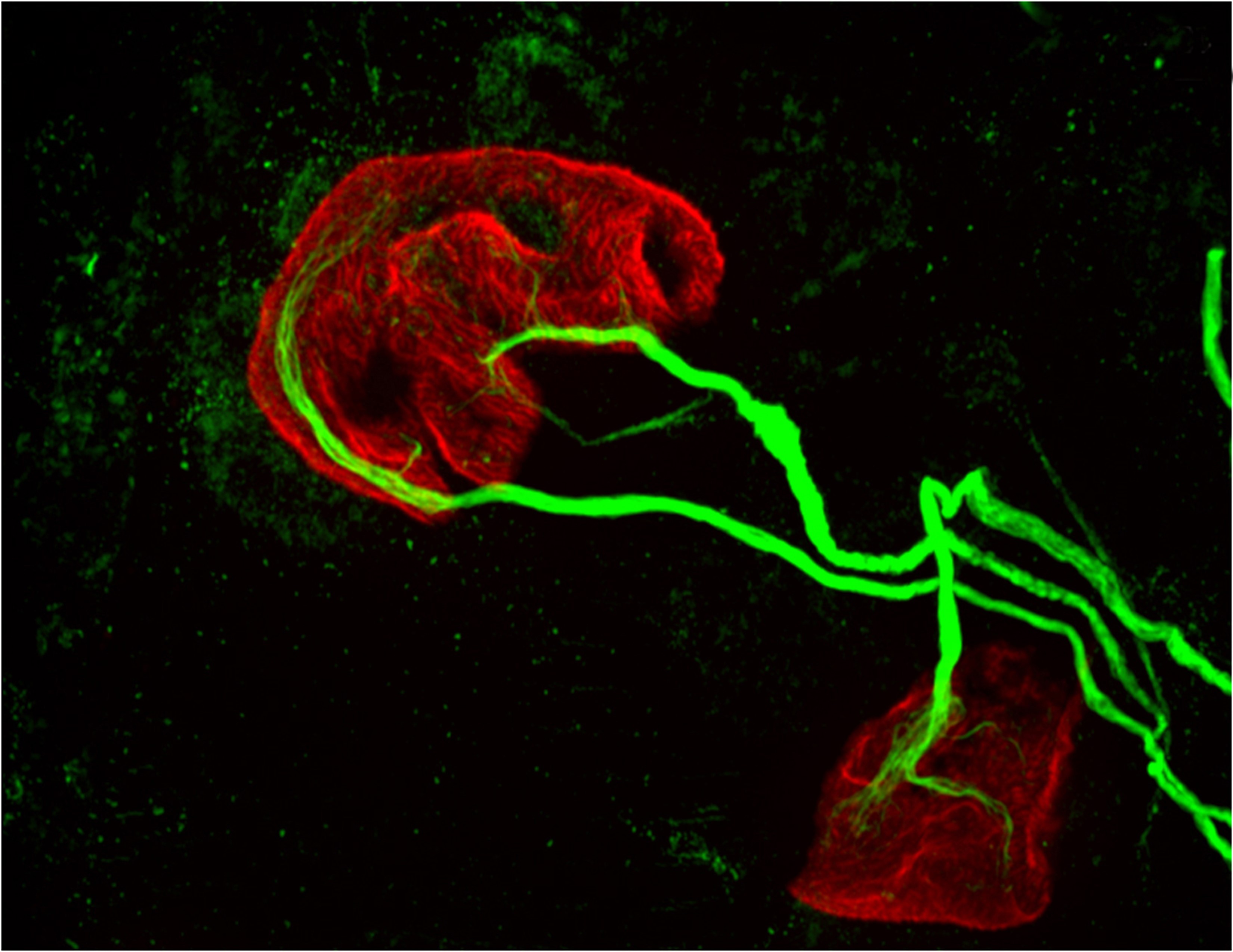
Princeton University researchers found that proteins in the MHCI, or major histocompatibility complex class I, family can “prune” the connections, or synapses, between motor neurons and muscle fibers, which is necessary during early development. But the researchers also found that MHCI levels can rise again in old age, and that the proteins may resume pruning nerve-muscle synapses. This image from a mouse bred to express less MHCI shows two motor neurons (green) connected to a single muscle fiber (red) at an age when only one connection should remain. (Image by Lisa Boulanger, Princeton Neuroscience Institute, and Mazell Tetruashvily, Department of Molecular Biology)
By Morgan Kelly, Office of Communications
Princeton University researchers have found that a family of proteins with important roles in the immune system may be responsible for fine-tuning a person’s motor control as they grow — and for their gradual loss of muscle function as they age. The research potentially reveals a biological cause of weakness and instability in older people, as well as a possible future treatment that would target the proteins specifically.
The researchers reported in the journal Brain, Behavior, and Immunity that proteins in the family MHCI, or major histocompatibility complex class I, “prune” the connections, or synapses, between motor neurons and muscle fibers. Pruning is necessary during early development because at birth each muscle fiber in humans, mice and other vertebrates receives signals from dozens of neural connections. Proper motor control, however, requires that each muscle fiber receive signals from only a single motor neuron, so without the pruning carried out by MHCI proteins, fine motor control would never emerge.
But the researchers also found that MHCI levels can rise again in old age, and that the proteins may resume pruning nerve-muscle synapses — except that in a mature organism there are no extra synapses. The result is that individual muscle fibers become completely “denervated,” or detached from nervous system control. Denervated muscle fibers cannot be recruited during muscle contraction, which can leave older people weaker and more susceptible to devastating falls, making independent living difficult.
However, the Princeton researchers discovered that when MHCI levels were reduced in mice, denervation during aging was largely prevented. These findings could help scientists identify and treat the neurological causes of denervation and muscle weakness in the elderly.
Corresponding author Lisa Boulanger, an assistant professor in the Princeton Neuroscience Institute, explained that in infants, motor neurons initially make far too many connections to muscle fibers, which is part of why infants lack fine motor control. Synapse overproduction followed by pruning occurs in many different regions of the vertebrate nervous system, and the neuromuscular junction has often been used as a model for studying this process.
It is not known why more synapses are made during development than are needed.
One possibility is that it allows the wiring diagram of the nervous system to be precisely tuned based on the way the circuit is used, Boulanger said. MHCI proteins help limit the final number of connections so that communication between neurons and muscles is more precise and efficient than would be possible using just a molecular code that produced a set number of connections.
“Molecules might get you to the right zip code, but pruning can make sure you arrive at the right house,” Boulanger said. “During development, it’s essential to get rid of extra synapses. But when you up-regulate MHCI when you’re older and start pruning synapses again, but you don’t have any extras to replace them.”
Boulanger worked with first author Mazell Tetruashvily, who received her doctorate in molecular biology from Princeton in 2015 and is now completing her M.D. training at University of Medicine and Dentistry of New Jersey; Marin McDonald, who received her doctorate in neuroscience from the University of California-San Diego (UCSD) in 2010, and is now a radiology resident at UCSD; and Karla Frietze, a doctoral student in Princeton’s Department of Molecular Biology. Boulanger was on the UCSD faculty before moving her lab to Princeton in 2009.
In the immune system, MHCI proteins present protein fragments, or peptides, to T cells, which are white blood cells with a central role in the body’s response to infection. This peptide presentation allows T cells to recognize and kill infected and cancerous cells, which present abnormal or foreign peptides on their MHCI proteins. It is unknown if the proteins’ ability to help recognize and destroy infected or cancerous cells is mechanistically related to the proteins’ ability to help eliminate excess synapses that the Princeton researchers discovered.
In the nervous system, MHCI proteins stop pruning synapses early in life. Why they may resume their synapse-eliminating activity later in life is unknown, Boulanger said. As immune-system proteins, MHCI levels increase with inflammation, she said. Aging is associated with chronic inflammation, which could explain the observed increase in MHCI levels and the reactivation of its former role.
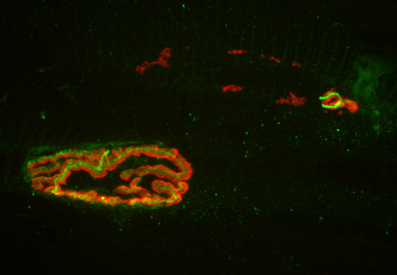
The researchers found that MHCI the proteins may resume pruning nerve-muscle synapses in older organisms, an age when there are no extra synapses. Instead, muscle fibers become completely “denervated,” or detached from nervous system control, which could lead to weakness and instability in older people. This image from a 2-year-old (elderly) normal mouse shows denervation in the upper-right synapse, as noted by the lack of overlap of the red and green fluorescent markers used to indicate cells where the neuron and muscle fiber meet. At lower left, a healthy-looking synapse displays good overlap. (Image by Lisa Boulanger, Department of Molecular Biology)
The Princeton researchers found that mice bred to express less MHCI proteins had “more youthful” patterns of muscle innervation, since they were protected from denervation as they aged, Boulanger said. The mice actually lacked a protein known as beta-2 microglobulin, which forms a complex with MHCI and is necessary for MHCI expression on the surface of cells. This could be beneficial from a clinical perspective because beta-2 microglobulin is a soluble protein and can be removed from the blood, Boulanger said.
“If a rise in MHCI is the problem, having less beta-2 microglobulin might be protective,” Boulanger said. Recent results from a lab at Stanford University showed that reducing beta-2 microglobulin also helped with cognitive aging because of its effects on MHCI proteins. “Our studies raise the possibility that targeting one protein could help with both motor and cognitive aspects of aging,” Boulanger said.
Because MHCI proteins are important in the immune system, however, such an approach could result in compromised immunity, Boulanger said. The mice bred to not express beta-2 microglobulin had weakened immune systems, as a result of their lower levels of MHCI proteins. Future work will include exploring the effectiveness of other approaches to reducing the proteins’ synapse-eliminating activity in older nervous systems, ideally while leaving their immune functions intact, she said.
The research was supported by the Princeton Department of Molecular Biology and the Princeton Neuroscience Institute (grant no. 1F30AG046044-01A1), the UCSD School of Medicine, the Alfred P. Sloan Foundation, the Whitehall Foundation, and the Princeton Neuroscience Institute Innovation Fund.
Mazell M. Tetruashvily, Marin A. McDonald, Karla K. Frietze and Lisa M. Boulanger. “MHCI promotes developmental synapse elimination and aging-related synapse loss at the vertebrate neuromuscular junction.” Brain, Behavior, and Immunity, in press. DOI: 10.1016/j.bbi.2016.01.008epartment of Molecular Biology)
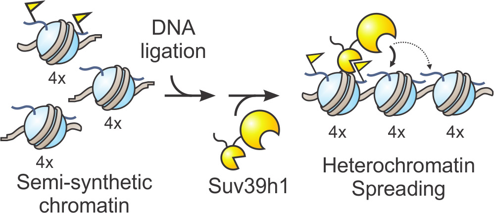
Graphical representation of designer chromatin experiments. Image courtesy of the Muir lab.
By Tien Nguyen, Department of Chemistry
Researchers have discovered the two-step process that activates an essential human enzyme, called Suv39h1, which is responsible for organizing large portions of the DNA found in every living cell.
For any particular cell, such as a skin or brain cell, much of this genetic information is extraneous and must be packed away to allow sufficient space and resources for more important genes. Failure to properly pack DNA jeopardizes the stability of chromosomes and can result in severe diseases. Suv39h1 is one of the main enzymes that chemically mark the irrelevant regions of DNA to be compacted by cellular machinery, but little is known about how it installs its tag.
Now, scientists at Princeton have used ‘designer chromatin’ templates – highly customized replicas of cellular DNA and histone proteins, the scaffolding proteins around which DNA is wrapped – to reveal new details about Suv39h1’s mechanism. The researchers investigated how Suv39h1 employs a positive feedback loop to chemically tag thousands of adjacent histones, thus signaling the cell to stow away these underlying, unnecessary DNA sequences. The work was published in in the journal Nature Chemical Biology.
“One of the things that has always fascinated me about feedback loops is that they’re super dangerous. If you make a mistake once, you end up getting reinforcement through the feedback loop,” said Manuel Müller, a postdoctoral researcher in the Muir lab and lead author on the study. “So how does Suv39h1 keep itself in check?”
Suv39h1 had been known to possess two distinct parts, but the new research revealed how they work together in order to ‘switch on’ the enzyme. One part of the enzyme, known as the chromodomain, is constantly exposed and seeks out specific chemical tags, known as a methyl groups, located at predetermined sites on histones. When the chromodomain finds these groups in the genome, it locks onto the spot and allows the other part, the enzymatic core, to install more methyl tags at adjacent histones.
“The second, anchoring step wasn’t really known before. It provides an extra level of control and allows the process to be extremely fine-tuned,” Müller said. A similar mechanism may be employed by many other enzymes operating on chromatin, given that they contain similar components of a feedback loop.
To understand how the enzyme carries out this process, the researchers synthesized complex chromatin templates that were three times larger than previously reported models. They divided the template into three blocks that could each be manipulated in various ways. For example, a block could be prepared with the chemical tag present, absent or mutated such that tagging can’t occur. “The different blocks should signal to the enzyme either start here or feel free to spread here or absolutely stop here,” said Glen Liszczak, a co-author and postdoctoral researcher in the Muir lab.
By rearranging the various domains, the research team observed where the enzyme spread its mark across the genome. They found that Suv39h1 preferred to spread across small distances, but that it could reach sequences further along if chromatin folding decreased the physical distance in space.
“We’ve learned something new about this enzyme, something that we couldn’t have without the pinpoint precision that the designer chromatin offers,” Liszczak said. “There are a lot of questions that our lab has been interested in that we can now start to answer.”
The research was funded by the Swiss National Science Foundation (postdoctoral fellowships) and the US National Institutes of Health (R01-GM107047).
Müller, M. M.; Fierz, B.; Bittova, L.; Liszczak, G.; Muir, T. W. “A two-state activation mechanism controls the histone methyltransferase Suv39h1.” Nature Chem. Bio. Available online January 25, 2016.

You must be logged in to post a comment.