Same immune-system proteins may first giveth, then taketh away motor control (Brain, Behavior, and Immunity)

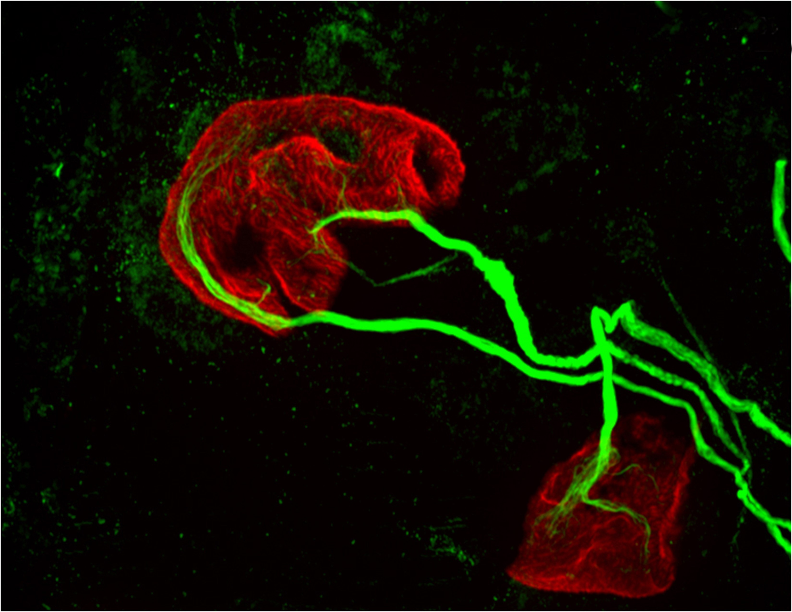
two motor neurons (green) connected to a single muscle fiber (red)
Princeton University researchers found that proteins in the MHCI, or major histocompatibility complex class I, family can “prune” the connections, or synapses, between motor neurons and muscle fibers, which is necessary during early development. But the researchers also found that MHCI levels can rise again in old age, and that the proteins may resume pruning nerve-muscle synapses. This image from a mouse bred to express less MHCI shows two motor neurons (green) connected to a single muscle fiber (red) at an age when only one connection should remain. (Image by Lisa Boulanger, Princeton Neuroscience Institute, and Mazell Tetruashvily, Department of Molecular Biology)

By Morgan Kelly, Office of Communications

Princeton University researchers have found that a family of proteins with important roles in the immune system may be responsible for fine-tuning a person’s motor control as they grow — and for their gradual loss of muscle function as they age. The research potentially reveals a biological cause of weakness and instability in older people, as well as a possible future treatment that would target the proteins specifically.

The researchers reported in the journal Brain, Behavior, and Immunity that proteins in the family MHCI, or major histocompatibility complex class I, “prune” the connections, or synapses, between motor neurons and muscle fibers. Pruning is necessary during early development because at birth each muscle fiber in humans, mice and other vertebrates receives signals from dozens of neural connections. Proper motor control, however, requires that each muscle fiber receive signals from only a single motor neuron, so without the pruning carried out by MHCI proteins, fine motor control would never emerge.

But the researchers also found that MHCI levels can rise again in old age, and that the proteins may resume pruning nerve-muscle synapses — except that in a mature organism there are no extra synapses. The result is that individual muscle fibers become completely “denervated,” or detached from nervous system control. Denervated muscle fibers cannot be recruited during muscle contraction, which can leave older people weaker and more susceptible to devastating falls, making independent living difficult.

However, the Princeton researchers discovered that when MHCI levels were reduced in mice, denervation during aging was largely prevented. These findings could help scientists identify and treat the neurological causes of denervation and muscle weakness in the elderly.

Corresponding author Lisa Boulanger, an assistant professor in the Princeton Neuroscience Institute, explained that in infants, motor neurons initially make far too many connections to muscle fibers, which is part of why infants lack fine motor control.  Synapse overproduction followed by pruning occurs in many different regions of the vertebrate nervous system, and the neuromuscular junction has often been used as a model for studying this process.

It is not known why more synapses are made during development than are needed.

One possibility is that it allows the wiring diagram of the nervous system to be precisely tuned based on the way the circuit is used, Boulanger said. MHCI proteins help limit the final number of connections so that communication between neurons and muscles is more precise and efficient than would be possible using just a molecular code that produced a set number of connections.

“Molecules might get you to the right zip code, but pruning can make sure you arrive at the right house,” Boulanger said. “During development, it’s essential to get rid of extra synapses. But when you up-regulate MHCI when you’re older and start pruning synapses again, but you don’t have any extras to replace them.”

Boulanger worked with first author Mazell Tetruashvily, who received her doctorate in molecular biology from Princeton in 2015 and is now completing her M.D. training at University of Medicine and Dentistry of New Jersey; Marin McDonald, who received her doctorate in neuroscience from the University of California-San Diego (UCSD) in 2010, and is now a radiology resident at UCSD; and Karla Frietze, a doctoral student in Princeton’s Department of Molecular Biology. Boulanger was on the UCSD faculty before moving her lab to Princeton in 2009.

In the immune system, MHCI proteins present protein fragments, or peptides, to T cells, which are white blood cells with a central role in the body’s response to infection. This peptide presentation allows T cells to recognize and kill infected and cancerous cells, which present abnormal or foreign peptides on their MHCI proteins. It is unknown if the proteins’ ability to help recognize and destroy infected or cancerous cells is mechanistically related to the proteins’ ability to help eliminate excess synapses that the Princeton researchers discovered.

In the nervous system, MHCI proteins stop pruning synapses early in life. Why they may resume their synapse-eliminating activity later in life is unknown, Boulanger said. As immune-system proteins, MHCI levels increase with inflammation, she said. Aging is associated with chronic inflammation, which could explain the observed increase in MHCI levels and the reactivation of its former role.
Neuron image
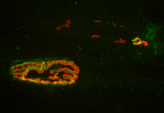
The researchers found that MHCI the proteins may resume pruning nerve-muscle synapses in older organisms, an age when there are no extra synapses. Instead, muscle fibers become completely “denervated,” or detached from nervous system control, which could lead to weakness and instability in older people. This image from a 2-year-old (elderly) normal mouse shows denervation in the upper-right synapse, as noted by the lack of overlap of the red and green fluorescent markers used to indicate cells where the neuron and muscle fiber meet. At lower left, a healthy-looking synapse displays good overlap. (Image by Lisa Boulanger, Department of Molecular Biology)

The Princeton researchers found that mice bred to express less MHCI proteins had “more youthful” patterns of muscle innervation, since they were protected from denervation as they aged, Boulanger said. The mice actually lacked a protein known as beta-2 microglobulin, which forms a complex with MHCI and is necessary for MHCI expression on the surface of cells. This could be beneficial from a clinical perspective because beta-2 microglobulin is a soluble protein and can be removed from the blood, Boulanger said.

“If a rise in MHCI is the problem, having less beta-2 microglobulin might be protective,” Boulanger said. Recent results from a lab at Stanford University showed that reducing beta-2 microglobulin also helped with cognitive aging because of its effects on MHCI proteins. “Our studies raise the possibility that targeting one protein could help with both motor and cognitive aspects of aging,” Boulanger said.

Because MHCI proteins are important in the immune system, however, such an approach could result in compromised immunity, Boulanger said. The mice bred to not express beta-2 microglobulin had weakened immune systems, as a result of their lower levels of MHCI proteins. Future work will include exploring the effectiveness of other approaches to reducing the proteins’ synapse-eliminating activity in older nervous systems, ideally while leaving their immune functions intact, she said.

The research was supported by the Princeton Department of Molecular Biology and the Princeton Neuroscience Institute (grant no. 1F30AG046044-01A1), the UCSD School of Medicine, the Alfred P. Sloan Foundation, the Whitehall Foundation, and the Princeton Neuroscience Institute Innovation Fund.

Read more.

Mazell M. Tetruashvily, Marin A. McDonald, Karla K. Frietze and Lisa M. Boulanger. “MHCI promotes developmental synapse elimination and aging-related synapse loss at the vertebrate neuromuscular junction.” Brain, Behavior, and Immunity, in press. DOI: 10.1016/j.bbi.2016.01.008epartment of Molecular Biology)

Genes for age-related cognitive decline found in adult worm neurons (Nature)

By Staff

Research image
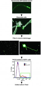
A research team from Princeton University led by Coleen Murphy, professor of Molecular Biology and the Lewis-Sigler Institute for Integrative Genomics, has developed a new method for isolating neurons from adult C. elegans worms. The first panel shows worms containing neurons labeled with green fluorescent protein (GFP). Using rapid, chilled chemomechanical disruption, the neuron cells were extracted and purified, then sorted using fluorescence-activated cell sorting (FACS).

Researchers from Princeton University have identified genes important for age-related cognitive declines in memory in adult worm neurons, which had not been studied previously. The research, published in the journal Nature, could eventually point the way toward therapies to extend life and enhance health in aging human populations.

“The newly discovered genes regulate enhanced short-term memory as well as the ability to repair damaged neurons, two factors that play an important role in healthy aging,” said Coleen Murphy, a professor of Molecular Biology and the Lewis-Sigler Institute for Integrative Genomics, director of the Glenn Center for Quantitative Aging Research at Princeton, and senior author on the study. “Identifying the individual factors involved in neuron health in the worm is the first step to understanding human neuronal decline with age.”

The small soil-dwelling roundworm, Caenorhabditis elegans, contains genes that determine the rate of aging and overall health during aging. Mutations in one of these genetic pathways, the insulin/IGF-1 signaling (IIS) pathway, can double worm lifespan. Similar mutations in humans have been found in long-lived humans.

But studying the IIS mutation in adult worm neurons was difficult because the adults have a thick, durable covering that protects the neurons.

Using a new technique they developed to break up the tough outer covering, researchers at Princeton succeeded in isolating adult neurons, which enabled the detection of the new set of genes regulated by the insulin/IGF-1 signaling pathway.

“Our technique enabled us to study gene expression in adult neurons, which are the cells that govern cognitive aspects such as memory and nerve-regeneration,” said Murphy, whose research on aging is funded in part by the National Institutes of Health. “Prior to this work, researchers were only able to examine gene regulation either using adult worms or individual tissues from young worms.”

The work allowed co-first authors Rachel Kaletsky and Vanisha Lakhina to explore why long-lived IIS mutants maintain memory and neuron-regeneration abilities with age. Until now, the known targets of the insulin longevity pathway were located mostly in the intestine and skin of the worm rather than the neurons. Kaletsky is a postdoctoral research fellow and Lakhina is a postdoctoral research associate in the Lewis-Sigler Institute.

Kaletsky worked out the new way to isolate neurons from adult worms, and with Lakhina, proceeded to profile the gene activity in adult C. elegans neurons for the first time. They discovered that the IIS mutant worms express genes that keep neurons working longer, and that these genes are completely different from the previously known longevity targets. They also discovered a new factor that is responsible for nerve cell (axon) regeneration in adult worms, which could have implications for human traumatic brain injury.

“Kaletsky and Lakhina developed a new technique that is going to be used by the entire worm community, so it really opens up new avenues of research even beyond the discoveries we describe in the paper,” Murphy said.

One of the newly identified genes, fkh-9, regulates both enhanced memory and neuronal regeneration in IIS mutants. Previous studies have detected only one other gene that regulates neuronal regeneration in the mutants, demonstrating the power of the technique to identify new gene regulators. The researchers also found that fkh-9 gene expression is required for long lifespan in many IIS mutants, but it did not play that role in neurons, suggesting the gene governs multiple outcomes in the worm.

Murphy’s lab is now working to understand how fkh-9 works to influence memory, axon regeneration, and lifespan. The gene codes for a protein, FKH-9, that acts as a transcription factor, meaning it controls the expression of other genes and is likely part of a larger regulatory network. FKH-9 also appears to regulate different processes in different tissues: It is required in neurons for memory and axon repair, but not for lifespan. Murphy’s group is working to figure out how FKH-9 acts in distinct tissues to regulate such different processes.

The study provides a more complete picture of how IIS mutants control gene expression in different tissues to promote healthy aging, Murphy said.

“fkh-9 is likely only one of the exciting genes that will emerge from using this technique,” Murphy said. “By identifying the suite of IIS-regulated neuronal genes, there are many candidates for follow-up, only a fraction of which have been characterized in any great detail,” she said.

Other contributors to the study included Rachel Arey, a postdoctoral research fellow; former graduate students April Williams and Jessica Landis; and Jasmine Ashraf, a research specialist in the Lewis-Sigler Institute.

Additional funding for the study was provided by the Keck Foundation, the Ruth L. Kirschstein National Research Service Awards, the National Science Foundation and the New Jersey Commission on Brain Injury Research.

Read the abstract.

The article, The C. elegans adult neuronal IIS/FOXO transcriptome reveals adult phenotype regulators, by Rachel Kaletsky, Vanisha Lakhina, Rachel Arey, April Williams, Jessica Landis, Jasmine Ashraf and Coleen T. Murphy, was published in the journal Nature online ahead of print on December 14, 2015. doi:10.1038/nature16483.

Genome-wide search reveals new genes involved in long-term memory (Neuron)

By Catherine Zandonella, Office of the Dean for Research

Whole genome expression data reveals new genes involved in long-term memory formation in worms. (Image source: Murphy lab)

A new study has identified genes involved in long-term memory in the worm as part of research aimed at finding ways to retain cognitive abilities during aging.

The study, which was published in the journal Neuron, identified more than 750 genes involved in long-term memory, including many that had not been found previously and that could serve as targets for future research, said senior author Coleen Murphy, an associate professor of molecular biology and the Lewis-Sigler Institute for Integrative Genomics at Princeton University.

“We want to know, are there ways to extend memory?” Murphy said. “And eventually, we would like to ask, are there compounds that could maintain memory with age?”

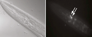
Long-term memory training in worms (left) led to induction of the transcription factor CREB in AIM neurons (shown by arrows in right). CREB-induced genes were shown to be involved in forming long-term memories in worm neurons. (Image source: Murphy lab)

The newly pinpointed genes are “turned on” by a molecule known as CREB (cAMP-response element-binding protein), a factor known to be required for long-term memory in many organisms, including worms and mice.

“There is a pretty direct relationship between CREB and long-term memory,” Murphy said, “and many organisms lose CREB as they age.” By studying the CREB-activated genes involved in long-term memory, the researchers hope to better understand why some organisms lose their long-term memories as they age.

To identify the genes, the researchers first instilled long-term memories in the worms by training them to associate meal-time with a butterscotch smell. Trained worms were able to remember that the butterscotch smell means dinner for about 16 hours, a significant amount of time for the worm.

The researchers then scanned the genomes of both trained worms and non-trained worms, looking for genes turned on by CREB.

The researchers detected 757 CREB-activated genes in the long-term memory-trained worms, and showed that these genes were turned on primarily in worm cells called the AIM interneurons.

They also found CREB-activated genes in non-trained worms, but the genes were not turned on in AIM interneurons and were not involved in long-term memory. CREB turns on genes involved in other biological functions such as growth, immune response, and metabolism. Throughout the worm, the researchers noted distinct non-memory (or “basal”) genes in addition to the memory-related genes.

The next step, said Murphy, is to find out what these newly recognized long-term memory genes do when they are activated by CREB. For example, the activated genes may strengthen connections between neurons.

Worms are a perfect system in which to explore that question, Murphy said. The worm Caenorhabditis elegans has only 302 neurons, whereas a typical mammalian brain contains billions of the cells.

“Worms use the same molecular machinery that higher organisms, including mammals, use to carry out long-term memory,” said Murphy. “We hope that other researchers will take our list and look at the genes to see whether they are important in more complex organisms.”

Murphy said that future work will involve exploring CREB’s role in long-term memory as well as reproduction in worms as they age.

The team included co-first-authors Postdoctoral Research Associate Vanisha Lakhina, Postdoctoral Research Associate Rachel Arey, and Associate Research Scholar Rachel Kaletsky of the Lewis-Sigler Institute for Integrative Genomics. Additional research was performed by Amanda Kauffman, who earned her Ph.D. in Molecular Biology in 2010; Geneva Stein, who earned her Ph.D. in Molecular Biology in 2014; William Keyes, a laboratory assistant in the Department of Molecular Biology; and Daniel Xu, who earned his B.A. in Molecular Biology in 2014.

Funding for the research was provided by the National Institutes of Health and the Paul F. Glenn Laboratory for Aging Research at Princeton University.

Read the abstract

Citation: Vanisha Lakhina, Rachel N. Arey, Rachel Kaletsky, Amanda Kauffman, Geneva Stein, William Keyes, Daniel Xu, and Coleen T. Murphy. Genome-wide Functional Analysis of CREB/Long-Term Memory-Dependent Transcription Reveals Distinct Basal and Memory Gene Expression Programs, Neuron (2015), http://dx.doi.org/10.1016/j.neuron.2014.12.029.

Small declines in agility, facial features may predict risk of dying (Epidemiology)

Photo source: Shutter Stock

By B. Rose Huber, Woodrow Wilson School of Public and International Affairs

A new study from Princeton University shows that health assessments made by medically untrained interviewers may predict the mortality of individuals better than those made by physicians or the individuals themselves.

Features like forehead wrinkles and lack of agility may reflect a person’s overall health and risk of dying, according to recent health research. But do physicians consider such details when assessing patients’ overall health and functioning?

In a survey of approximately 1,200 Taiwanese participants, Princeton University researchers found that interviewers — who were not health professionals but were trained to administer the survey — provided health assessments that were related to a survey participant’s risk of dying, in part because they were attuned to facial expressions, responsiveness and overall agility.

The researchers report in the journal Epidemiology that these assessments were even more accurate predictors of dying than assessments made by physicians or even the individuals themselves. The findings show that survey interviewers, who typically spend a fair amount of time observing participants, can glean important information regarding participants’ health through thorough observations.

“Your face and body reveal a lot about your life. We speculate that a lot of information about a person’s health is reflected in their face, movements, speech and functioning, as well as in the information explicitly collected during interviews,” said Noreen Goldman, Hughes-Rogers Professor of Demography and Public Affairs in the Woodrow Wilson School.

Together with lead author of the paper, Princeton Ph.D. candidate Megan Todd, Goldman analyzed data collected by the Social Environment and Biomarkers of Aging Study (SEBAS). This study was designed by Goldman and co-investigator Maxine Weinstein at Georgetown University to evaluate the linkages among the social environment, stress and health. Beginning in 2000, SEBAS conducted extensive home interviews, collected biological specimens and administered medical examinations with middle-aged and older adults in Taiwan. Goldman and Todd used the 2006 wave of this study, which included both interviewer and physician assessments, for their analysis. They also included death registration data through 2011 to ascertain the survival status of those interviewed.

The survey used in the study included detailed questions regarding participants’ health conditions and social environment. Participants’ physical functioning was evaluated through tasks that determined, for example, their walking speed and grip strength. Health assessments were elicited from participants, interviewers and physicians on identical five-point scales by asking “Regarding your/the respondent’s current state of health, do you feel it is excellent (5), good (4), average (3), not so good (2) or poor (1)?”

Participants answered this question near the beginning of the interview, before other health questions were asked. Interviewers assessed the participants’ health at the end of the survey, after administering the questionnaire and evaluating participants’ performance on a set of tasks, such as walking a short distance and getting up and down from a chair. And physicians — who were hired by the study and were not the participants’ primary care physicians — provided their assessments after physical exams and reviews of the participants’ medical histories. (Study investigators did not provide special guidance about how to rate overall health to any group.)

In order to understand the many variables that go into predicting mortality, Goldman and Todd factored into their statistical models such socio-demographic variables as gender, place of residence, education, marital status, and participation in social activities. They also considered chronic conditions, psychological wellbeing (such as depressive symptoms) and physical functioning to account for a fuller picture of health.

“Mortality is easy to measure because we have death records indicating when a person has died,” Goldman said. “Overall health, on the other hand, is very complicated to measure but obviously very important for addressing health policy issues.”

Two unexpected results emerged from Goldman and Todd’s analysis. The first: physicians’ ratings proved to be weak predictors of survival. “The physicians performed a medical exam equivalent to an annual physical exam, plus an abdominal ultrasound; they have specialized knowledge regarding health conditions,” Goldman explained. “Given access to such information, we anticipated stronger, more accurate predictions of death,” she said. “These results call into question previous studies’ assumptions that physicians’ ‘objective health’ ratings are superior to ‘subjective’ ratings provided by the survey participants themselves.”

In a second surprising finding, the team found that interviewers’ ratings were considerably more powerful for predicting mortality than self-ratings. This is likely, Goldman said, because interviewers considered respondents’ movements, appearance and responsiveness in addition to the detailed health information gathered during the interviews. Also, Goldman posits, interviewer ratings are probably less affected by bias than self-reports.

“The ‘self-rated health’ question is religiously used by health researchers and social scientists, and, although it has been shown to predict mortality, it suffers from many biases. People use it because it’s easy and simple,” Goldman continued. “But the problem with self-rated health is that we have no idea what reference group the respondent is using when evaluating his or her own health. Different ethnic and racial groups respond differently as do varying socioeconomic groups. We need other simple ways to rate individual health instead of relying so heavily on self-rated health.”

One way, Goldman suggests, is by including interviewer ratings in surveys along with self-ratings: “This is a straightforward and cost-free addition to a questionnaire that is likely to improve our measurement of health in any population,” Goldman said.

The paper, “Do Interviewer and Physician Health Ratings Predict Mortality? A Comparison with Self-Rated Health,” first appeared online in Epidemology in August 2013. The article also will be featured in the November print edition. The research was conducted with the assistance of colleagues at Princeton’s Office of Population Research, Georgetown University and the Bureau of Health Promotion in the Taiwan Department of Health.

Read the abstract.

Todd MA, Goldman N. Do interviewer and physician health ratings predict mortality?: a comparison with self-rated health. Epidemiology. 2013 Nov;24(6):913-20. doi: 10.1097/EDE.0b013e3182a713a8.


DNA Gridlock – Cells undo glitches to prevent mutations (Nature)

By Catherine Zandonella, Office of the Dean for Research

G4 Quadruplex
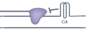
The diagram shows a G-quadruplex (G4) on the upper of the two strands that make up DNA. The purple shape represents DNA polymerase, which is blocked by the G4 in its attempt to copy DNA. Regions of the genome that are especially susceptible to forming G-quadruplexes are ones rich in guanine, which is one of the four nucleotides, designated by the letters G, A, C, and T, in DNA. Adapted from Nature Genetics, 2012.

Roughly six feet of DNA are packed into every human cell, so it is not surprising that our genetic material occasionally folds into odd shapes such as hairpins, crosses and clover leafs. But these structures can block the copying of DNA during cell division, leading to gene mutations that could have implications in cancer and aging.

Now researchers based at Princeton University have uncovered evidence that cells contain a built-in system for eliminating one of the worst of these roadblocks, a structure known as a G-quadruplex. In a paper published earlier this month in Nature, a group of researchers led by Princeton’s Virginia Zakian reported that an enzyme known as the Pif1 helicase can unfold these structures both in test tubes and in cells, bringing DNA replication back on track.

Given that Pif1 mutations have been associated with an increased risk of breast cancer, Zakian said, the study of how Pif1 ensures proper DNA replication could be relevant to human health. Zakian is Princeton’s Harry C. Wiess Professor in the Life Sciences.

Most DNA is made of up of two strands twisted about each other in a way that resembles a spiral staircase. Every time a cell divides, each DNA molecule must be duplicated, a process that involves unwinding the staircase so that an enzyme known as DNA polymerase can work down each strand, copying each letter in the DNA code. During this exposed period, regions of the unwound single strands can fold into G-quadruplexes (see diagram).

Like a car that encounters a pile-up on an Interstate, the DNA polymerase halts when it encounters a G-quadruplex, explained Matthew Bochman, a postdoctoral researcher who was a co-first author with Katrin Paeschke, now an independent investigator at University of Würzburg in Germany. The work also included Princeton graduate student Daniela Garcia.

“The DNA that is folded into a G-quadruplex cannot be replicated, so essentially it is skipped,” Bochman said. “Failure to copy specific areas of DNA that you really need is a serious problem, especially in regions that control genes that either suppress or contribute to cancer,” Bochman said.

Last year, the Zakian group in collaboration with human geneticists at the University of Washington reported that a mutation in human Pif1 is associated with an increased risk of breast cancer, suggesting that the ability to unwind G-quadruplexes could be important for protecting against cancer. The finding was published in the journal PLoS One.

G-quadruplexes could also be implicated in the process of aging, according to the researchers. The structures are thought to form at the ends of chromosomes in regions called telomeres, said Zakian, an expert on telomere biology.  Damaged or shortened telomeres are associated with premature aging and cancer.

To explore the role of Pif1 helicases in tackling G-quadruplexes, Bochman and Paeschke purified Pif1 helicases from yeast and bacteria and found that in test tubes, all of the Pif1 helicases unwind G-quadruplex structures extremely fast and very efficiently, much better than other helicases tested in the same way.

Next, these investigators set up an experiment to determine if Pif1 acts on G-quadruplexes inside cells. Using a system that could precisely evaluate the effects of G-quadruplex structures on the integrity of chromosomes, the researchers found that normal cells had no problem with the addition of a G-quadruplex structure, but when cells lack Pif1 helicases, the G-quadruplex induced a high amount of genome instability.

“To me, the most remarkable aspect of the study was the demonstration that Pif1-like helicases taken from species ranging from bacteria to humans and placed in yeast cells can suppress G-quadruplex-induced DNA damage,” Zakian said. “This finding suggests that resolving G-quadruplexes is an evolutionarily conserved function of Pif1 helicases.”

The Zakian lab also found that replicating through G-quadruplexes in the absence of Pif1 helicases results not only in mutations of the DNA at the site of the G-quadruplex but also in intriguing “epigenetic” effects on expression of nearby genes that were totally unexpected. Epigenetic events cause changes in gene expression that are inherited, yet they do not involve loss or mutation of DNA. Graduate student Daniela Garcia has proposed that the epigenetic silencing of gene expression that occurs near G-quadruplexes in the absence of Pif1 helicases is a result of the addition or removal of molecular tags on histones, which are proteins that bind DNA and regulate gene expression. This hypothesis is currently being studied.

The study involved contributions from Petr Cejka and Stephen C. Kowalczykowski of the University of California-Davis, and Katherine Friedman of Vanderbilt University.

Read the abstract.

Paeschke, Katrin, Matthew L. Bochman, P. Daniela Garcia, Petr Cejka, Katherine L. Friedman, Stephen C. Kowalczykowski & Virginia A. Zakian. Pif1 family helicases suppress genome instability at G-quadruplex motifs. Nature. 2013. doi:10.1038/nature12149.

This research was supported by the National Institutes of Health (V.A.Z., GM026938-34; S.C.K.GM041347), the National Science Foundation (K.L.F., MCB-0721595), the German Research Foundation (DFG), the New Jersey Commission on Cancer Research (K.P.) and the American Cancer Society (M.L.B., PF-10-145-02-01).