Researchers find an alternative mode of bacterial quorum sensing

By the Department of Molecular Biology

Whether growing in a puddle of dirty water or inside the human body, large groups of bacteria must coordinate their behavior to perform essential tasks that they would not be able to carry out individually. Bacteria achieve this coordination through a process called quorum sensing in which the microorganisms produce and secrete small molecules called autoinducers that can be detected by neighboring bacterial cells. Only when a large number of bacteria are present can the levels of secreted autoinducer build up to the point where the community can detect them and respond as a coordinated group.

In a paper published last month in PLoS Pathogens, a team of researchers led by postdoctoral research associate Sampriti Mukherjee and professor Bonnie Bassler of the Department of Molecular Biology at Princeton University revealed the existence of a new quorum-sensing molecule that increases the virulence of the pathogenic bacterium Pseudomonas aeruginosa. The finding could help researchers develop new antimicrobial drugs to treat the serious infections caused by this bacterium.

P. aeruginosa is an incredibly adaptable organism that can grow in environments ranging from soil and freshwater to the tissues of plants and animals. It thrives on the surfaces of medical equipment and is therefore a major cause of hospital-acquired infections, causing life-threatening conditions such as pneumonia and sepsis in vulnerable patients. The bacterium has become resistant to commonly used antibiotics, making the development of new antimicrobials a priority for both the Centers for Disease Control and Prevention and the World Health Organization.

Quorum sensing is crucial for P. aeruginosa‘s adaptability. The process regulates the development of biofilms, the three-dimensional structures formed by large bacterial communities that promote their ability to establish and maintain infections. “P. aeruginosa strains harboring mutations in the quorum-sensing machinery are attenuated for virulence, and thus, interfering with quorum sensing holds promise for the development of novel anti-microbial therapies,” said Bassler, the Squibb Professor in Molecular Biology at Princeton University and a Howard Hughes Medical Institute Investigator.

P. aeruginosa possesses similar quorum-sensing machinery to other species of bacteria. For example, it produces an enzyme called RhlI that synthesizes an autoinducer molecule known as N-butanoyl-L-homoserine lactone, or C4-HSL. This molecule can then bind and activate a protein called RhlR that regulates the expression of multiple genes that P. aeruginosa needs to form a biofilm and/or infect a host.
Biofilms
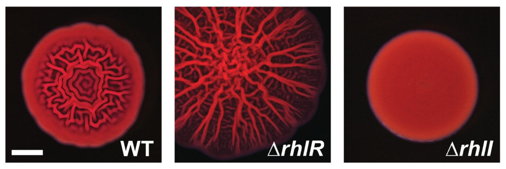
Caption: Compared to a typical, or wild-type (WT) colony (left), P. aeruginosa cells lacking RhlR form a much more wrinkled biofilm (middle), while cells lacking RhlI form a biofilm that is abnormally smooth (right). Credit: Mukherjee et al., 2017.

In theory, removing RhlI or RhlR should have the same effect on P. aeruginosa cells, since the latter protein shouldn’t be able to work without the autoinducer produced by the former. But the researchers, led by postdoctoral fellow Sampriti Mukherjee, noticed that bacterial colonies lacking RhlI formed unusually smooth biofilms, whereas strains lacking RhlR formed biofilms that were much more wrinkled than normal.

The researchers went on to show that in biofilms, many genes only depended on RhlR, not on RhlI. “That suggested that RhlR can be activated by an alternative molecule, in addition to C4-HSL,” Bassler said.

The researchers found that bacteria lacking RhlI, which are therefore unable to synthesize the C4-HSL autoinducer, still secrete a molecule capable of activating RhlR. Bassler and colleagues don’t yet know what this molecule is, but it seems to be quite different from C4-HSL. “We are currently working to purify and identify this molecule,” Bassler said.

Researchers at Princeton discovered that a molecule called RhlR, which is important for the ability of P. aeruginosa to infect animals, can be activated by an alternative molecule, in addition to C4-HSL. Image credit: Mukherjee et al., 2017.

Crucially, the activation of RhlR by this unknown molecule may be important for P. aeruginosa‘s ability to infect animals. Mukherjee and the team found that bacteria lacking RhlI were just as effective as wild-type bacteria in infecting both roundworms and mice. But bacteria lacking RhlR were much less virulent and far less able to grow inside these animals. “Targeting RhlR with small-molecule inhibitors could provide an exciting path forward for the development of novel antimicrobial drugs,” Bassler said.

The ability of RhlR to be activated by distinct molecules might also help explain P. aeruginosa‘s adaptability. Bassler and colleagues speculate that different environments could stimulate discrete levels of production of the different autoinducer molecules, each of which could activate RhlR, or a related protein called LasR, to induce expression of the specific genes the bacteria need to thrive in that particular location.

The work was supported by the Howard Hughes Medical Institute, the National Institutes of Health (grant 2R37GM065859), the National Science Foundation (grant MCB-0948112), and a Life Science Research Foundation Postdoctoral Fellowship through the Gordon and Betty Moore Foundation (grant GBMF2550.06).

In addition to Mukherjee and Bassler, the team consisted of postdoctoral fellow Dina Moustafa and professor Joanna Goldberg in the Department of Pediatrics at Emory University School of Medicine, and Chari Smith, a research consultant at Princeton University.

The study, “The RhlR quorum-sensing receptor controls Pseudomonas aeruginosa pathogenesis and biofilm development independently of its canonical homoserine lactone autoinducer,” by Sampriti Mukherjee, Dina Moustafa, Chari D. Smith, Joanna B. Goldberg, and Bonnie L. Bassler, was published in the journal PLoS Pathogens on July 17, 2017. DOI:10.1371/journal.ppat.1006504.

Study questions the prescription for drug resistance (Proceedings of the Royal Society B)

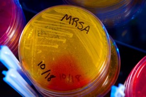
A drug-resistant strain of bacteria known as MRSA. Photo by James Gathany
A new study examines the question of aggressive versus moderate drug treatment on the emergence of drug-resistant pathogens. Shown is a strain of bacteria known as methicillin-resistant Staphylococcus aureus (MRSA). Photo by James Gathany

By Catherine Zandonella, Office of the Dean for Research

In response to the rise of drug-resistant pathogens, doctors are routinely cautioned against overprescribing antimicrobials. But when a patient has a confirmed bacterial infection, the advice is to treat aggressively to quash the infection before the bacteria can develop resistance.

A new study questions the accepted wisdom that aggressive treatment with high drug dosages and long durations is always the best way to stem the emergence and spread of resistant pathogens. The review of nearly 70 studies of antimicrobial resistance, which was authored by researchers at Princeton and other leading institutions and published last week in the journal Proceedings of the Royal Society B, reveals the lack of evidence behind the practice of aggressive treatment in many cases.

“We found that while there are many studies that test for resistance emergence between different drug regimes, surprisingly few have looked at the topic of how varying drug dosage might affect the emergence and spread of resistance,” said Ruthie Birger, a Princeton graduate student who works with C. Jessica Metcalf, an assistant professor of ecology and evolutionary biology and public affairs at Princeton’s Woodrow Wilson School, and Bryan Grenfell, the Kathryn Briger and Sarah Fenton Professor of Ecology and Evolutionary Biology and Public Affairs in Princeton’s Woodrow Wilson School. Birger, Metcalf and Grenfell coauthored the paper with colleagues from 16 universities. “We are a long way from having the evidence for the best treatment decisions with respect to resistance for a range of diseases,” Birger said.

Microbes such as bacteria and parasites can evade today’s powerful drugs by undergoing genetic mutations that enable them to avoid being killed by the drug. For example, bacteria can develop enzymes that degrade certain antibiotics. The logic behind aggressive treatment goes something like this: kill off as many microbes as you can so that few will be around to evolve into resistant forms.

But some scientists have observed a different outcome in mice infected with both an already-resistant strain of malaria and a non-resistant strain. The high-dose drug treatment killed off the non-resistant malarial parasites, leaving the resistant strains to multiply and make the mice even sicker.

The idea that aggressive treatment may backfire against malarial parasites led the authors of the current study to comb the scientific literature to examine whether the same may be true for other types of microbes such as bacteria. The few studies that they found — mostly in laboratory cell cultures rather than animal models or patients — suggest that the picture is complicated, and depends on whether the resistance is new or existing, how many mutations are necessary for the pathogen to become resistant, and how long the drugs have been in use. “It’s remarkable how little we know about this topic,” said Metcalf. “The malaria study conducted by Silvie Huijben and colleagues at Pennsylvania State University is an inspiring step towards developing an evidence base for these important issues.”

In the current analysis, the study authors found that drug resistance is governed by two factors: the abundance of the pathogen and the strength of the selection pressure that drives the pathogen to evolve. Aggressive treatment deals with the first factor by killing off as much pathogen as possible, while moderate treatment may, for some pathogens, reduce the ability for the resistant pathogen to thrive (for example, by maintaining the competitive advantage of a co-infecting drug-sensitive strain of the pathogen) but still reduce total pathogen levels sufficiently that the patient can recover.

Finding the ideal dose and duration of treatment, one that cures the patient without aiding the spread of resistance, will likely be done on a disease by disease basis, the authors found.

One possibility is that moderate treatment might be best used against already-resistant microbes to prevent their spread. Moderate treatment may also be best for drugs that have been on the market for several years with plenty of time for resistant strains to develop.

Aggressive treatment might be best for pathogens that develop resistance slowly, over the course of several mutations. High doses early in the process could be effective at heading off the development of resistance.

Read the abstract.

Roger D. Kouyos, C. Jessica E. Metcalf, Ruthie Birger, Eili Y. Klein, Pia Abel zur Wiesch, Peter Ankomah, Nimalan Arinaminpathy, Tiffany L. Bogich, Sebastian Bonhoeffer, Charles Brower, Geoffrey Chi-Johnston, Ted Cohen, Troy Day, Bryan Greenhouse, Silvie Huijben, Joshua Metlay, Nicole Mideo, Laura C. Pollitt, Andrew F. Read, David L. Smith, Claire Standley, Nina Wale and Bryan Grenfell. Proc. R. Soc. B: Biological Sciences, 281, 20140566. Published Sept. 24, 2014

The work emerged from two workshops held at Princeton University and funded by the RAPIDD program of the Science and Technology Directorate, Department of Homeland Security and the Fogarty International Center, National Institutes of Health; Science and Technology Directorate, Department of Homeland Security; contract HSHQDC-12-C-00058

Conservation versus innovation in the fight against antibiotic resistance (Science)

Pills (Image source: NIH)
(Image source: NIH)

“Antibiotic resistance is a problem of managing an open-access resource, such as fisheries or oil,” writes Ramanan Laxminarayan, a research scholar at Princeton University and the director of the Center for Disease Dynamics, Economics & Policy in Washington, D. C., in today’s issue of the journal Science. He goes on to say that individuals have little incentive to use antibiotics wisely, just as people have little incentive to conserve oil when it is plentiful.

As with many other natural resources, maintaining the effectiveness of antibiotics requires two approaches: conserving the existing resource and exploring new sources, Laxminarayan says. These two approaches are linked, however. “Just as incentives for finding new sources of oil reduce incentives to conserve oil,” Laxminarayan writes, “large public subsidies for new drug development discourage efforts to improve how existing antibiotics are used.” Yet new antibiotics tend to cost more than existing ones due to the expense of clinical trials and the fact that the easiest-to-find drugs may have already been discovered.

Laxminarayan’s analysis reveals that the benefits of conserving existing drugs are significant, and argues that the proposed increases in public subsidies for new antibiotics should be matched by greater spending on conservation of antibiotic effectiveness through public education, research and surveillance.

Ramanan Laxminarayan is a research scholar at the Princeton Environmental Institute. His perspective, “Antibiotic effectiveness: Balancing conservation against innovation,” appeared in the September 12, 2014 issue of Science.

Read the article.

Unlocking the potential of bacterial gene clusters to discover new antibiotics (Proc. Natl. Acad. Sci.)

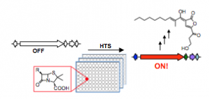
High-throughput screening for the discovery of small molecules that activate silent bacterial gene clusters
High-throughput screening for the discovery of small molecules that activate silent bacterial gene clusters. Image courtesy of Mohammad Seyedsayamdost.

by Tien Nguyen, Department of Chemistry

Resistance to antibiotics has been steadily rising and poses a serious threat to the stronghold of existing treatments. Now, a method from Mohammad Seyedsayamdost, an assistant professor of chemistry at Princeton University, may open the door to the discovery of a host of potential drug candidates.

The vast majority of anti-infectives on the market today are bacterial natural products, made by biosynthetic gene clusters. Genome sequencing of bacteria has revealed that these active gene clusters are outnumbered approximately ten times by so-called silent gene clusters.

“Turning these clusters on would really expand our available chemical space to search for new antibiotic or otherwise therapeutically useful molecules,” Seyedsayamdost said.

In an article published last week in the journal Proceedings of the National Academy of Sciences, Seyedsayamdost reported a strategy to quickly screen whole libraries of compounds to find elicitors, small molecules that can turn on a specific gene cluster. He used a genetic reporter that fluoresces or generates a color when the gene cluster is activated to easily identify positive hits. Using this method, two silent gene clusters were successfully activated and a new metabolite was discovered.

Application of this work promises to uncover new bacterial natural products and provide insights into the regulatory networks that control silent gene clusters.

Read the abstract.

Seyedsayamdost, M. R. “High-throughput platform for the discovery of elicitors of silent bacterial gene clusters.” Proc. Natl. Acad. Sci. 2014, Early edition.

Why a bacterium got its curve — and why biologists should know (Nature Communications)

Art by Laura Ancona
Princeton University researchers found that the banana-like curve of the bacteria Caulobacter crescentus provides stability and helps them flourish as a group in the moving water they experience in nature. The findings suggest a new way of studying the evolution of bacteria that emphasizes using naturalistic settings. The illustration shows how C. crescentus divides asymmetrically into a “stalked” mother cell that anchors to a bacterium’s home surface, and an upper unattached portion that forms a new, juvenile cell known as a “swarmer.” Swarmer cells later morph into stalked cells and lay down roots nearby. They repeat the life cycle with their own swarmer cell and the bacterial colony grows. The Princeton researchers found that in moving water, curvature points the swarmer cell toward the surface to which it needs to attach. This ensures that the bacteria’s next generation does not stray too far from its progenitors. (Image by Laura Ancona)

By Morgan Kelly, Office of Communications

Drawing from his engineering background, Princeton University researcher Alexandre Persat had a notion as to why the bacteria Caulobacter crescentus are curved — a hunch that now could lead to a new way of studying the evolution of bacteria, according to research published in the journal Nature Communications.

Commonly used in labs to study cell division, C. crescentus naturally take on a banana-like curve, but they also can undergo a mutation in which they grow to be perfectly straight. The problem was that in a laboratory there was no apparent functional difference between the two shapes. So a question among biologists was, why would nature bother?

Then Persat, who is a postdoctoral researcher in the group of Associate Professor of Molecular Biology Zemer Gitai, considered that the bacteria dwell in large groups attached to surfaces in lakes, ponds and streams. That means that their curvature could be an adaptation that allows C. crescentus to better develop in the water currents the organisms experience in nature.

In the new paper, first author Persat, corresponding author Gitai and Howard Stone, Princeton’s Donald R. Dixon ’69 and Elizabeth W. Dixon Professor of Mechanical and Aerospace Engineering, report that curvature does more than just help C. crescentus hold their ground in moving fluid. The researchers monitored C. crescentus growth on surfaces in flow and found that the bacteria’s arched anatomy is crucial to flourishing as a group.

“It didn’t take a long time to figure out how flow brought out the advantages of curvature,” Persat said. “The obvious thing to me as someone with a fluid-dynamics background was that this shape had something to do with fluid flow.”

The findings emphasize the need to study bacteria in a naturalistic setting, said Gitai, whose group focuses on how bacterial shapes are genetically determined. While a petri dish generally suffices for this line of study, the functionality of bacterial genes and anatomy can be elusive in most lab settings, he said. For instance, he said, 80 percent of the genes in C. crescentus are seemingly disposable — but they might not be in nature.

“We now see there can be benefits to bacterial shapes that are only seen in a growth environment that is close to the bacteria’s natural environment,” Gitai said.

“For C. crescentus, the ecology was telling us there is an advantage to being curved, but nothing we previously did in the lab could detect what that was,” he said. “We need to not only think of the chemical environment of the bacteria — we also need to think of the physical environment. I think of this research as opening a whole new axis of studying bacteria.”

While most bacteria grow and divide as two identical “daughter” cells, C. crescentus divides asymmetrically. A “stalked” mother cell anchors to a bacterium’s home surface while the upper unattached portion forms a new, juvenile version of the stalked cell known as a “swarmer” cell. The swarmer cells later morph into stalked cells then eventually detach before laying down roots nearby. They repeat the life cycle with their own swarmer cell and the bacterial colony grows.

The Princeton researchers found that in moving water, curvature points the swarmer cell toward the surface to which it needs to attach. This ensures that the bacteria’s next generation does not stray too far from its progenitors, as well as from the nutrients that prompted cell division in the first place, Gitai said. On the other hand, the upper cells of straight bacteria — which are comparatively higher from the ground — are more likely to be carried far away as they are to stay near home.

But the advantage of curvature only goes so far. The researchers found that when the water current was too strong, both curved and straight bacteria were pressed flat against the surface, eliminating the curved cells’ colonization advantage.

These findings put some interesting boundaries on what is known about C. crescentus, starting with the upper limits of the current in which the organism can thrive, Gitai said. He and Persat also plan to pursue whether the bacteria are able to straighten out and cast offspring downstream when the home colony faces a decline in available nutrients.

At the same time, understanding why C. crescentus got its curve helps in figuring out the evolution of other bacteria, he said. Close relatives of the bacteria, for example, are not curved — could it have to do with the severity of their natural environment, such as the powerful turbulence of an ocean? Harmful bacteria such as Vibrio cholerae, strains of which cause cholera, are curved, though the reason is unclear. It’s possible this shape could be related to the organism’s environment in a way that might help treat those infected by it, Gitai said.

Whatever the reason for a specific bacteria’s shape, the Princeton research shows that exploring the influence of its natural habitat could be worthwhile, Gitai said.

“It was clear with C. crescentus that we needed to try something different,” Gitai said. “People didn’t really think of flow as a major driver of this bacteria’s evolution. That really is a new idea.”

Read the article..

Persat, Alexandre, Howard A. Stone, Zemer Gitai. 2014. The curved shape of Caulobacter crescentus enhances surface colonization in flow. Nature Communications. Article published online May 8, 2014. DOI: 10.1038/ncomms4824

The work was supported by the Gordon and Betty Moore Foundation (grant no. GBMF 2550.02), the National Science Foundation (grant no. CBET-1234500), and the National Institutes of Health Director’s New Investigator Innovator Award (grant no. 1DP2OD004389).

How do bacteria clog medical devices? Very quickly. (PNAS)

stone-figure-2D_540A new study has examined how bacteria clog medical devices, and the result isn’t pretty. The microbes join to create slimy ribbons that tangle and trap other passing bacteria, creating a full blockage in a startlingly short period of time.

The finding could help shape strategies for preventing clogging of devices such as stents — which are implanted in the body to keep open blood vessels and passages — as well as water filters and other items that are susceptible to contamination. The research was published in Proceedings of the National Academy of Sciences.

stone-figure-2D_540
Click on the image to view movie. Over a period of about 40 hours, bacterial cells (green) flowed through a channel, forming a green biofilm on the walls. Over the next ten hours, researchers sent red bacterial cells through the channel. The red cells became stuck in the sticky biofilm and began to form thin red streamers. Once stuck, these streamers in turn trapped additional cells, leading to rapid clogging. (Image source: Knut Drescher)

Using time-lapse imaging, researchers at Princeton University monitored fluid flow in narrow tubes or pores similar to those used in water filters and medical devices. Unlike previous studies, the Princeton experiment more closely mimicked the natural features of the devices, using rough rather than smooth surfaces and pressure-driven fluid instead of non-moving fluid.

The team of biologists and engineers introduced a small number of bacteria known to be common contaminants of medical devices. Over a period of about 40 hours, the researchers observed that some of the microbes — dyed green for visibility — attached to the inner wall of the tube and began to multiply, eventually forming a slimy coating called a biofilm. These films consist of thousands of individual cells held together by a sort of biological glue.

Over the next several hours, the researchers sent additional microbes, dyed red, into the tube. These red cells became stuck to the biofilm-coated walls, where the force of the flowing liquid shaped the trapped cells into streamers that rippled in the liquid like flags rippling in a breeze. During this time, the fluid flow slowed only slightly.

At about 55 hours into the experiment, the biofilm streamers tangled with each other, forming a net-like barrier that trapped additional bacterial cells, creating a larger barrier which in turn ensnared more cells. Within an hour, the entire tube became blocked and the fluid flow stopped.

The study was conducted by lead author Knut Drescher with assistance from technician Yi Shen. Drescher is a postdoctoral research associate working with Bonnie Bassler, Princeton’s Squibb Professor in Molecular Biology and a Howard Hughes Medical Institute Investigator, and Howard Stone, Princeton’s Donald R. Dixon ’69 and Elizabeth W. Dixon Professor of Mechanical and Aerospace Engineering.

“For me the surprise was how quickly the biofilm streamers caused complete clogging,” said Stone. “There was no warning that something bad was about to happen.”

By constructing their own controlled environment, the researchers demonstrated that rough surfaces and pressure driven flow are characteristics of nature and need to be taken into account experimentally. The researchers used stents, soil-based filters and water filters to prove that the biofilm streams indeed form in real scenarios and likely explain why devices fail.

The work also allowed the researchers to explore which bacterial genes contribute to biofilm streamer formation. Previous studies, conducted under non-realistic conditions, identified several genes involved in formation of the biofilm streamers. The Princeton researchers found that some of those previously identified genes were not needed for biofilm streamer formation in the more realistic habitat.

Read the abstract.

Drescher, Knut, Yi Shen, Bonnie L. Bassler, and Howard A. Stone. 2013. Biofilm streamers cause catastrophic disruption of flow with consequences for environmental and medical systems. Proceedings of the National Academy of Sciences. Published online February 11.

This work was supported by the Howard Hughes Medical
Institute, National Institutes of Health grant 5R01GM065859, National Science Foundation (NSF) grant MCB-0343821, NSF grant MCB-1119232, and the Human Frontier Science Program.

Transition from individual to group behavior in bacteria (Journal of Bacteriology)

Bacteria use a chemical communication process called quorum sensing to control transitions between individual and group behaviors. In the bacteria known as Vibrio harveyi, two master “switches” of gene regulation, or transcription factors, coordinate the quorum-sensing response.The researchers found that one of the regulators, LuxR, acts as a sort of master switch that regulates quorum-sensing, while the other regulator, AphA, does the fine-tuning. Together the two regulators generate a precise pattern of activity as bacteria transition from acting as individuals to acting as a group.

Julia C. van Kessel, Steven T. Rutherford, Yi Shao, Alan F. Utria, and Bonnie L. Bassler. The master regulators AphA and LuxR control the Vibrio harveyi quorum-sensing regulon: analysis of their individual and combined effects
J. Bacteriol. published 30 November 2012, 10.1128/JB.01998-12

Read the abstract.