Researchers’ Sudoku strategy democratizes powerful tool for genetics research (Nature Communications)

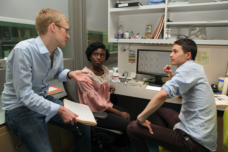
Princeton University researchers Buz Barstow (left), graduate student Kemi Adesina and undergraduate researcher Isao Anzai ’17,
Princeton University researchers Buz Barstow (left), graduate student Kemi Adesina and undergraduate researcher Isao Anzai, Class of 2017, with colleagues at Harvard Universiy, have developed a strategy called “Knockout Sodoku” for figuring out gene function.

By Tien Nguyen, Department of Chemistry

Researchers at Princeton and Harvard Universities have developed a way to produce the tools for figuring out gene function faster and cheaper than current methods, according to new research in the journal Nature Communications.

The function of sizable chunks of many organisms’ genomes is a mystery, and figuring out how to fill these information gaps is one of the central questions in genetics research, said study author Buz Barstow, a Burroughs-Wellcome Fund Research Fellow in Princeton’s Department of Chemistry. “We have no idea what a large fraction of genes do,” he said.

One of the best strategies that scientists have to determine what a particular gene does is to remove it from the genome, and then evaluate what the organism can no longer do. The end result, known as a whole-genome knockout collection, provides full sets of genomic copies, or mutants, in which single genes have been deleted or “knocked out.” Researchers then test the entire knockout collection against a specific chemical reaction. If a mutant organism fails to perform the reaction that means it must be missing the particular gene responsible for that task.

It can take several years and millions of dollars to build a whole-genome knockout collection through targeted gene deletion. Because it’s so costly, whole-genome knockout collections only exist for a handful of organisms such as yeast and the bacterium Escherichia coli. Yet, these collections have proven to be incredibly useful as thousands of studies have been conducted on the yeast gene-deletion collection since its release.

The Princeton and Harvard researchers are the first to create a collection quickly and affordably, doing so in less than a month for several thousand dollars. Their strategy, called “Knockout Sudoku,” relies on a combination of randomized gene deletion and a powerful reconstruction algorithm. Though other research groups have attempted this randomized approach, none have come close to matching the speed and cost of Knockout Sudoku.

“We sort of see it as democratizing these powerful tools of genetics,” said Michael Baym, a co-author on the work and a Harvard Medical School postdoctoral researcher. “Hopefully it will allow the exploration of genetics outside of model organisms,” he said.

Their approach began with steep pizza bills and a technique called transposon mutagenesis that ‘knocks out’ genes by randomly inserting a single disruptive DNA sequence into the genome. This technique is applied to large colonies of microbes to ensure the likelihood that every single gene is disrupted. For example, the team started with a colony of about 40,000 microbes for the bacterium Shewanella oneidensis, which has approximately 3,600 genes in its genome.

Barstow recruited undergraduates and graduate students to manually transfer 40,000 mutants out of laboratory Petri dishes into separate wells using toothpicks. He offered pizza as an incentive, but after a full day of labor, they only managed to move a couple thousand mutants. “I thought to myself, ‘Wait a second, this pizza is going to ruin me,’” Barstow said.

Instead, they decided to rent a colony-picking robot. In just two days, the robot was able to transfer each mutant microbe to individual homes in 96-well plates, 417 plates in total.

But the true challenge and opportunity for innovation was in identifying and cataloging the mutants that could comprise a whole-genome knockout collection in a fast and practical way.

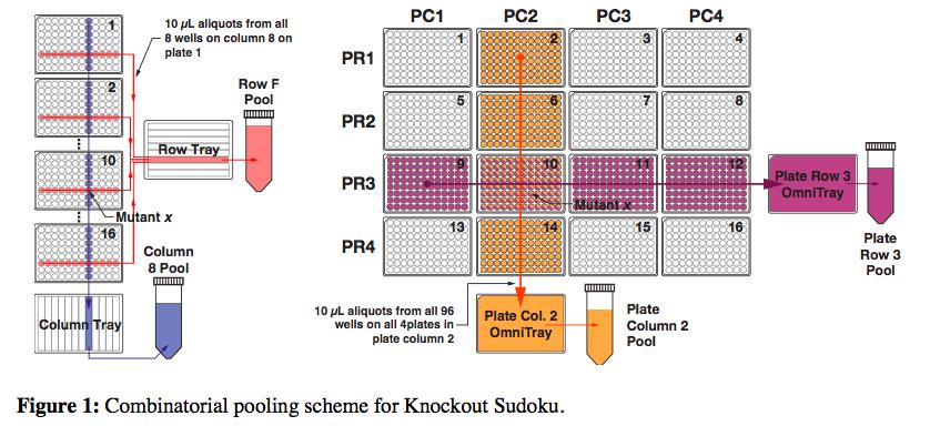
DNA amplification and sequencing is a straightforward way to identify each mutant, but doing it individually quickly gets very expensive and time-consuming. So the researchers’ proposed a scheme in which mutants could be combined into groups that would only require 61 amplification reactions and a single sequencing run.

But still, after sequencing each of the pools, the researchers had an incredible amount of data. They knew the identities of all the mutants, but now they had to figure exactly where each mutant came from in the grid of plates. This is where the Sudoku aspect of the method came in. The researchers built an algorithm that could deduce the location of individual mutants through its repeated appearance in various row, column, plate-row and plate-column pools.

Knockout sodoku helps find genes' functions.

But there’s a problem. Because the initial gene-disruption process is random, it’s possible that the same mutant is formed more than once, which means that playing Sudoku wouldn’t be simple. To find a solution for this issue, Barstow recalled watching the movie, “The Imitation Game,” about Alan Turing’s work on the enigma code, for inspiration.

“I felt like the problem in some ways was very similar to code breaking,” he said. There are simple codes that substitute one letter for another that can be easily solved by looking at the frequency of the letter, Barstow said. “For instance, in English the letter A is used 8.2 percent of the time. So, if you find that the letter X appears in the message about 8.2 percent of the time, you can tell this is supposed to be decoded as an A. This is a very simple example of Bayesian inference.”

With that same logic, Barstow and colleagues developed a statistical picture of what a real location assignment should look like based on a mutant that only appeared once and used that to rate the likelihood of possible locations being real.

“One of the things I really like about this technique is that it’s a prime example of designing a technique with the mathematics in mind at the outset which lets you do much more powerful things than you could do otherwise,” Baym said. “Because it was designed with the mathematics built in, it allows us to get much, much more data out of much less experiments,” he said.

Using their expedient strategy, the researchers created a collection for microbe Shewanella oneidensis. These microbes are especially good at transferring electrons and understanding their powers could prove highly valuable for developing sustainable energy sources, such as artificial photosynthesis, and for environmental remediation in the neutralization of radioactive waste.

Using the resultant collection, the team was able to recapitulate 15 years of research, Barstow said, bolstering their confidence in their method. In an early validation test, they noticed a startlingly poor accuracy rate. After finding no fault with the math, they looked at the original plates to realize that one of the researchers had grabbed the wrong sample. “The least reliable part of this is the human,” Barstow said.

The work was supported by a Career Award at the Scientific Interface from the Burroughs Wellcome Fund and Princeton University startup funds and Fred Fox Class of 1939 funds.

Read the full article here:

Baym, M.; Shaker, L.; Anzai, I. A.; Adesina, O.; Barstow, B. “Rapid construction of a whole-genome transposon insertion collection for Shewanella oneidensis by Knockout Sudoku.” Nature Comm. Available online on Nov. 10, 2016.

Revisiting the mechanics of the action potential (Nature Communications)

By Staff

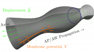
AW_Pic
The action potential (AP) and the accompanying action wave (AW) constitute an electromechanical pulse traveling along the axon.

The action potential is widely understood as an electrical phenomenon. However, a long experimental history has documented the existence of co-propagating mechanical signatures.

In a new paper in the journal Nature Communications, two Princeton University researchers have proposed a theoretical model to explain these mechanical signatures, which they term “action waves.” The research was conducted by Ahmed El Hady, a visiting postdoctoral research associate at the Princeton Neuroscience Institute and a postdoctoral fellow at the Howard Hughes Medical Institute, and Benjamin Machta, an associate research scholar at the Lewis-Sigler Institute for Integrative Genomics and a lecturer in physics and the Lewis-Sigler Institute for Integrative Genomics.

In the model, the co-propagating waves are driven by changes in charge separation across the axonal membrane, just as a speaker uses charge separation to drive sound waves through the air. The researchers argue that these forces drive surface waves involving both the axonal membrane and cytoskeleton as well as its surrounding fluid. Their model may help shed light on the functional role of the surprisingly structured axonal cytoskeleton that recent super-resolution techniques have uncovered, and suggests a wider role for mechanics in neuronal function.

Read the paper.

Ahmed El Hady & Benjamin B. Machta. Mechanical surface waves accompany action potential propagation. Nature Communications 6, No. 6697 doi:10.1038/ncomms7697

Letting go of the (genetic) apron strings (Cell)

Researchers explore the shift from maternal genes to the embryo’s genes during development

By Catherine Zandonella, Office of the Dean for Research

Fruit fly embryo
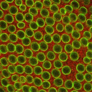
Cells in an early-stage fruit fly embryo. (Image courtesy of NIGMS image gallery).

A new study from Princeton University researchers sheds light on the handing over of genetic control from mother to offspring early in development. Learning how organisms manage this transition could help researchers understand larger questions about how embryos regulate cell division and differentiation into new types of cells.

The study, published in the March 12 issue of the journal Cell, provides new insight into the mechanism for this genetic hand-off, which happens within hours of fertilization, when the newly fertilized egg is called a zygote.

“At the beginning, everything the embryo needs to survive is provided by mom, but eventually that stuff runs out, and the embryo needs to start making its own proteins and cellular machinery,” said Princeton postdoctoral researcher in the Department of Molecular Biology and first author Shelby Blythe. “We wanted to find out what controls that transition.”

Blythe conducted the study with senior author Eric Wieschaus, Princeton’s Squibb Professor in Molecular Biology, Professor of Molecular Biology and the Lewis-Sigler Institute for Integrative Genomics, a Howard Hughes Medical Institute investigator, and a Nobel laureate in physiology or medicine.

Researchers have known that in most animals, a newly fertilized egg cell divides rapidly, producing exact copies of itself using gene products supplied by the mother. After a short while, this rapid cell division pauses, and when it restarts, the embryonic DNA takes control and the cells divide much more slowly, differentiating into new cell types that are needed for the body’s organs and systems.

To find out what controls this maternal to zygotic transition, also called the midblastula transition (MBT), Blythe conducted experiments in the fruit fly Drosophila melanogaster, which has long served as a model for development in higher organisms including humans.

These experiments revealed that the slower cell division is a consequence of an upswing in DNA errors after the embryo’s genes take over. Cell division slows down because the cell’s DNA-copying machinery has to stop and wait until the damage is repaired.

Blythe found that it wasn’t the overall amount of embryonic DNA that caused this increase in errors. Instead, his experiments indicated that the high error rate was due to molecules that bind to DNA to activate the reading, or “transcription,” of the genes. These molecules stick to the DNA strands at thousands of sites and prevent the DNA copying machinery from working properly.

To discover this link between DNA errors and slower cell replication, Blythe used genetic techniques to create Drosophila embryos that were unable to repair DNA damage and typically died shortly after beginning to use their own genes. He then blocked the molecules that initiate the process of transcription of the zygotic genes, and found that the embryos survived, indicating that these molecules that bind to the DNA strands, called transcription factors, were triggering the DNA damage. He also discovered that a protein involved in responding to DNA damage, called Replication Protein A (RPA), appeared near the locations where DNA transcription was being initiated. “This provided evidence that the process of awakening the embryo’s genome is deleterious for DNA replication,” he said.

The study also demonstrates a mechanism by which the developing embryo ensures that cell division happens at a pace that is slow enough to allow the repair of damage to DNA during the switchover from maternal to zygotic gene expression. “For the first time we have a mechanistic foothold on how this process works,” Blythe said.

The work also enables researchers to explore larger questions of how embryos regulate DNA replication and transcription. “This study allows us to think about the idea that the ‘character’ of the DNA before and after the MBT has something to do with the DNA acquiring the architectural features of chromatin [the mix of DNA and proteins that make up chromosomes] that allow us to point to a spot and say ‘this is a gene’ and ‘this is not a gene’,” Blythe said. “Many of these features are indeed absent early in embryogenesis, and we suspect that the absence of these features is what allows the rapid copying of the DNA template early on. Part of what is so exciting about this is that early embryos may represent one of the only times when this chromatin architecture is missing or ‘blank’. Additionally, these early embryos allow us to study how the cell builds and installs these features that are so essential to the fundamental processes of cell biology.”

This work was supported in part by grant 5R37HD15587 from the Eunice Kennedy Shriver National Institute of Child Health and Human Development.

Read the abstract

Blythe, Shelby A. & Eric R. Wieschaus. Zygotic Genome Activation Triggers the DNA Replication Checkpoint at the Midblastula Transition. Cell. Published online on March 5, 2015. doi:10.1016/j.cell.2015.01.050. http://www.sciencedirect.com/science/article/pii/S0092867415001282


Genome-wide search reveals new genes involved in long-term memory (Neuron)

By Catherine Zandonella, Office of the Dean for Research

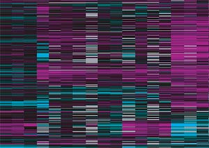
Whole genome expression data reveals new genes involved in long-term memory formation in worms. (Image source: Murphy lab)

A new study has identified genes involved in long-term memory in the worm as part of research aimed at finding ways to retain cognitive abilities during aging.

The study, which was published in the journal Neuron, identified more than 750 genes involved in long-term memory, including many that had not been found previously and that could serve as targets for future research, said senior author Coleen Murphy, an associate professor of molecular biology and the Lewis-Sigler Institute for Integrative Genomics at Princeton University.

“We want to know, are there ways to extend memory?” Murphy said. “And eventually, we would like to ask, are there compounds that could maintain memory with age?”

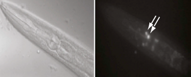
Long-term memory training in worms (left) led to induction of the transcription factor CREB in AIM neurons (shown by arrows in right). CREB-induced genes were shown to be involved in forming long-term memories in worm neurons. (Image source: Murphy lab)

The newly pinpointed genes are “turned on” by a molecule known as CREB (cAMP-response element-binding protein), a factor known to be required for long-term memory in many organisms, including worms and mice.

“There is a pretty direct relationship between CREB and long-term memory,” Murphy said, “and many organisms lose CREB as they age.” By studying the CREB-activated genes involved in long-term memory, the researchers hope to better understand why some organisms lose their long-term memories as they age.

To identify the genes, the researchers first instilled long-term memories in the worms by training them to associate meal-time with a butterscotch smell. Trained worms were able to remember that the butterscotch smell means dinner for about 16 hours, a significant amount of time for the worm.

The researchers then scanned the genomes of both trained worms and non-trained worms, looking for genes turned on by CREB.

The researchers detected 757 CREB-activated genes in the long-term memory-trained worms, and showed that these genes were turned on primarily in worm cells called the AIM interneurons.

They also found CREB-activated genes in non-trained worms, but the genes were not turned on in AIM interneurons and were not involved in long-term memory. CREB turns on genes involved in other biological functions such as growth, immune response, and metabolism. Throughout the worm, the researchers noted distinct non-memory (or “basal”) genes in addition to the memory-related genes.

The next step, said Murphy, is to find out what these newly recognized long-term memory genes do when they are activated by CREB. For example, the activated genes may strengthen connections between neurons.

Worms are a perfect system in which to explore that question, Murphy said. The worm Caenorhabditis elegans has only 302 neurons, whereas a typical mammalian brain contains billions of the cells.

“Worms use the same molecular machinery that higher organisms, including mammals, use to carry out long-term memory,” said Murphy. “We hope that other researchers will take our list and look at the genes to see whether they are important in more complex organisms.”

Murphy said that future work will involve exploring CREB’s role in long-term memory as well as reproduction in worms as they age.

The team included co-first-authors Postdoctoral Research Associate Vanisha Lakhina, Postdoctoral Research Associate Rachel Arey, and Associate Research Scholar Rachel Kaletsky of the Lewis-Sigler Institute for Integrative Genomics. Additional research was performed by Amanda Kauffman, who earned her Ph.D. in Molecular Biology in 2010; Geneva Stein, who earned her Ph.D. in Molecular Biology in 2014; William Keyes, a laboratory assistant in the Department of Molecular Biology; and Daniel Xu, who earned his B.A. in Molecular Biology in 2014.

Funding for the research was provided by the National Institutes of Health and the Paul F. Glenn Laboratory for Aging Research at Princeton University.

Read the abstract

Citation: Vanisha Lakhina, Rachel N. Arey, Rachel Kaletsky, Amanda Kauffman, Geneva Stein, William Keyes, Daniel Xu, and Coleen T. Murphy. Genome-wide Functional Analysis of CREB/Long-Term Memory-Dependent Transcription Reveals Distinct Basal and Memory Gene Expression Programs, Neuron (2015), http://dx.doi.org/10.1016/j.neuron.2014.12.029.

Caught in the act: Video system for mapping behaviors (Royal Society: Interface)

By Catherine Zandonella, Office of the Dean for Research

Studies of animal behavior have come a long way from the days when scientists followed their subjects around with pen and notepad. But although cameras have replaced clipboards, evaluating the resulting videos is still a cumbersome process.

To make that job easier and more comprehensive, researchers at Princeton have built a computerized system that analyzes videos to reveal what animals do, how often and for how long, and then generates an easy-to-understand map of the behaviors.

By looking at the map, the researchers can tell what sorts of movements the animal did without having to spend time watching the video. They used the system to track the behavior of fruit flies (Drosophila melanogaster) and found that it accurately detected behaviors such as grooming a leg or waggling a wing.

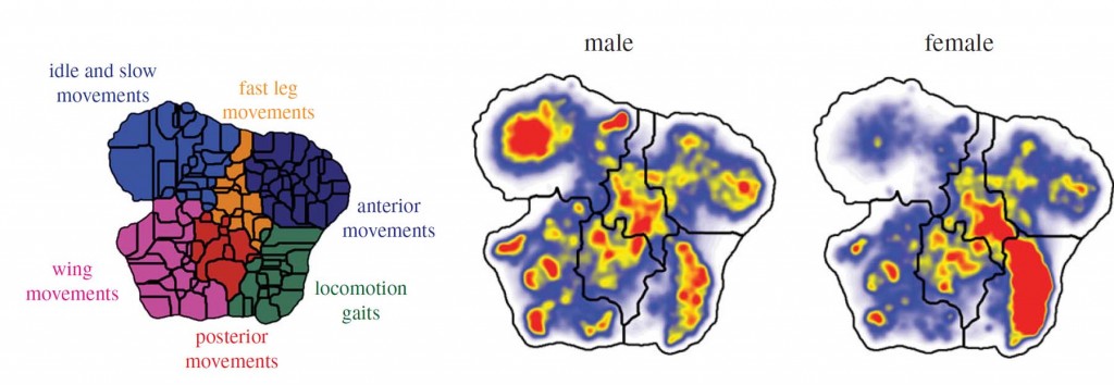
Map that indicates the type of movements of a fly.
Princeton researchers have developed a computerized system for evaluating animal behavior. A video system records an animal, in this case a fruit fly, for one hour. Then a computer program analyzes the video to create a map of the behaviors. The map can reveal, for example, the differences in movements between male and female fruit flies. (Image source: Joshua Shaevitz)

The researchers’ goal was a system that could create accurate records of behaviors for use in studies of the mechanisms behind those behaviors – in other words, the genes and brain circuits that govern movements. Knowing which genes and neural circuits govern behavior will help answer basic scientific questions and could shed light on the mechanisms behind conditions such as autism.

Described in the journal The Royal Society Interface, the system was built for monitoring Drosophila flies, which are commonly used in studies of genes, but the researchers say that the system can also analyze the movements of other creatures, including worms, mice and humans.

The new system is a significant advance over current approaches because it takes note of all the animal’s activities, not just behaviors that seem important to researchers, said Joshua Shaevitz, associate professor of physics and the Lewis-Sigler Institute for Integrative Genomics, who led the study. “We don’t know which behaviors are important to a fruit fly, or a mouse,” he said. “Instead, we ask the computer to find behaviors that are frequently repeated, which tend to be the ones that are worth studying.”

Researchers need reliable ways to catalog behaviors in studies of how genes and neural pathways control behavior. A common experiment involves deleting a gene in an organism such as a fruit fly or worm to look for any resulting loss of function or change in the organism’s behavior. But without reliable behavioral data, researchers cannot make firm conclusions about the role of the deleted gene.

With the new system, a high-speed camera records the animal for a given period of time, and the computer sorts the resulting video frames. For the current study, the researchers recorded individual fruit flies for a period of one hour, resulting in an enormous amount of data. The challenge was how to sort the frames and translate the images into data that could be evaluated.

“We have to figure out how the body parts of the fly are positioned in relation to each other at each point in time, and then find a way to tell how those body parts are moving, while keeping the amount of data manageable using the available computing power,” said Gordon Berman, an associate research scholar in the Lewis-Sigler Institute for Integrative Genomics. “Then you have to project the information into some form that allows you to understand the data.”

The researchers accomplished this goal by writing a computer program that converts the information about a fly’s position and movements into mathematical descriptions that are then grouped according to their similarities. The computer places these groupings on a two-dimensional map, so that different regions of the map represent different types of movements.

A computerized system for evaluating animal behavior.
Individual fruit flies were filmed for about one hour, resulting in over 300,000 video frames. Then, a computer program converts the fly’s position in each frame into mathematical descriptions that represent movements. The computer then groups the movements on a map, so that one region of the map represents leg movements while another represents wing movements, and so on. By looking at the map, the researchers can tell what sorts of movements the fly did, such as grooming its front leg, grooming its head, or waggling its wing. (Image source: Joshua Shaevitz)

To verify that the system worked, the researchers collected data from 50 male and 50 female flies. They identified hundreds of behaviors, including ones that had never been documented and others that were consistent with findings from human observers. For example, some of the findings consistent with previous observations were that male fruit flies kick out their legs when grooming, whereas females don’t, and that young females are more active than young males. “Males and females do things slightly differently, and we can pick up on that right away,” said Shaevitz.

Male fruit flies kick out with their legs when grooming:
http://youtu.be/66__OKybOuI

whereas females do not:
http://youtu.be/FYVslcYSYsk

The team is now expanding the capabilities of the method, Shaevitz said. “The technique that we have developed can also be used to study patterns of behavior in humans,” Shaevitz said. “There are several conditions that are diagnosed on the basis of behavior, such as autism. Having a more quantitative way of measuring behavior could improve the accuracy of diagnoses.”

The study co-authors included William Bialek, Princeton’s John Archibald Wheeler/Battelle Professor in Physics and the Lewis-Sigler Institute for Integrative Genomics, and Daniel Choi, a graduate student working with Shaevitz.

The work was funded through awards from the National Institutes of Health (GM098090, GM071508), the National Science Foundation (PHY 0957573, PHY 1066293), the Pew Charitable Trusts, the Swartz Foundation and the Alfred P. Sloan Foundation.

Read the abstract

Gordon J. Berman, Daniel M. Choi, William Bialek, Joshua W. Shaevitz. Mapping the stereotyped behaviour of freely moving fruit flies. The Journal of the Royal Society Interface (2014). DOI: 10.1098/rsif.2014.0672 Published online 20 August 2014 http://rsif.royalsocietypublishing.org/content/11/99/20140672

Unlocking the potential of bacterial gene clusters to discover new antibiotics (Proc. Natl. Acad. Sci.)

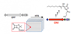
High-throughput screening for the discovery of small molecules that activate silent bacterial gene clusters
High-throughput screening for the discovery of small molecules that activate silent bacterial gene clusters. Image courtesy of Mohammad Seyedsayamdost.

by Tien Nguyen, Department of Chemistry

Resistance to antibiotics has been steadily rising and poses a serious threat to the stronghold of existing treatments. Now, a method from Mohammad Seyedsayamdost, an assistant professor of chemistry at Princeton University, may open the door to the discovery of a host of potential drug candidates.

The vast majority of anti-infectives on the market today are bacterial natural products, made by biosynthetic gene clusters. Genome sequencing of bacteria has revealed that these active gene clusters are outnumbered approximately ten times by so-called silent gene clusters.

“Turning these clusters on would really expand our available chemical space to search for new antibiotic or otherwise therapeutically useful molecules,” Seyedsayamdost said.

In an article published last week in the journal Proceedings of the National Academy of Sciences, Seyedsayamdost reported a strategy to quickly screen whole libraries of compounds to find elicitors, small molecules that can turn on a specific gene cluster. He used a genetic reporter that fluoresces or generates a color when the gene cluster is activated to easily identify positive hits. Using this method, two silent gene clusters were successfully activated and a new metabolite was discovered.

Application of this work promises to uncover new bacterial natural products and provide insights into the regulatory networks that control silent gene clusters.

Read the abstract.

Seyedsayamdost, M. R. “High-throughput platform for the discovery of elicitors of silent bacterial gene clusters.” Proc. Natl. Acad. Sci. 2014, Early edition.

Nano-dissection identifies genes involved in kidney disease (Genome Research)

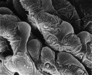
Scanning electron microscope (SEM) micrograph of podocytes
Researchers at Princeton and the University of Michigan have created a computer-based method for separating and identifying genes from diseased kidney cells known as podocytes, pictured above. (Image courtesy of Matthias Kretzler)

By Catherine Zandonella, Office of the Dean for Research

Understanding how genes act in specific tissues is critical to our ability to combat many human diseases, from heart disease to kidney failure to cancer.  Yet isolating individual cell types for study is impossible for most human tissues.

A new method developed by researchers at Princeton University and the University of Michigan called “in silico nano-dissection” uses computers rather than scalpels to separate and identify genes from specific cell types, enabling the systematic study of genes involved in diseases.

The team used the new method to successfully identify genes expressed in cells known as podocytes — the “work-horses” of the kidney — that malfunction in kidney disease. The investigators showed that certain patterns of activity of these genes were correlated with the severity of kidney impairment in patients, and that the computer-based approach was significantly more accurate than existing experimental methods in mice at identifying cell-lineage-specific genes. The study was published in the journal Genome Research.

Using this technique, researchers can now examine the genes from a section of whole tissue, such as a biopsied section of the kidney, for specific signatures associated with certain cell types. By evaluating patterns of gene expression under different conditions in these cells, a computer can use machine-learning techniques to deduce which types of cells are present. The system can then identify which genes are expressed in the cell type in which they are interested.  This information is critical both in defining novel disease biomarkers and in selecting potential new drug targets.

By applying the new method to kidney biopsy samples, the researchers identified at least 136 genes as expressed specifically in podocytes. Two of these genes were experimentally shown to be able to cause kidney disease. The authors also demonstrated that in silico nano-dissection can be used for cells other than those found in the kidney, suggesting that the method is useful for the study of a range of diseases.

The computational method was significantly more accurate than another commonly used technique that involves isolating specific cell types in mice. The nano-dissection method’s accuracy was 65% versus 23% for the mouse method, as evaluated by a time-consuming process known as immunohistochemistry which involves staining each gene of interest to study its expression pattern.

The research was co-led by Olga Troyanskaya, a professor of computer science and the Lewis-Sigler Institute for Integrative Genomics at Princeton, and Matthias Kretzler, a professor of computational medicine and biology at the University of Michigan. The first authors on the study were Wenjun Ju, a research assistant professor at the University of Michigan, and Casey Greene, now at the Geisel School of Medicine at Dartmouth and a former postdoctoral fellow at Princeton.

The research was supported in part by National Institutes of Health (NIH) R01 grant GM071966 to OGT and MK, by NIH grants RO1 HG005998 and DBI0546275 to OGT, by NIH center grant P50 GM071508, and by NIH R01 grant DK079912 and P30 DK081943 to MK. OGT also receives support from the Canadian Institute for Advanced Research.

Read the abstract.

Wenjun Ju, Casey S Greene, Felix Eichinger, Viji Nair, Jeffery B Hodgin, Markus Bitzer, Young-suk Lee, Qian Zhu, Masami Kehata, Min Li, Song Jiang, Maria Pia Rastaldi, Clemens D Cohen, Olga G Troyanskaya and Matthias Kretzler. 2013. Defining cell-type specificity at the transcriptional level in human disease. Genome Research. Published in Advance August 15, 2013, doi: 10.1101/gr.155697.113.

A global post-transcriptional map of the elements that modulate RNA behavior (Nature)

Read the abstract here:
Systematic discovery of structural elements governing stability of mammalian messenger RNAs.
Goodarzi H, Najafabadi HS, Oikonomou P, Greco TM, Fish L, Salavati R, Cristea IM, Tavazoie S. Nature. 2012 Apr 8. doi: 10.1038/nature11013. [Epub ahead of print]