Dopamine neurons have a role in movement, new study finds (Nature Neuroscience)

Dopamine neurons. Image credit: Witten et al., Nature Neuroscience

By Catherine Zandonella, Office of the Dean for Research

Princeton University researchers have found that dopamine – a brain chemical involved in learning, motivation and many other functions – also has a direct role in representing or encoding movement. The finding could help researchers better understand dopamine’s role in movement-related disorders such as Parkinson’s disease.

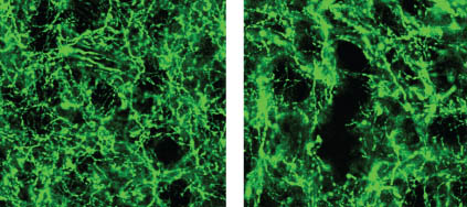
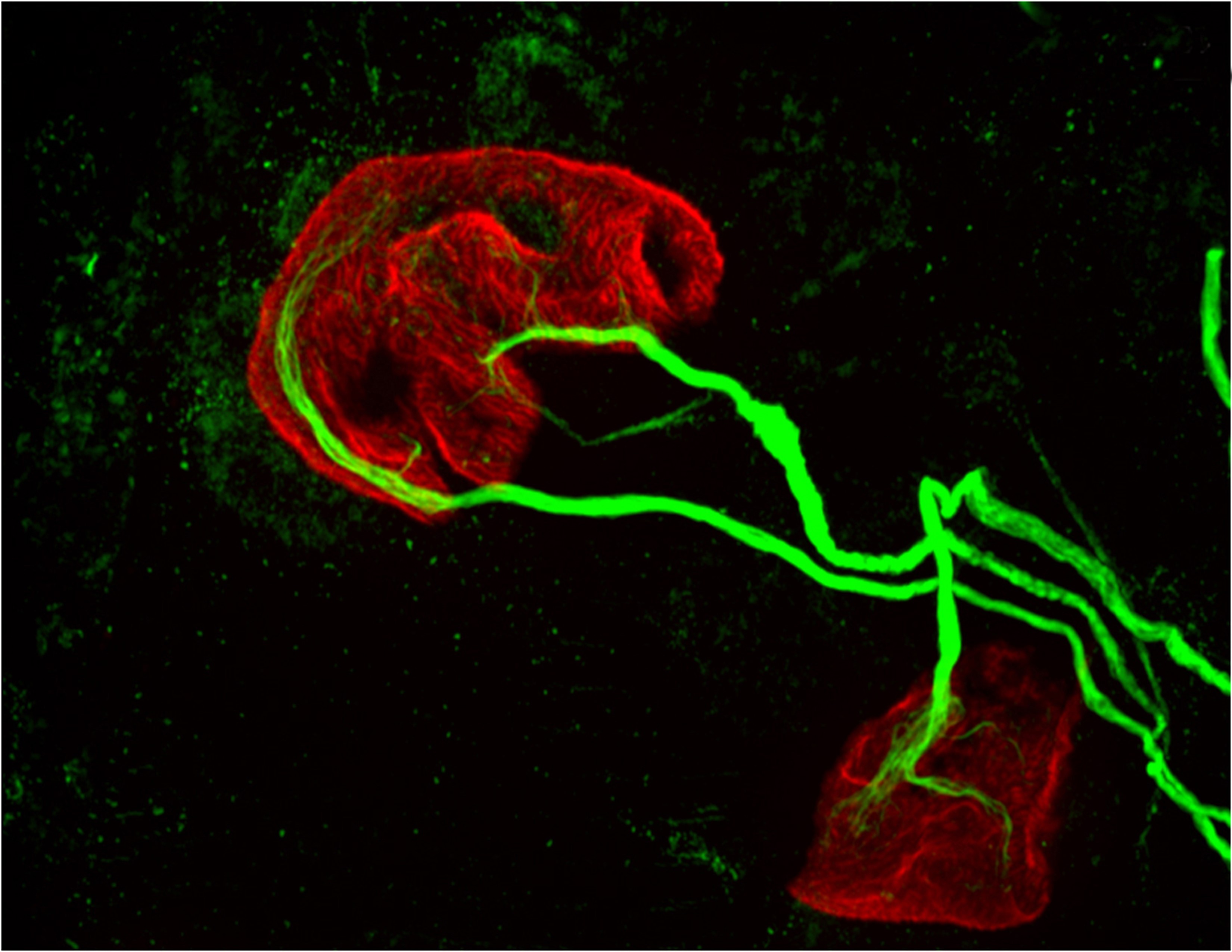
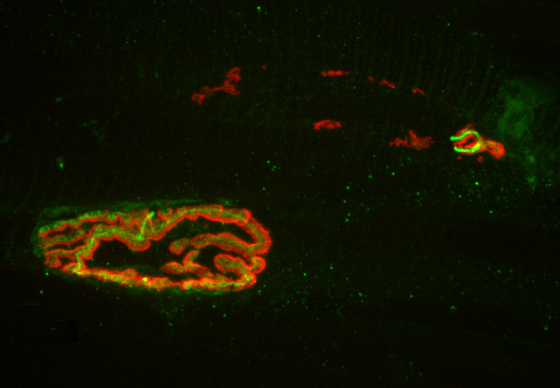
The researchers used a new, more precise technique to record the activity of dopamine neurons at two regions within a part of the brain known as the striatum, which oversees action planning, motivation and reward perception. The researchers found that while all of the neurons carried signals needed to learn and plan movement, one of the nerve bundles, the one that went to the region called the dorsomedial striatum, also carried a signal that could be used to control movements.

The work was published online in the journal Nature Neuroscience this week.

“What we learned from this study is that dopamine neurons that go to one part of the brain act differently than dopamine neurons that go to another part of the brain,” said Ilana Witten, assistant professor of psychology and the Princeton Neuroscience Institute. “This is contrary to what has been the mainstream view of dopamine neurons.”

The research may shed light on how Parkinson’s disease, which involves the destruction of dopamine neurons in the dorsomedial striatum, deprives patients of the ability to move. Previous studies have failed to find a direct link between dopamine neuron activity and the control of movement or actions. Instead, the mainstream view suggested an indirect role for dopamine: the neurons make it possible for us to learn which actions are likely to lead to a rewarding experience, which in turn enables us to plan to take that action. When dopamine neurons are destroyed, the individual cannot learn to plan actions and thus cannot move.

The new study affirmed the role of dopamine in reward-based learning, but also found that in the dorsomedial striatum, dopamine neurons can play a direct role in movement. The researchers used a method for measuring neuron activity at very precise locations in the brain. They measured the activity at the ends of neurons – the terminals where dopamine is released into the junction, or synapse, between two cells – in two locations in the striatum: the nucleus accumbens, known to be involved in processing reward, and the dorsomedial striatum, known for evaluating and generating actions.

Until recently, it has been difficult to measure dopamine neuron activity in these regions due to the small size of the regions and the fact that there are many other neurons present that are delivering other brain chemicals, or neurotransmitters, to the same areas of the brain.

To restrict their measurements to only dopamine-carrying neurons, the researchers used mice whose brains carry genetically altered cells that glow green when active. The mice also contain a second gene that ensured that the glowing could only occur when dopamine was present.

The researchers then recorded neuron activity from either the nucleus accumbens or the dorsomedial striatum by inserting a very thin optical fiber into each region to record the fluorescing dopamine cells in only the desired regions.

Once the ability to measure neuron activity was in place, the researchers gave the mice a task that involved both reward-based learning as well as movement.

The task involved presenting the mice with two levers, one of which, when pressed, gave a drink of sweetened water. Through trial and error, the mice learned which lever would give the reward. During the task, the researchers recorded their brain activity.

The task is analogous to playing slot machines at a casino. Picture yourself at a casino with two slot machines in front of you. You pull the lever on the machine to your left and it spits out some coins. Your brain learns that the left lever leads to a reward, so you plan and execute an action: you pull the left lever again. After a few more pulls on the left lever without a reward, you switch to the machine on the right.

When an action is rewarding, you are likely to remember it, an important step in learning. The difference between how much reward you expect, and how much you get, is also important, because it tells you whether or not something is new and how much you should pay attention to it. Researchers call this gap between your predicted reward and the reward you actually get the “reward-prediction error” and consider it an important teaching signal.

By matching the mice’s actions to the dopamine activity in their brains during these tasks, the researchers could determine which parts of the brain were active during reward-based learning, and which parts were active when choosing to press a lever. Assistance with computational modeling of the mice’s behaviors was provided by Nathaniel Daw, a professor of the Princeton Neuroscience Institute and Psychology.

The researchers found that the dopamine neurons that innervate the nucleus accumbens and the dorsomedial striatum did indeed encode reward-prediction cues, which is consistent with previous findings. But they also found that in the dorsomedial striatum, the dopamine neurons carried information about what actions the animal is going to take.

“This idea was that dopamine neurons carry this reward-prediction error signal, and that could indirectly affect movement or actions, because if you don’t have this, you won’t correctly learn which actions to perform,” Witten said. “We show that while this is true, it is certainly not the whole story. There is also a layer where dopamine is directly coding movement or actions.”

Nathan Parker, a graduate student in the Witten lab who designed and conducted the experiments and is first author on the paper, added that new findings were made possible both by the improvements in recording of neurons and by the experimental design, which gave researchers a detailed evaluation of neuron activity during a relatively complex task.

Additional research assistance was provided by Princeton postdoctoral research associates Courtney Cameron and Junuk Lee, and graduate student Jung Yoon Choi. Research Specialist Joshua Taliaferro, Class of 2015, begin working on the project as part of his senior thesis. The study also involved contributions from Thomas Davidson, a postdoctoral researcher at Stanford University.

The study also addresses the general question of how dopamine can be involved in so many functions in the brain, Witten said. “We think that some of the way that dopaminergic neurons achieve such diverse functions in the brain is by having specific roles based on their anatomical target.”

Naoshige Uchida, a professor of molecular and cellular biology at Harvard University who was not involved in the study, said the results challenge long-held views and open up new directions of research. “This study by the Witten lab elegantly shows that the activity of some dopamine neurons is modulated by the direction of motion,” Uchida said. “More importantly, they found some of the clearest evidence indicating the heterogeneity of dopamine neurons: A specific population of dopamine neurons projecting to the dorsomedial striatum encodes movement direction more so compared to another population projecting to the ventral striatum.”

Uchida continued, “A similar phenomenon has also been reported in an independent study in non-human primates (Kim, et al., Cell, 2015), suggesting that the Witten lab finding is more universal and not specific to mice. This is particularly important because dopamine has been implicated in Parkinson’s disease but how dopamine regulates movement remains a large mystery.”

Funding for the study was provided by the Pew Charitable Trusts, the McKnight Foundation, the Brain & Behavior Research (NARSAD) Foundation, the Alfred P. Sloan Foundation, the National Institutes of Health, the National Science Foundation, and Princeton’s Stuart M. Essig ’83 and Erin S. Enright ’82 Fund for Innovation in Engineering and Neuroscience.

Read the abstract.

The study, “Reward and choice encoding in terminals of midbrain dopamine neurons depends on striatal target,” by Nathan Parker, Courtney Cameron, Joshua Taliaferro, Junuk Lee, Jung Yoon Choi, Thomas Davidson, Nathaniel Daw and Ilana Witten, was published online in the journal Nature Neuroscience (Advance Online Publication, http://dx.doi.org/10.1038/nn.4287).

Chemical tracers reveal oxygen-dependent switch in cellular pathway to fat (Nature Chemical Biology)

By Tien Nguyen, Department of Chemistry

Using tracer compounds, scientists have been able to track the cellular production of NADPH, a key coenzyme for making fat, through a pathway that has never been measured directly before.

By tracking this pathway, known as malic enzyme metabolism, which is one of a few recognized routes to make NADPH, researchers from Rabinowitz lab discovered a novel switch in the way fat cells make NADPH depending on the presence of oxygen. The findings were published in Nature Chemical Biology.

Ling Liu (left) and Joshua Rabinowitz (right)

“No one had ever shown an environmental dependent switch in any NADPH production pathway,” said Joshua Rabinowitz, Professor of Chemistry and the Lewis-Sigler Institute for Integrative Genomics at Princeton and principal investigator of the work. “No one had the tools to look,” he said.

NADPH is critical to not only fat synthesis, but also protein and DNA synthesis, and antioxidant defense, implicating it in many diseases such as cancer and diabetes. By understanding and monitoring the pathways through which NADPH is made, scientists can work towards influencing these processes using therapeutic compounds.

The Rabinowitz lab first applied their tracer method in 2014 to study the most well known NADPH production pathway, the oxidative pentose phosphate pathway (oxPPP). The method relied on compounds labeled with deuterium atoms, hydrogen’s heavier cousin, which can be deployed in the cell and measured by a technique called mass spectrometry.

In this work, the researchers extended their method to probe the lesser-known malic enzyme pathway by developing two new, orthogonal tracer compounds specific to this pathway. One tracer, a deuterated succinate compound, enters the cycle more directly but is somewhat challenging for the cell to uptake, while the other, a deuterated glucose molecule, is taken up by the cell readily but takes an extra step to enter the pathway.

The research team investigated the malic enzyme pathway under various concentrations of oxygen. Low oxygen environments, which are found in fat cells in obesity, are of particular clinical interest. They found that in a low oxygen environment, the oxidative pentose phosphate pathway produced more NADPH than the malic enzyme pathway, but in a higher oxygen environment, the pathway contributions completely flipped.

“It’s like the cells are quite clever. They choose the pathway depending on what they want to make, and what nutrients they can access,” said Ling Liu, a graduate student in the Rabinowitz lab and lead author on the work.

One advantage of this method is that it tracks NADPH made specifically in the cytosolic compartment of the cell, whereas the previous leading technique, which relied on tracer compounds with carbon-13 atoms, is unable to differentiate between malic enzyme activity in the cytosol and mitochondria.

NADPH involvement in essential cellular processes has a direct impact on diseases such as diabetes, obesity and cancer. “All of these central biomedical questions depend on an understanding of NADPH pathways, and if you can’t quantify how a metabolite is made and used, you can’t understand what’s going on,” Rabinowitz said. “Ultimately, we’re trying to understand the fundamental chemistry that’s leading to these important biological outcomes,” he said.

Read the full article or abstract:

Liu, L.; Shah, S.; Fan, J.; Park, J. O.; Wellen, K. E.; Rabinowitz, J. D. “Malic enzyme tracers reveal hypoxia-induced switch in adipocyte NADPH pathway usage.Nat. Chem. Bio. Published online March 21, 2016.

This work was supported by the US National Institutes of Health grants R01CA163591, R01AI097382 and P30DK019525 (to the University of Pennsylvania Diabetes Research Center).

Electrons slide through the hourglass on surface of bizarre material (Nature)

An illustration of the hourglass fermion predicted to lie on the surface of crystals of potassium mercury antimony. (Bernevig et al., Princeton University)
An illustration of the hourglass fermion predicted to lie on the surface of crystals of potassium mercury antimony. (Image credit: Laura R. Park and Aris Alexandradinata)

By Staff

A team of researchers at Princeton University has predicted the existence of a new state of matter in which current flows only through a set of surface channels that resemble an hourglass. These channels are created through the action of a newly theorized particle, dubbed the “hourglass fermion,” which arises due to a special property of the material. The tuning of this property can sequentially create and destroy the hourglass fermions, suggesting a range of potential applications such as efficient transistor switching.

In an article published in the journal Nature this week, the researchers theorize the existence of these hourglass fermions in crystals made of potassium and mercury combined with either antimony, arsenic or bismuth. The crystals are insulators in their interiors and on their top and bottom surfaces, but perfect conductors on two of their sides where the fermions create hourglass-shaped channels that enable electrons to flow.

The research was performed by Princeton University postdoctoral researcher Zhi Jun Wang and former graduate student Aris Alexandradinata, now a postdoctoral researcher at Yale University, working with Robert Cava, Princeton’s Russell Wellman Moore Professor of Chemistry, and Associate Professor of Physics B. Andrei Bernevig.

The new hourglass fermion exists – theoretically for now, until detected experimentally – in a family of materials broadly called topological insulators, which were first observed experimentally in the mid-2000s and have since become one of the most active and interesting branches of quantum physics research. The bulk, or interior, acts as an insulator, which means it prohibits the travel of electrons, but the surface of the material is conducting, allowing electrons to travel through a set of channels created by particles known as Dirac fermions.

Fermions are a family of subatomic particles that include electrons, protons and neutrons, but they also appear in nature in many lesser known forms such as the massless Dirac, Majorana and Weyl fermions. After years of searching for these particles in high-energy accelerators and other large-scale experiments, researchers found that they can detect these elusive fermions in table-top laboratory experiments on crystals. Over the past few years, researchers have used these “condensed matter” systems to first predict and then confirm the existence of Majorana and Weyl fermions in a wide array of materials.

The next frontier in condensed matter physics is the discovery of particles that can exist in the so-called “material universe” inside crystals but not in the universe at large. Such particles come about due to the properties of the materials but cannot exist outside the crystal the way other subatomic particles do. Classifying and discovering all the possible particles that can exist in the material universe is just beginning. The work reported by the Princeton team lays the foundations of one of the most interesting of these systems, according to the researchers.

In the current study, the researchers theorize that the laws of physics prohibit current from flowing in the crystal’s bulk and top and bottom surfaces, but permit electron flow in completely different ways on the side surfaces through the hourglass-shaped channels. This type of channel, known more precisely as a dispersion, was completely unknown before.

The researchers then asked whether this dispersion is a generic feature found in certain materials or just a fluke arising from a specific crystal model.

It turned out to be no fluke.

A long-standing collaboration with Cava, a material science expert, enabled Bernevig, Wang, and Alexandradinata to uncover more materials exhibiting this remarkable behavior.

“Our hourglass fermion is curiously movable but unremovable,” said Bernevig. “It is impossible to remove the hourglass channel from the surface of the crystal.”

Bernevig explained that this robust property arises from the intertwining of spatial symmetries, which are characteristics of the crystal structure, with the modern band theory of crystals. Spatial symmetries in crystals are distinguished by whether a crystal can be rotated or otherwise moved without altering its basic character.

In a paper published in Physical Review X this week to coincide with the Nature paper, the team detailed the theory behind how the crystal structure leads to the existence of the hourglass fermion.

An illustration of the complicated dispersion of the surface fermion arising from a background of mercury and bismuth atoms (blue and red). (Image credit: Mingyee Tsang and Aris Alexandradinata)

“Our work demonstrates how this basic geometric property gives rise to a new topology in band insulators,” Alexandradinata said. The hourglass is a robust consequence of spatial symmetries that translate the origin by a fraction of the lattice period, he explained. “Surface bands connect one hourglass to the next in an unbreakable zigzag pattern,” he said.

The team found esoteric connections between their system and high-level mathematics. Origin-translating symmetries, also called non-symmorphic symmetries, are described by a field of mathematics called cohomology, which classifies all the possible crystal symmetries in nature. For example, cohomology gives the answer to how many crystal types exist in three spatial dimensions: 230.

In the cohomological perspective, there are 230 ways to combine origin-preserving symmetries with real-space translations, known as the “space groups.” The theoretical framework to understand the crystals in the current study requires a cohomological description with momentum-space translations.

“The hourglass theory is the first of its kind that describes time-reversal-symmetric crystals, and moreover, the crystals in our study are the first topological material class which relies on origin-translating symmetries,” added Wang.

Out of the 230 space groups in which materials can exist in nature, 157 are non-symmorphic, meaning they can potentially host interesting electronic behavior such as the hourglass fermion.

“The exploration of the behavior of these interesting fermions, their mathematical description, and the materials where they can be observed, is poised to create an onslaught of activity in quantum, solid state and material physics,” Cava said. “We are just at the beginning.”

The study was funded by the National Science Foundation, the Office of Naval Research, the David and Lucile Packard Foundation, the W. M. Keck Foundation, and the Eric and Wendy Schmidt Transformative Technology Fund at Princeton University.

The paper, “Hourglass fermions” by Zhijun Wang, A. Alexandradinata, R. J. Cava and B. Andrei Bernevig, was published in the April 14, 2016 issue of Nature, 532,189–194, doi:10.1038/nature17410. Read the preprint.

The paper, “Topological insulators from group cohomology” by A. Alexandradinata, Zhijun Wang, and B. Andrei Bernevig, was published in the April 15, 2016 issue of Phys. Rev. X 6, 021008.

Same immune-system proteins may first giveth, then taketh away motor control (Brain, Behavior, and Immunity)

two motor neurons (green) connected to a single muscle fiber (red)
Princeton University researchers found that proteins in the MHCI, or major histocompatibility complex class I, family can “prune” the connections, or synapses, between motor neurons and muscle fibers, which is necessary during early development. But the researchers also found that MHCI levels can rise again in old age, and that the proteins may resume pruning nerve-muscle synapses. This image from a mouse bred to express less MHCI shows two motor neurons (green) connected to a single muscle fiber (red) at an age when only one connection should remain. (Image by Lisa Boulanger, Princeton Neuroscience Institute, and Mazell Tetruashvily, Department of Molecular Biology)

By Morgan Kelly, Office of Communications

Princeton University researchers have found that a family of proteins with important roles in the immune system may be responsible for fine-tuning a person’s motor control as they grow — and for their gradual loss of muscle function as they age. The research potentially reveals a biological cause of weakness and instability in older people, as well as a possible future treatment that would target the proteins specifically.

The researchers reported in the journal Brain, Behavior, and Immunity that proteins in the family MHCI, or major histocompatibility complex class I, “prune” the connections, or synapses, between motor neurons and muscle fibers. Pruning is necessary during early development because at birth each muscle fiber in humans, mice and other vertebrates receives signals from dozens of neural connections. Proper motor control, however, requires that each muscle fiber receive signals from only a single motor neuron, so without the pruning carried out by MHCI proteins, fine motor control would never emerge.

But the researchers also found that MHCI levels can rise again in old age, and that the proteins may resume pruning nerve-muscle synapses — except that in a mature organism there are no extra synapses. The result is that individual muscle fibers become completely “denervated,” or detached from nervous system control. Denervated muscle fibers cannot be recruited during muscle contraction, which can leave older people weaker and more susceptible to devastating falls, making independent living difficult.

However, the Princeton researchers discovered that when MHCI levels were reduced in mice, denervation during aging was largely prevented. These findings could help scientists identify and treat the neurological causes of denervation and muscle weakness in the elderly.

Corresponding author Lisa Boulanger, an assistant professor in the Princeton Neuroscience Institute, explained that in infants, motor neurons initially make far too many connections to muscle fibers, which is part of why infants lack fine motor control.  Synapse overproduction followed by pruning occurs in many different regions of the vertebrate nervous system, and the neuromuscular junction has often been used as a model for studying this process.

It is not known why more synapses are made during development than are needed.

One possibility is that it allows the wiring diagram of the nervous system to be precisely tuned based on the way the circuit is used, Boulanger said. MHCI proteins help limit the final number of connections so that communication between neurons and muscles is more precise and efficient than would be possible using just a molecular code that produced a set number of connections.

“Molecules might get you to the right zip code, but pruning can make sure you arrive at the right house,” Boulanger said. “During development, it’s essential to get rid of extra synapses. But when you up-regulate MHCI when you’re older and start pruning synapses again, but you don’t have any extras to replace them.”

Boulanger worked with first author Mazell Tetruashvily, who received her doctorate in molecular biology from Princeton in 2015 and is now completing her M.D. training at University of Medicine and Dentistry of New Jersey; Marin McDonald, who received her doctorate in neuroscience from the University of California-San Diego (UCSD) in 2010, and is now a radiology resident at UCSD; and Karla Frietze, a doctoral student in Princeton’s Department of Molecular Biology. Boulanger was on the UCSD faculty before moving her lab to Princeton in 2009.

In the immune system, MHCI proteins present protein fragments, or peptides, to T cells, which are white blood cells with a central role in the body’s response to infection. This peptide presentation allows T cells to recognize and kill infected and cancerous cells, which present abnormal or foreign peptides on their MHCI proteins. It is unknown if the proteins’ ability to help recognize and destroy infected or cancerous cells is mechanistically related to the proteins’ ability to help eliminate excess synapses that the Princeton researchers discovered.

In the nervous system, MHCI proteins stop pruning synapses early in life. Why they may resume their synapse-eliminating activity later in life is unknown, Boulanger said. As immune-system proteins, MHCI levels increase with inflammation, she said. Aging is associated with chronic inflammation, which could explain the observed increase in MHCI levels and the reactivation of its former role.

Neuron image
The researchers found that MHCI the proteins may resume pruning nerve-muscle synapses in older organisms, an age when there are no extra synapses. Instead, muscle fibers become completely “denervated,” or detached from nervous system control, which could lead to weakness and instability in older people. This image from a 2-year-old (elderly) normal mouse shows denervation in the upper-right synapse, as noted by the lack of overlap of the red and green fluorescent markers used to indicate cells where the neuron and muscle fiber meet. At lower left, a healthy-looking synapse displays good overlap. (Image by Lisa Boulanger, Department of Molecular Biology)

The Princeton researchers found that mice bred to express less MHCI proteins had “more youthful” patterns of muscle innervation, since they were protected from denervation as they aged, Boulanger said. The mice actually lacked a protein known as beta-2 microglobulin, which forms a complex with MHCI and is necessary for MHCI expression on the surface of cells. This could be beneficial from a clinical perspective because beta-2 microglobulin is a soluble protein and can be removed from the blood, Boulanger said.

“If a rise in MHCI is the problem, having less beta-2 microglobulin might be protective,” Boulanger said. Recent results from a lab at Stanford University showed that reducing beta-2 microglobulin also helped with cognitive aging because of its effects on MHCI proteins. “Our studies raise the possibility that targeting one protein could help with both motor and cognitive aspects of aging,” Boulanger said.

Because MHCI proteins are important in the immune system, however, such an approach could result in compromised immunity, Boulanger said. The mice bred to not express beta-2 microglobulin had weakened immune systems, as a result of their lower levels of MHCI proteins. Future work will include exploring the effectiveness of other approaches to reducing the proteins’ synapse-eliminating activity in older nervous systems, ideally while leaving their immune functions intact, she said.

The research was supported by the Princeton Department of Molecular Biology and the Princeton Neuroscience Institute (grant no. 1F30AG046044-01A1), the UCSD School of Medicine, the Alfred P. Sloan Foundation, the Whitehall Foundation, and the Princeton Neuroscience Institute Innovation Fund.

Read more.

Mazell M. Tetruashvily, Marin A. McDonald, Karla K. Frietze and Lisa M. Boulanger. “MHCI promotes developmental synapse elimination and aging-related synapse loss at the vertebrate neuromuscular junction.” Brain, Behavior, and Immunity, in press. DOI: 10.1016/j.bbi.2016.01.008epartment of Molecular Biology)

Researchers propose surveillance system for Zika virus and other infectious diseases (The Lancet)

Test tube image courtesy of the NIH
Image courtesy of the National Institutes of Health

By Catherine Zandonella, Office of the Dean for Research

A group of prominent researchers from seven institutions including Princeton University are calling for the establishment of a worldwide program to collect and test blood and other human bodily fluids to aid in the study and prevention of emerging infectious diseases such as the mosquito-borne Zika fever, which is caused by the Zika virus and has spread throughout Latin America and the Caribbean since early 2015.

In an article published in The Lancet April 5, the authors call for the creation of a World Serology Bank that would include storage repositories located around the world that use a common set of best practices to collect and evaluate blood and other bodily fluids from people infected by various pathogens. The resulting data would help scientists better understand the susceptibility of humans to emerging diseases such as Zika fever. The information could be shared widely among scientists who track disease.

The authors include Princeton’s C. Jessica Metcalf, an assistant professor of Ecology and Evolutionary Biology and Public Affairs in the Woodrow Wilson School for Public and International Affairs, and Bryan Grenfell, the Kathryn Briger and Sarah Fenton Professor of Ecology and Evolutionary Biology and Public Affairs. In the interview below, Metcalf explains more about the World Serology Bank proposal.

Why is it important to create a World Serology Bank?

Serology is the study of bodily fluids including serum, the part of the blood that contains antibodies, with the aim of detecting the body’s immune response to pathogens. Serology provides us with the clearest window we have onto the landscape of susceptibility to pathogens across human populations, and the consequent risk of outbreaks. A World Serology Bank would shed light on the global risk of infectious disease outbreaks, and would be of tremendous public health benefit.

Why do you feel it is important to address this issue now?

With the emergence of pathogens like Zika virus, it becomes ever more important to understand what enhances or limits the global spread of these pathogens, and what the consequences of such spread may be across our pathogen communities. A World Serology Bank would provide a powerful mechanism toward such a global perspective.

What are the challenges involved in creating the Bank?

Challenges range from developing systems for collecting fluids, which can be done on a regular schedule or during specific disease events, to methods for sera storage and sera testing. Other challenges include defining who will administer the World Serology Bank, and what global data-sharing agreements will be put in place. Finally, we will need to develop new methods to translate what we learn from the evaluation of sera, such as patterns of susceptibility to specific pathogens, or protection from those pathogens. These methods will be driven by the underlying biology, and are likely to require an array of analytical innovations.

Read more

The article, “Use of serological surveys to generate key insights into the changing global landscape of infectious disease,” by C. Jessica E Metcalf, Jeremy Farrar, Felicity T. Cutts, Nicole E. Basta, Andrea L. Graham, Justin Lessler, Neil M. Ferguson, Donald S. Burke and Bryan T. Grenfell was published online in the journal The Lancet on April 5, 2016. http://dx.doi.org/10.1016/S0140-6736(16)30164-7.