Compressing turbulence to improve inertial confinement fusion experiments (PRL)

Compression of a turbulent plasma. Credit: Seth Davidovits

By John Greenwald, Princeton Plasma Physics Laboratory

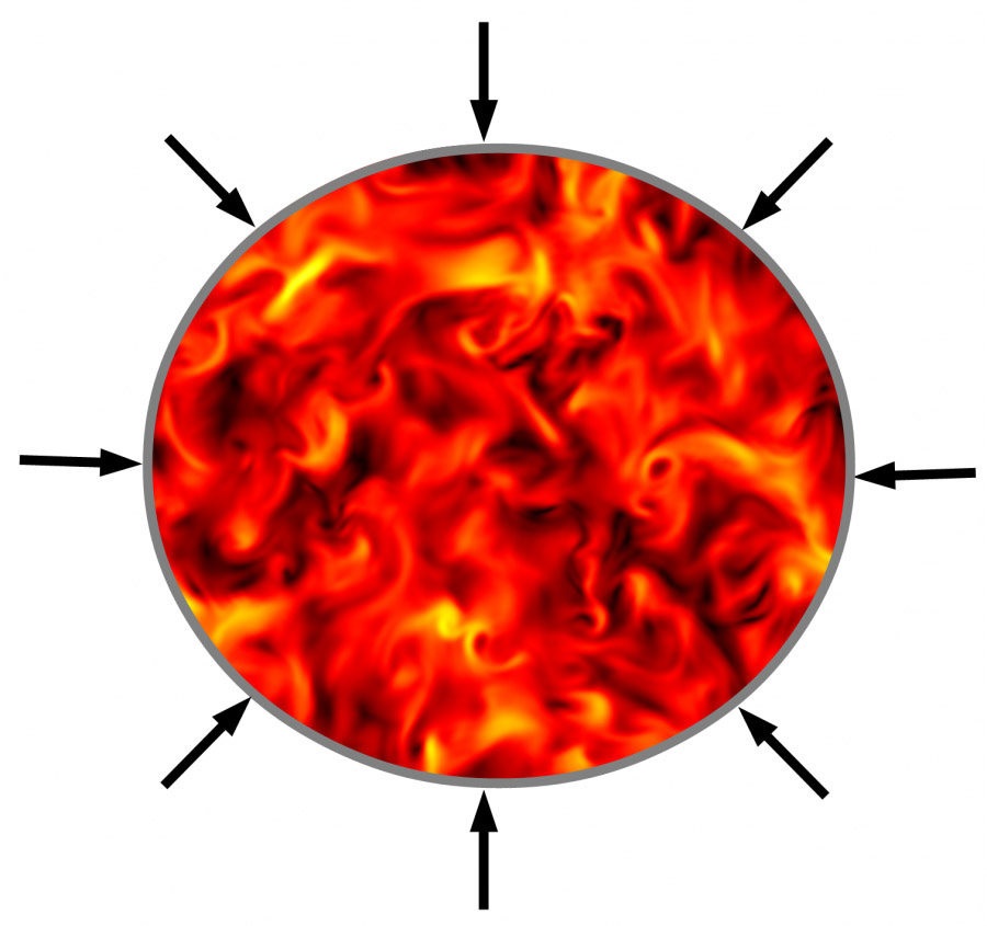
Physicists have long regarded plasma turbulence as unruly behavior that can limit the performance of fusion experiments. But new findings by researchers associated with the U.S. Department of Energy’s (DOE) Princeton Plasma Physics Laboratory (PPPL) and the Department of Astrophysical Sciences at Princeton University indicate that turbulent swirls of plasma could benefit one of the two major branches of such research.   The editors of Physical Review Letters highlighted these findings — a distinction given to one of every six papers per issue — when they published the results last week on March 11.

Lead author Seth Davidovits, a Princeton University graduate student, and Professor of Astrophysical Sciences Nathaniel Fisch, his thesis adviser and Associate Director for Academic Affairs at PPPL, produced the findings. They modeled the compression of fluid turbulence, showing effects that suggested a surprising positive impact of turbulence on inertial confinement fusion (ICF) experiments.

Stimulating this work were experiments conducted by Professor Yitzhak Maron at the Weizmann Institute of Science in Israel. Those experiments, on a Z-pinch inertial confinement machine, showed turbulence that contained a surprising amount of energy, which caught Fisch’s attention during a recent sabbatical at Weizmann.

In a Z-pinch and other inertial confinement (ICF) machines, plasma is compressed to create fusion energy. The method contrasts with the research done at PPPL and other laboratories, which controls plasma with magnetic fields and heats it to fusion temperatures in doughnut-shaped devices called tokamaks. The largest Z-pinch device in the United States is at the DOE’s Sandia National Laboratory. Other inertial confinement approaches are pursued at, among other places, the DOE’s Lawrence Livermore National Laboratory.

Present ICF approaches use compression to steadily heat the plasma. Methods range from squeezing plasma with magnetic fields at Sandia to firing lasers at capsules filled with plasma at Livermore’s National Ignition Facility. The presence of turbulence in the plasma is widely thought to increase the difficulty of achieving fusion.

But there could be advantages to turbulence if handled properly, the authors point out, since energy contained in turbulence does not radiate away. This compares with hotter plasmas in which heat radiates away quickly, making fusion harder to achieve. By storing the energy of the compression in turbulence rather than temperature, the authors suppress the energy lost to radiation during the compression.

The turbulent energy also does not immediately lead to fusion, which requires high temperature. This means a mechanism is needed to change the turbulence into the temperature required for fusion once the plasma has been compressed.

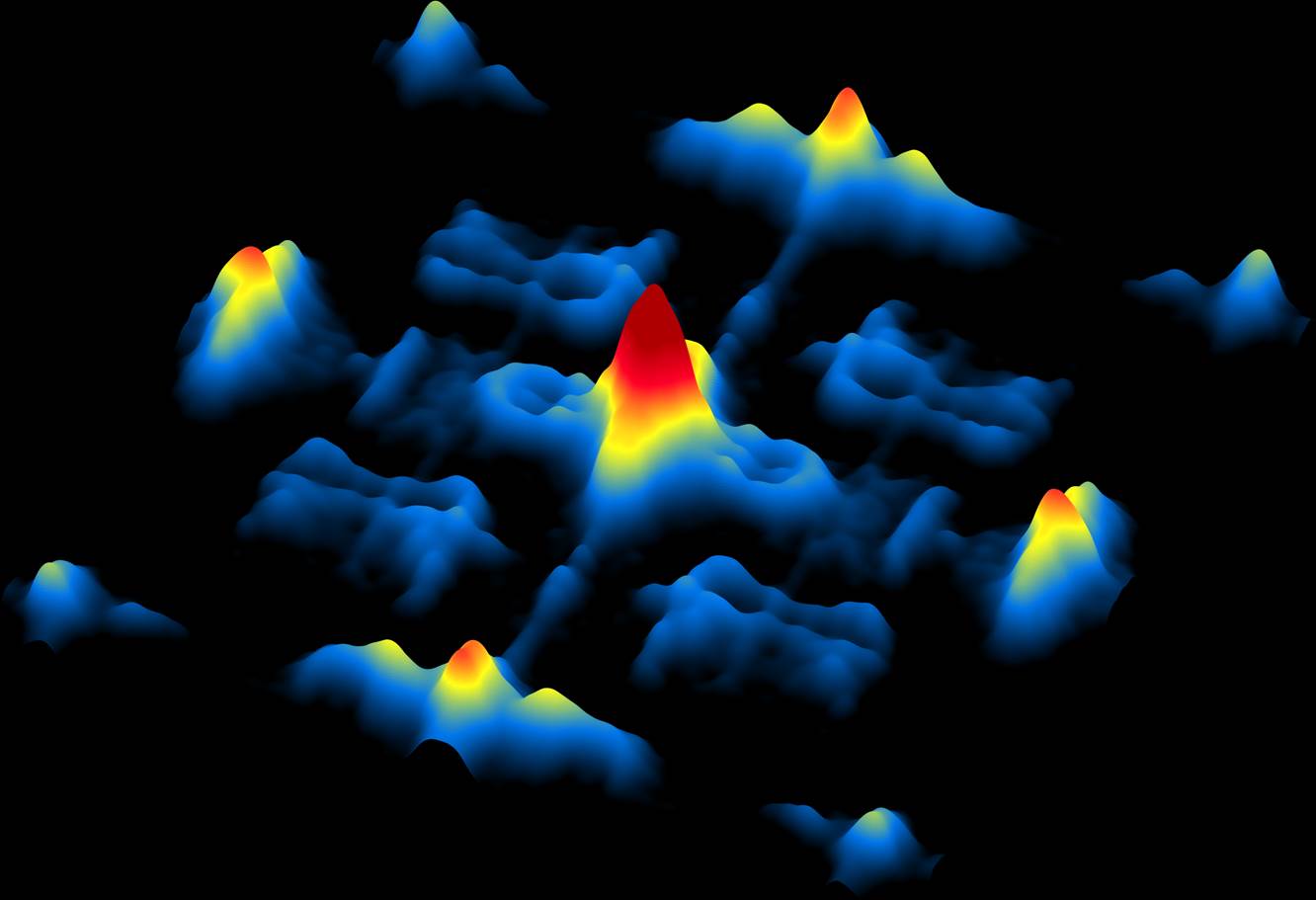
Davidovits used a software code called Dedalus to show that turbulent energy is increased during the compression, but then suddenly transformed into heat. As external forces in his simulation compress the turbulence to increase the energy stored within it, they also gradually raise the temperature and viscosity of the plasma. The viscosity, which describes how “thick” or resistant to flow a fluid is, acts to slow the turbulence and convert its energy to temperature. The viscosity started small so that the turbulence was initially unhindered. The rapid compression then kept the viscosity growing until it suddenly catalyzed the transfer of energy from the turbulence to the temperature.

In an experiment, this process would create the conditions for nuclear fusion in a plasma composed of the hydrogen isotopes deuterium and tritium. “This suggests a fundamentally different design for compression-based fusion experiments,” Davidovits said, “and a new paradigm for the inertial technique of producing fusion energy.”

He warns, however, that the simulation includes caveats that could diminish the findings. For example, the model doesn’t consider any possible interaction between the plasma and the containing capsule, and highly energetic turbulence might mix parts of the capsule into the plasma and contaminate the fusion fuel.

Nonetheless, the authors call the rapid transfer of turbulent energy into temperature during ICF experiments a “tantalizing” prospect that could benefit such research. And they note that their findings could lead to new understanding of the evolution of the relationship between the pressure, volume and temperature of a gas that is substantially turbulent. Determining this will be quite challenging, they say, “but the understanding will be important not only for the new fusion approach, but also for many situations involving the behavior of low viscosity compressible fluids and gases.”

This research was initiated through a grant by the Defense Threat Reduction Agency, a unit of the U.S. Department of Defense, and has been supported also by the DOE’s National Nuclear Security Administration through a consortium with Cornell University. Recently, the National Science Foundation and the Israel Binational Science Foundation combined funding opportunities to ensure further experiments at Weizmann on this topic and continued collaboration with the Princeton researchers.

PPPL, on Princeton University’s Forrestal Campus in Plainsboro, N.J., is devoted to creating new knowledge about the physics of plasmas — ultra-hot, charged gases — and to developing practical solutions for the creation of fusion energy. Results of PPPL research have ranged from a portable nuclear materials detector for anti-terrorist use to universally employed computer codes for analyzing and predicting the outcome of fusion experiments. The Laboratory is managed by the University for the U.S. Department of Energy’s Office of Science, which is the largest single supporter of basic research in the physical sciences in the United States, and is working to address some of the most pressing challenges of our time. For more information, please visit science.energy.gov.

Read the abstract or paper here.

S. Davidovits, N. Fisch, Sudden Viscous Dissipation of Compressing Turbulence. Phys. Rev. Lett. 116, 105004 – Published 11 March 2016.

This work was supported by DOE through Contracts No. DE-AC02-09CH1-1466 and NNSA 67350-9960 (Prime No. DOE DE-NA0001836), by DTRA HDTRA1-11-1-0037, and by NSF Contract No. PHY-1506122.


Down the rabbit hole: how electrons travel through exotic new material (Science)

Electrons on the surface of a Weyl semi-metal
Three-dimensional image using scanning tunneling electron microscopy of electrons on the surface of a Weyl semi-metal, a kind of crystal with unusual conducting and insulating properties. Image credit: Yazdani et al, Princeton University.

By Catherine Zandonella, Office of the Dean for Research

Researchers at Princeton University have observed a bizarre behavior in a strange new crystal that could hold the key for future electronic technologies. Unlike most materials in which electrons travel on the surface, in these new materials the electrons sink into the depths of the crystal through special conductive channels.

“It is like these electrons go down a rabbit hole and show up on the opposite surface,” said Ali Yazdani, the Class of 1909 Professor of Physics. “You don’t find anything else like this in other materials.”

The research was published in the journal Science.

Tantalum arsenide
Crystal structure of tantalum arsenide (TaAs), with Ta in blue and As in green. Image credit: Yazdani et al., Princeton University.

Yazdani and his colleagues discovered the odd behavior while studying electrons in a crystal made of layers of tantalum and arsenic. The material, called a Weyl semi-metal, behaves both like a metal, which conducts electrons, and an insulator, which blocks them. A better understanding of these and other “topological” materials someday could lead to new, faster electronic devices.

The team’s experimental results suggest that the surface electrons plunge into the crystal only when traveling at a certain speed and direction of travel called the Weyl momentum, said Yazdani. “It is as if you have an electron on one surface, and it is cruising along, and when it hits some special value of momentum, it sinks into the crystal and appears on the opposite surface,” he said.

These special values of momentum, also called Weyl points, can be thought of as portals where the electrons can depart from the surface and be conducted to the opposing surface. The theory predicts that the points come in pairs, so that a departing electron will make the return trip through the partner point.

Schematic of the connections between the top and bottom surface of a crystal
New research from Princeton University demonstrates the bizarre movement of electrons through a novel material called a Weyl semi-metal. The image shows a schematic of the connections at special values of electron momentum, which come in pairs and are called Weyl points (red and blue dots). An electron that leaves the surface on a red point can return through its partner blue point, and vice versa. This bizarre behavior is due to the “topological” connections through the bulk of the material. Image credit: Yazdani et al., Princeton University.

The team decided to explore the behavior of these electrons following research, published in Science last year by another Princeton team and separately by two independent groups, revealing that electrons in Weyl semi-metals are quite unusual. For example, their experiments implied that while most surface electrons create a wave pattern that resembles the spreading rings that ripple out when a stone is thrown into a pond, the surface electrons in the new materials should make only a half circle, earning them the name “Fermi arcs.”

To get a more direct look at the patterns of electron flow in Weyl semi-metals, postdoctoral researcher Hiroyuki Inoue and graduate student András Gyenis in Yazdani’s lab, with help from graduate student Seong Woo Oh, used a highly sensitive instrument called a scanning tunneling microscope, one of the few tools that can observe electron waves on a crystal surface.

They obtained the tantalum arsenide crystals from graduate student Shan Jiang and assistant professor Ni Ni at the University of California-Los Angeles.

The results were puzzling. “Some of the interference patterns that we expected to see were missing,” Yazdani said.

Comparison of experiment (left) to theory (right)
Comparison of experiment (left) to theory (right): The image on the left shows a pattern of waves imaged with scanning tunneling microscopy, revealing all the possible ways in which electrons can interfere with each other on the surface. This pattern is dictated by the connection of the surface electrons with the interior of the crystal, which is determined by the Weyl momentum of electrons, the special momentum when the electrons sink easily through the sample. The observed pattern closely matches the pattern predicted by theoretical calculations (right). Image credit: Yazdani et al., Princeton University.

To help explain the phenomenon, Yazdani consulted B. Andrei Bernevig, associate professor of physics at Princeton, who has expertise in the theory of topological materials and whose group was involved in the first predictions of Weyl semi-metals in a 2015 paper published in Physical Review X.

Bernevig, with help from postdoctoral researchers Jian Li and Zhijun Wang, realized that the observed pattern made sense if the electrons in these unusual materials were sinking into the bulk of the crystal. “Nobody had predicted that there would be signals of this type of transport from a scanning tunneling microscope, so it came as a bit of a surprise,” said Bernevig.

The next step, said Bernevig, is to look for the behavior in other crystals.

The research at Princeton was supported by the Army Research Office Multidisciplinary University Research Initiative program (W911NF-12-1-0461), the Gordon and Betty Moore Foundation as part of EPiQS initiative (GBMF4530), the National Science Foundation Materials Research Science and Engineering Centers programs through the Princeton Center for Complex Materials (DMR-1420541, NSF-DMR-1104612, NSF CAREER DMR-0952428), the David and Lucile Packard Foundation, and the W. M. Keck Foundation. This project was also made possible through use of the facilities at Princeton Nanoscale Microscopy Laboratory (ARO-W911NF-1-0262, ONR-N00014-14-1-0330, ONR-N00014-13-10661), the U.S. Department of Energy Basic Energy Sciences (DOE-BES) Defense Advanced Research Projects Agency, the U.S. Space and Naval Warfare Systems Command Meso program (N6601-11-1-4110, LPS and ARO-W911NF-1-0606), and the Eric and Wendy Schmidt Transformative Technology Fund at Princeton. Work at University of California–Los Angeles was supported by the DOE-BES (DE-SC0011978).

Read the abstract.

The article, “Quasiparticle interference of the Fermi arcs and surface-bulk connectivity of a Weyl semimetal,” by Hiroyuki Inoue, András Gyenis, Zhijun Wang, Jian Li, Seong Woo Oh, Shan Jiang, Ni Ni, B. Andrei Bernevig,and Ali Yazdani, was published in the March 11, 2016 issue of the journal Science.

Further reading:

M. Weng, C. Fang, Z. Fang, B. A. Bernevig, X. Dai, Phys. Rev. X 5, 011029 (2015)

Q. Lv et al., Phys. Rev. X 5, 1–8 (2015)

X. Yang et al., Nat. Phys. 11, 728–732 (2015)

Y. Xu et al., Science 349, 613–617 (2015)

How an artificial protein rescues dying cells (PNAS)

By Tien Nguyen, Department of Chemistry

A new study from Princeton has revealed how a synthetic protein revives E. coli cells that lack a life-sustaining gene, offering insight into how life can adapt to survive and potentially be reinvented.

Michael Hecht and Katie Digianantonio

Researchers in the Hecht lab discovered the unexpected way in which a synthetic protein called SynSerB promotes the growth of cells that lack the natural SerB gene, which encodes an enzyme responsible for the last step in the production of the essential amino acid serine. The findings were published in the Proceedings of the National Academy of Sciences.

The Hecht group first discovered SynSerB’s ability to rescue serine-depleted E. Coli cells in 2011. At that time, they also discovered several other de novo proteins capable of rescuing the deletions of three other essential proteins in E. coli. “These are novel proteins that have never existed on Earth, and aren’t related to anything on Earth yet they enable life to grow where it otherwise would not,” said Michael Hecht, professor of chemistry at Princeton and corresponding author on the article.

Natural proteins are complex molecular machines constructed from a pool of twenty different amino acids. Typically they range from several dozen to several hundred amino acids in length.  In principle, there are more possible protein sequences than atoms in the universe, but through evolution Nature has selected just a small fraction to carry out the cellular functions that make life possible.

“Those proteins must be really special,” Hecht said. “The driving question was, ‘Can we do that in the laboratory? Can we come up with non-natural sequences that are that special, from an enormous number of possibilities?”

To address this question, the Hecht lab developed a library of non-natural proteins guided by a concept called binary design. The idea was to narrow down the number of possible sequences by choosing from eleven select amino acids that were divided into two groups: polar and non-polar. By using only the polar or non-polar characteristics of those amino acids, the researchers could design a plethora of novel proteins to fold into a particular shape based on their affinity to and repulsion from water. Then, by allowing the specific positions to have different amino acids, the researchers were able to produce a diverse library of about one million proteins, each 102 amino acids long.

“We had to focus on certain subsets of proteins that we knew would fold and search there first for function,” said Katie Digianantonio, a graduate student in the Hecht lab and first author on the paper. “It’s like instead of searching the whole universe for life, we’re looking in specific solar systems.”

Having found several non-natural proteins that could rescue specific cell lines, this latest work details their investigation specifically into how SynSerB promotes cell growth. The most obvious explanation, that SynSerB simply catalyzed the same reaction performed by the deleted SerB gene, was discounted by an early experiment.

To discern SynSerB’s mechanism among the multitude of complex biochemical pathways in the cell, the researchers turned to a technique called RNA sequencing. This technique allowed them to take a detailed snapshot of the serine-depleted E. Coli cells with and without their synthetic protein and compare the differences.

“Instead of guessing and checking, we wanted to look at the overall environment to see what was happening,” Digianantonio said. The RNA sequencing experiment revealed that SynSerB induced overexpression of a protein called HisB, high levels of which have been shown to promote the key reaction normally performed by the missing gene. By enlisting the help of HisB, the non-natural protein was able to induce the production of serine, which ultimately allowed the cell to survive.

“Life is opportunistic. Some proteins are going to work by acting similarly to what they replaced and some will find another pathway,” Hecht said. “Either way it’s cool.”

Read the full articles here:

Digianantonio, K. M.; Hecht, M. H. “A protein constructed de novo enables cell growth by altering gene regulation.Proc. Natl. Acad. Sci. Published online before print on Feb. 16, 2016.

Fisher, M. A.; McKinley, K. L.; Bradley, L. H; Viola, S. R; Hecht, M. H. “De Novo designed proteins from a library of artificial sequences function in Escherichia Coli and enable cell growth.” PLoS One 2011, 6(1): e15364.

This research was funded by the US National Science Foundation (NSF) Grant MCB-1050510.

“Kurly” protein keeps cilia moving, oriented in the right direction (Cell Reports)

Cilia with a mutant form of the Kurly protein are wild and disorganized.

By Catherine Zandonella, Office of the Dean for Research

A new study of a protein found in cilia – the hair-like projections on the cell surface – may help explain how genetic defects in cilia play a role in developmental abnormalities, kidney disease and a number of other disorders.

The researchers at Princeton University and Northwestern University found that the protein, which goes by the name C21orf59 or “Kurly,” is needed for cilia to undulate to keep fluid moving over the surface of cells. They also found that the protein is needed during development to properly orient the cilia so that they are facing the right direction to move the fluid.

“It’s extremely exciting that we’ve found a single protein that is responsible for these two functions – orientation and motility – in cilia,” said Rebecca Burdine, an associate professor of molecular biology at Princeton University. “Despite their importance in human disease, very little is known about how cilia motility and orientation are coordinated, so this protein will provide an important gateway into looking at this process.” The finding is published online and in the March 1 issue of the journal Cell Reports.

Kurly panels
Caption: Staining of cilia (hair-like projections in green and nuclei in blue) in zebrafish kidney tubules show cilia are disorganized and oriented incorrectly in fish with mutated Kurly protein (bottom panel) versus normal Kurly (top panel). Image courtesy of the Burdine lab.

The studies were conducted in zebrafish at Princeton and in African clawed frogs (Xenopus laevis) at Northwestern. In the zebrafish kidney, the researchers found that the Kurly protein enabled cilia to orient themselves in a uniform direction, and most importantly, in the proper direction to facilitate the flow of fluid along the narrow channels in the kidney. In frogs, the cilia on skin cells help move fluid along the surface of the animal during its larval stage. In both cases, knocking out the gene for Kurly caused the cilia to orient incorrectly thereby losing their ability to move in the waving fashion that helps push fluid along.

The discovery of Kurly’s role in cilia movement and orientation stemmed from work in the Burdine lab on fetal organ development, specifically an investigation of mutations that alter the left-right asymmetric orientation of the heart. Such mutations can result in an organ that is working properly but is an exact mirror image of a normal heart. During a search for genes involved in this left-right patterning, the Burdine team discovered that mutations in a gene they called kur, which codes for the Kurly protein, were linked to errors in left-right orientation in zebrafish heart.

When the kurly protein is mutated, the cilia cannot orient and move properly.
Image credit: Burdine lab

As the team investigated kur, they noted that the mutation also affected the function of cilia. It has been known for some time that cilia are important for a number of jobs, from sensing the environment to facilitating fluid flow, to ensuring that the lungs excrete inhaled contaminants. Cilia genetic defects are linked to a number of human diseases, including polycystic kidney disease, respiratory distress, hearing loss, infertility, and left-right patterning disorders such as the one Burdine studies.

Researchers in Burdine’s laboratory found that Kurly’s role in cilia movement stems from its ability to ensure proteins called dynein arms are correctly located in the cilia. When the researchers knocked out the kur gene, the dynein proteins failed to form in the proper location.

The finding that a single protein is involved in both movement and orientation is surprising, said co-first author Daniel Grimes, a postdoctoral research associate in the Burdine lab. “These are two aspects that are both required to generate fluid flow, and we’d like to know how they are linked molecularly. This work adds a new gene that aids this discovery.”

The gene for Kurly has also been detected in relation to human cilia disorders, so the work may have an impact on understanding the mechanisms of human disease, Grimes added. The researchers also found that the mutation they discovered rendered the Kurly protein sensitive to temperature, and used this trait to find that the Kurly protein may be involved in initiating movement rather than keeping the cilia moving once they’ve started.

The team also explored proteins that interact with Kurly. The Northwestern team showed that when the kur gene was inactivated using a gene-editing technique called CRISPR-Cas9, the lack of a functioning Kurly protein led to the mis-positioning of a second protein on the cell surface called Prickle2, which helps cells know which direction they face. Without proper Prickle2 positioning, the cilia pushed fluid in the wrong direction.

The study of the Kurly protein involved Grimes as well as two additional co-authors, Kimberly Jaffe and Jodi Schottenfeld-Roames, a former postdoctoral researcher and graduate student respectively, in the Burdine lab. The initial studies on the Kurly protein were conducted as part of an undergraduate research project by Tse-shuen (Jade) Ku, Class of 2007. Additional work was contributed by Nicholas Morante and José Pelliccia, graduate students in the Burdine lab.

The work at Northwestern University was performed in the laboratory of Brian Mitchell with the assistance of Michael Werner and Sun Kim.

The research was supported by a National Institutes of Health (NIH) Ruth L. Kirschstein Institutional National Research Service Award grant to K. Jaffe (#1F32HD060396-01A1), an NIH National Institute of General Medical Sciences grant to B. Mitchell (#2R01GM089970), and an NIH Eunice Kennedy Shriver National Institute of Child Health and Human Development grant to R. Burdine (#2R01HD048584).

Read the article.

Kimberly M. Jaffe, Daniel T. Grimes, Jodi Schottenfeld-Roames, Michael E. Werner, Tse-Shuen J. Ku, Sun K. Kim, Jose L. Pelliccia, Nicholas F.C. Morante, Brian J. Mitchell, Rebecca D. Burdine.c21orf59/kurly controls both cilia motility and polarization. Cell Reports (2016), http://dx.doi.org/10.1016/j.celrep.2016.01.069. In Press Corrected Proof.


Study reveals mechanism behind enzyme that tags unneeded DNA (Nature Chem. Bio.)

Designer chromatin experiments
Graphical representation of designer chromatin experiments. Image courtesy of the Muir lab.

By Tien Nguyen, Department of Chemistry

Researchers have discovered the two-step process that activates an essential human enzyme, called Suv39h1, which is responsible for organizing large portions of the DNA found in every living cell.

For any particular cell, such as a skin or brain cell, much of this genetic information is extraneous and must be packed away to allow sufficient space and resources for more important genes. Failure to properly pack DNA jeopardizes the stability of chromosomes and can result in severe diseases. Suv39h1 is one of the main enzymes that chemically mark the irrelevant regions of DNA to be compacted by cellular machinery, but little is known about how it installs its tag.

Now, scientists at Princeton have used ‘designer chromatin’ templates – highly customized replicas of cellular DNA and histone proteins, the scaffolding proteins around which DNA is wrapped – to reveal new details about Suv39h1’s mechanism. The researchers investigated how Suv39h1 employs a positive feedback loop to chemically tag thousands of adjacent histones, thus signaling the cell to stow away these underlying, unnecessary DNA sequences. The work was published in in the journal Nature Chemical Biology.

“One of the things that has always fascinated me about feedback loops is that they’re super dangerous. If you make a mistake once, you end up getting reinforcement through the feedback loop,” said Manuel Müller, a postdoctoral researcher in the Muir lab and lead author on the study. “So how does Suv39h1 keep itself in check?”

Suv39h1 had been known to possess two distinct parts, but the new research revealed how they work together in order to ‘switch on’ the enzyme. One part of the enzyme, known as the chromodomain, is constantly exposed and seeks out specific chemical tags, known as a methyl groups, located at predetermined sites on histones. When the chromodomain finds these groups in the genome, it locks onto the spot and allows the other part, the enzymatic core, to install more methyl tags at adjacent histones.

“The second, anchoring step wasn’t really known before. It provides an extra level of control and allows the process to be extremely fine-tuned,” Müller said. A similar mechanism may be employed by many other enzymes operating on chromatin, given that they contain similar components of a feedback loop.

To understand how the enzyme carries out this process, the researchers synthesized complex chromatin templates that were three times larger than previously reported models. They divided the template into three blocks that could each be manipulated in various ways. For example, a block could be prepared with the chemical tag present, absent or mutated such that tagging can’t occur. “The different blocks should signal to the enzyme either start here or feel free to spread here or absolutely stop here,” said Glen Liszczak, a co-author and postdoctoral researcher in the Muir lab.

By rearranging the various domains, the research team observed where the enzyme spread its mark across the genome. They found that Suv39h1 preferred to spread across small distances, but that it could reach sequences further along if chromatin folding decreased the physical distance in space.

“We’ve learned something new about this enzyme, something that we couldn’t have without the pinpoint precision that the designer chromatin offers,” Liszczak said. “There are a lot of questions that our lab has been interested in that we can now start to answer.”

The research was funded by the Swiss National Science Foundation (postdoctoral fellowships) and the US National Institutes of Health (R01-GM107047).

Read the abstract or full article.

Müller, M. M.; Fierz, B.; Bittova, L.; Liszczak, G.; Muir, T. W. “A two-state activation mechanism controls the histone methyltransferase Suv39h1.” Nature Chem. Bio. Available online January 25, 2016.


Antibiotic’s killer strategy revealed (PNAS)

Marine algae
Satellite image of a E. huxleyi marine algae bloom. (Image: NASA)

By Tien Nguyen, Department of Chemistry

Using a special profiling technique, scientists at Princeton have determined the mechanism of action of a potent antibiotic, known as tropodithietic acid (TDA), leading them to uncover its hidden ability as a potential anticancer agent.

TDA is produced by marine bacteria belonging to the roseobacter family, which exist in a unique symbiosis with microscopic algae. The algae provide food for the bacteria, and the bacteria provide protection from the many pathogens of the open ocean.

“This molecule keeps everything out,” said Mohammad Seyedsayamdost, an assistant professor of chemistry at Princeton and corresponding author on the study published in the Proceedings of the National Academy of Science. “How could something so small be so broad spectrum? That’s what got us interested,” he said.

In collaboration with researchers in the laboratory of Zemer Gitai, an associate professor of molecular biology at Princeton, the team used a laboratory technique referred to as bacterial cytological profiling to investigate the mode of action of TDA. This method involves destroying bacterial cells with the antibiotic in the presence of a set of dyes, and then visually assessing the aftermath. “The key assumption is that dead cells that look the same probably died by the same mechanism,” he said.

marine algae
Scanning electron microscope image of E. huxleyi (Image credit. M. Seyedsayamdost)

The team used three dyes to evaluate 13 different features of the deceased cells, such as cell membrane thickness and nucleoid area, comprising TDA’s cytological profile. By comparing to profiles of known drugs, the researchers found a match with a class of compounds called polyethers, which possess anticancer activity.

Given their similar profiles, Seyedsayamdost and coworkers hypothesized that TDA might exhibit anticancer properties as well, and indeed observed its strong anticancer activity in a screen against 60 different cancer cell lines. “The strength of this profiling technique is that it tells you how to repurpose molecules,” Seyedsayamdost said.

The researchers were surprised by the compounds’ shared mode of action because unlike the small sized TDA, polyether compounds are quite large. But through different chemical reactions, they are both able to cause chemical disruptions in the cell membrane that render the bacterium unable to produce the energy needed to perform critical tasks, such as cell division and making proteins.

In addition to TDA’s killing mechanism, the researchers were interested in understanding the mechanism by which a bacterial strain could become resistant to the antibiotic. Particularly, they wondered how the marine roseobacter kept itself safe from the deadly antibiotic weapon that it produced.

The research team approached the task by probing the genes in roseobacter that synthesize TDA as well as the surrounding genes. They identified three nearby genes responsible for transport in and out of the cell, and upon transferring these specific genes to E. coli, were able to produce an elusive TDA resistant bacterial strain.

“We often look at natural products as black boxes,” said Seyedsayamdost, “but these molecules have evolved for millennia to fulfill a certain function. By linking the unusual structural features of TDA to its mode of action, we have begun to explain why TDA looks the way it does.”

Read the abstract:

Wilson, M. Z.; Wang, R.; Gitai, Z.; Seyedsayamdost, M. R. “Tropodithietic Acid: Mode of Action and Mechanism of Resistance.” Proc. Natl. Acad. Sci. 2016, Published online on January 22, 2016.

This work was supported by grants from the National Institutes of Health (GM 098299 and 1DPOD004389).

Fruit flies adjust their courtship song based on distance (Neuron)

A fly runs on an air-supported ball. The audio traces of the fly’s courtship song are shown.

Article courtesy of Joseph Caputo, Cell Press

Outside of humans, the ability to adjust the intensity of acoustic signals with distance has only been identified in songbirds. Research published February 3 in Neuron now demonstrates that the male fruit fly also displays this complex behavior during courtship, adjusting the amplitude of his song depending on how far away he is from a female. Studying this process in the fruit fly can help shed light on the building blocks for social interactions across the animal kingdom.

Mala Murthy, of Princeton University, and her colleagues have revealed an unanticipated level of control in insect acoustic communication by analyzing each stage of the neuronal pathway underlying male fruit flies’ ability to adjust their courtship song—from the visual cues that help estimate distance to the signals that pass through nervous system and cause changes in muscle activity that drive softer or louder song. The complexity is remarkable considering that the fruit fly has only 100,000 neurons, one-millionth that of a human brain.

During courtship, males chase females, extending and vibrating one wing at a time to produce a courtship song. Songs, which consist primarily of two modes: sine and pulse, are extremely quiet and must be recorded on sensitive microphones, then amplified more than 1 million times in order to be heard by humans. When amplified, the sine song sounds like the whine of an approaching mosquito, while the pulse song is more akin to a cat’s purr.

“Females listen to many minutes of male song before deciding whether to accept him,” says Murthy, of the Princeton Neuroscience Institute and Department of Molecular Biology. “There is thus enormous evolutionary pressure for males to optimize their song to match the female’s preference while simultaneously minimizing the energetic cost of singing for long periods of time.” Adjusting the amplitude of song to compensate for female distance allows males to conserve energy and thereby court for longer periods of time and better compete with other males.

“While the precise neural mechanisms underlying the generation and patterning of fly song may be distinct from humans or even songbirds, the fundamental problem is the same: how can a neural network produce such a complex and dynamic signal?” Murthy says. “For this reason, we anticipate that similar neural mechanisms will be employed in all systems, and the genetic model system of the fruit fly is an ideal starting point from which to dissect them.”

The researchers were funded by the Howard Hughes Medical Institute, the DAAD (German Academic Exchange Foundation), the Alfred P. Sloan Foundation, the Human Frontiers Science Program, a National Science Foundation CAREER award, a NIH New Innovator Award, the NSF BRAIN Initiative, an EAGER award, the McKnight Foundation, and the Klingenstein-Simons Foundation.

Read the abstract

Philip Coen, Marjorie Xie, Jan Clemens and Mala Murthy. Sensorimotor Transformations Underlying Variability in Song Intensity during Drosophila Courtship. Neuron. Vol. 89, Issue 3, p629–644, 3 February 2016.


‘Radiolabeling’ lets scientists track the breakdown of drugs (Nature)

Graduate student Renyuan Pony Yu
Renyuan Pony Yu, a graduate student working with Princeton Professor Paul Chirik, has discovered a new way to radiolabel compounds for use in drug development.

By Tien Nguyen, Department of Chemistry

A new method for labeling molecules with radioactive elements could let chemists more easily track how drugs under development are metabolized in the body.

Chemists consider thousands of compounds in the search for a new drug, and a candidate’s metabolism is a key factor that must be evaluated carefully and quickly. Researchers at Princeton University and pharmaceutical company Merck & Co., Inc. report in the journal Nature that scientists can selectively replace hydrogen atoms in molecules with tritium atoms — a radioactive form of hydrogen that possesses two extra neutrons — to “radiolabel” compounds. This technique can be done in a single step while preserving the biological properties of the parent compound.

While current state-of-the-art techniques are quite reliable, they only work when dissolved in specific solvents, ones that aren’t always capable of dissolving the drug compound of interest. The researchers’ method, however, used an iron-based catalyst that is tolerant to a wider variety of solvents, and it labels the molecules at the opposite positions as compared to existing methods.

“The fact that you can access other positions is what makes this reaction really special,” said corresponding author Paul Chirik, the Edwards S. Sanford Professor of Chemistry at Princeton. Previous methods only incorporate radioactive tritium atoms into the molecule directly next to an atom or a group of atoms called a directing group. The new iron-catalyzed method does not require a directing group, and instead places tritium at whatever positions in the molecules are the least crowded.

“Radiolabeled compounds help medicinal chemists get a better picture of what actually happens to the drug by showing how the drug is metabolized and cleared,” said David Hesk, a collaborator at Merck and co-author on the work. By rapidly assessing the compounds’ metabolism early on, scientists can shorten the time it takes to develop and bring a drug to market. “Having another labeling reaction is very powerful because it gives radiochemists another tool in the toolbox,” he said.

This unique reactivity was actually discovered unexpectedly. Renyuan Pony Yu, a graduate student in the Chirik lab, had originally set out to use their iron catalyst for a different reaction that they were collaborating on with Merck. To study the iron catalyst’s capabilities, Yu subjected it to a technique called proton nuclear magnetic resonance spectroscopy (NMR), which allows chemists to deduce the positions of hydrogen atoms in molecules.

“We started seeing this beautiful, very systematic pattern of signals in the NMR, but we didn’t really know what they were,” said Yu, who is first author on the new study. Particularly puzzling was the fact that the pattern of signals would disappear over time.

The researchers turned to Istvan Pelczer, Director of the NMR Facility at Princeton chemistry and co-author on the work, who developed a special technique that helped them analyze the signals with much greater confidence. Using this method, they realized that the iron catalyst was reacting with the liquid solvent used to dissolve the NMR sample. The solvent’s deuterium atoms, another form of hydrogen that has one extra neutron and is not radioactive, were replacing the hydrogen atoms.

It wasn’t until Yu presented his findings to Matt Tudge, the Princeton authors’ collaborator at Merck, that the catalyst’s potential to introduce tritium atoms into radiolabeled molecules was recognized. “This is a classic example where you really need both partners,” Chirik said. “We were the catalyst experts, but they were the applications experts.”

Though tritium-labeled compounds are used mostly in metabolism studies, they can also be helpful at the very outset of a drug-discovery project to identify a biological target that the potential drugs can be tested against. The biological target could be an enzyme or protein associated with a certain disease. For example, statins are a well-known class of cholesterol-lowering drugs that target a specific enzyme in the body called HMG-CoA reductase.

To explore the scope of the reaction, Yu first optimized the reaction to incorporate deuterium atoms, which is commonly accepted as a model system for tritium. He found that the iron catalyst was surprisingly robust and successfully labeled many different types of compounds, including some from Merck’s library of past drug candidates.

“It was a very exciting project for me because I got to work with real drugs that are fully functionalized and useful,” Yu said. One of their test substrates was Claritin, which Yu bought from a local store; he extracted its active ingredient back in the lab.

Finally, Yu traveled to Merck’s campus in Rahway, where he received radioactivity training — Chirik’s laboratory isn’t equipped to handle radioactivity — and performed the reactions using tritium gas. The reactions were run in a special apparatus that looks like a steel-lined box and releases radioactive tritium gas. The apparatus can capture any unspent gas to limit the amount of radioactive waste produced.

Chemists take care to handle radioactive compounds and waste very carefully, but tritium’s radioactivity is so weak that the particles it emits cannot penetrate simple glassware. For this reason, tritium-labeled compounds can’t be used in any human imaging studies such as PET scans, which require radiolabeled compounds that emit high-energy particles.

This past summer, Yu presented the preliminary results of the iron-catalyzed reaction at the 2015 International Isotope Society Symposia to researchers in the radiolabeling and pharmaceutical community. They were very excited about the research and eager to use the catalyst in their own studies, Yu said.

But the major challenge for the researchers is that the iron catalyst is extremely air and moisture sensitive, and it can only be handled inside a glovebox, a special chamber in which oxygen and water vapor have been excluded. The Chirik group is working to develop a more stable catalyst that can be made commercially available, and have recently entered into a partnership with Green Center Canada, a company that helps bring academic research to market.

In the meantime, the Chirik group has found that the iron catalyst can replace hydrogen atoms with other groups besides deuterium and tritium atoms and is extending this chemistry into many other projects in the lab.

“This project is always going to be a special one for me because it’s kind of a pivot point for the type of chemistry that our group can do,” Chirik said, “and there’s this really cool application.”

Read the abstract.

Yu, R. P.; Hesk, D.; Rivera, N.; Pelczer, I.; Chirik, P. J. “Iron-Catalyzed Tritiation of Pharmaceuticals.” Nature, 2016, DOI: 10.1038/nature16464.

This work was supported by Merck & Co. and Princeton University’s Intellectual Property Accelerator Fund.

In rainforests, battle for sunlight shapes forest structure (Science)

New finding helps explain rainforests’ influence on global climate

Rainforest photo
Rainforests around the globe have a remarkably consistent pattern of tree sizes. Now researchers have found that the reason for this structure has to do with the competition for sunlight after a large tree falls and leaves an opening in the canopy. Image credit: Caroline Farrior

By Catherine Zandonella, Office of the Dean for Research

Despite their diversity, the structure of most tropical rainforests is highly predictable. Scientists have described the various sizes of the trees by a simple mathematical relationship called a power law.

In a new study using data from a rainforest in Panama, researchers determined that competition for sunlight is the underlying cause of this common structure, which is observed in rainforests around the globe despite differences in plant species and geography. The new finding can be used in climate simulations to predict how rainforests absorb excess carbon dioxide from the atmosphere.

The study, conducted by researchers at Princeton University, the National Institute for Mathematical and Biological Synthesis, the Smithsonian Tropical Research Institute and collaborating institutions, was published Jan. 8 in the journal Science. The investigation was supported in large part by the National Science Foundation.

Diagram of rainforest trees
After a large tree falls, many small individuals are able to grow due to an increase in available sunlight (T=1). Once they have grown to touch one another (T=2), they begin to overtop one another and leave individuals behind in the understory (T=3). Image courtesy of Caroline Farrior.

The researchers found that the rainforest structure stems from what happens after a tall tree falls and creates a gap in the canopy. The gap enables sunlight to reach the forest floor and fuel the rapid growth of small trees. Over time, the trees’ crowns grow to fill the gap until the point where not all of the trees can fit in the sunlit patch. Some will be left behind in the shade of their competitors.

“This process of moving from fast growth in the sun to slow growth in the shade sets up this characteristic size structure that is common across tropical rainforests, despite the differences in their environments,” said Caroline Farrior, first author of the study who is a postdoctoral fellow at the National Institute for Mathematical and Biological Synthesis and will soon be an assistant professor of integrative biology at the University of Texas-Austin.

Farrior, who earned her Ph.D. in ecology and evolutionary biology from Princeton University in 2012, completed most of the work as a postdoctoral researcher in the Princeton Environmental Institute with co-author Stephen Pacala, Princeton’s Frederick D. Petrie Professor in Ecology and Evolutionary Biology.

(View a video interview with Dr. Farrior courtesy of the National Institute for Mathematical and Biological Synthesis.)

“Rainforests store about twice as much carbon as other forests,” Pacala said. “About half of that is due to huge trees, but the other half is all that stuff in the middle. It is not possible to build an accurate climate model without getting that right.”

To gain an understanding of how rainforests grow, Farrior and colleagues analyzed decades of tree census data from a 50-hectare plot on Barro Colorado Island in the Panama Canal. From these data, they identified the mechanism most important in driving the observed size structure in tropical rainforests.

“With this new understanding of tropical forests, we can go on to build better models, we can make more accurate estimates of the carbon storage that’s currently in tropical forests, and we can go on to more accurately predict the pace of climate change in the future,” Farrior said.

The research included work by Stephanie Bohlman, an assistant professor at the University of Florida and a research associate at the Smithsonian Tropical Research Institute (STRI), and Stephen Hubbell, a staff scientist at STRI.

The study was supported by Princeton’s Carbon Mitigation Initiative and the National Institute for Mathematical and Biological Synthesis (NSF grant no. DBI-1300426) at the University of Tennessee-Knoxville. The Barro Colorado Island forest dynamics research project was founded by Stephen Hubbell, Robin Foster and Richard Condit of STRI.

The study, “Dominance of the suppressed: Power-law size structure in tropical forests,” by Caroline Farrior, Stephanie Bohlman, Stephen Hubbell and Stephen Pacala, appeared in the journal Science on Jan. 8, 2016.

PPPL physicists simulate innovative method for starting up tokamaks without using a solenoid (Nuclear Fusion)

Francesca Poli
PPPL Scientist Francesca Poli. Photo Credit: Elle Starkman / PPPL Office of Communications. PPPL, located on Princeton University’s Forrestal Campus and managed by the University, is devoted to developing practical solutions for the creation of sustainable energy from fusion and to creating new knowledge about the physics of ultra-hot, charged gases known as plasmas.

By Raphael Rosen, PPPL Office of Communications

Scientists at the U.S. Department of Energy’s Princeton Plasma Physics Laboratory (PPPL) have produced self-consistent computer simulations that capture the evolution of an electric current inside fusion plasma without using a central electromagnet, or solenoid.

The computer simulations of the process, known as non-inductive current ramp-up, were performed using TRANSP, the gold-standard code developed at PPPL. The results were published in October 2015 in the journal Nuclear Fusion. The research was supported by the DOE Office of Science.

In traditional donut-shaped tokamaks, a large solenoid runs down the center of the reactor. By varying the electrical current in the solenoid scientists induce a current in the plasma. This current starts up the plasma and creates a second magnetic field that completes the forces that hold the hot, charged gas together.

But spherical tokamaks, a compact variety of fusion reactor that produces high plasma pressure with relatively low magnetic fields, have little room for solenoids. Spherical tokamaks look like cored apples and have a smaller central hole for the solenoid than conventional tokamaks do. Physicists, therefore, have been trying to find alternative methods for producing the current that starts the plasma and completes the magnetic field in spherical tokamaks.

One such method is known as coaxial helicity injection (CHI). During CHI, researchers switch on an electric coil that runs beneath the tokamak. Above this coil is a gap that opens into the tokamak’s vacuum vessel and circles the tokamak’s floor. The switched-on electrical current produces a magnetic field that connects metal plates on either side of the gap.

Researchers next puff gas through the gap and discharge a spark across the two plates. This process causes magnetic reconnection — the process by which the magnetic fields snap apart and reconnect. This reconnection creates a magnetic bubble that fills the tokamak and produces the vital electric current that starts up the plasma and completes the magnetic field.

This current must be nurtured and fed. According to lead author Francesca Poli, the new computer simulations show that the current can best be sustained by injecting high-harmonic radio-frequency waves (HHFWs) and neutral beams into the plasma.

HHFW’s are radio-frequency waves that can heat both electrons and ions. The neutral beams, which consist of streams of hydrogen atoms, become charged when they enter the plasma and interact with the ions. The combination of the HHFWs and neutral beams increases the current from 300 kiloamps to 1 mega amp.

But neither HHFWs nor neutral beams can be used at the start of the process, when the plasma is relatively cool and not very dense. Poli found that HHFWs would be more effective if the plasma were first heated by electron cyclotron waves, which transfer energy to the electrons that circle the magnetic field lines.

“With no electron cyclotron waves you would have to pump in four megawatts of HHFW power to create 400 kiloamps of current,” she said. “With these waves you can get the same amount of current by pumping in only one megawatt of power.

“All of this is important because it’s hard to control the plasma at the start-up,” she added. “So the faster you can control the plasma, the better.”

PPPL is managed by Princeton University for the U.S. Department of Energy’s Office of Science.

Read the abstract.

F.M. Poli, R.G. Andre, N. Bertelli, S.P. Gerhardt, D. Mueller and G. Taylor. “Simulations towards the achievement of non-inductive current ramp-up and sustainment in the National Spherical Torus Experiment Upgrade.” Nuclear Fusion. Published October 30, 2015. DOI: 10.1088/0029-5515/55/12/123011